Otto Deiters - Anatomical Neuropharmacology Unit
Otto Deiters - Anatomical Neuropharmacology Unit
Otto Deiters - Anatomical Neuropharmacology Unit
- No tags were found...

Create successful ePaper yourself
Turn your PDF publications into a flip-book with our unique Google optimized e-Paper software.
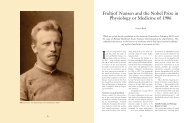
V.S. <strong>Deiters</strong> and R.W. GuilleryFigure 1. Portrait of <strong>Otto</strong> <strong>Deiters</strong> by August Bausch, 1858.doctoral thesis (<strong>Deiters</strong>, 2006) based largely on personalrecords kept in her family, and RWG wrote brieflyabout <strong>Otto</strong>’s work and life in three earlier papers. Onemarked the 100th anniversary of <strong>Otto</strong>’s death (<strong>Deiters</strong>and Guillery, 1964) and was written with his great aunt,who was a psychiatrist in D€usseldorf and the daughterof <strong>Otto</strong>’s Brother Hermann. The other two were aboutthe neuron doctrine that included comments on therelevance of <strong>Otto</strong>’s work (Guillery, 2005, 2007). Wecombine and extend these earlier studies now to markthe 150th anniversary of <strong>Otto</strong>’s death. He died showingsignificant promise of becoming one of the great neurohistologistsof his generation. Most of the material presentedin the early sections is based on VSD’s thesis,and the rest is largely based on <strong>Otto</strong>’s published work.EARLY LIFE AND EDUCATIONChildhood<strong>Otto</strong> <strong>Deiters</strong> was born on November 15, 1834, thesecond son of Franz Peter Ignaz <strong>Deiters</strong> and EmilieEleonore Henriette <strong>Deiters</strong> (born Bausch). The familytree in Figure 2 includes his siblings, two of which diedin their infancy; <strong>Otto</strong> and his younger brother Max diedbefore they were 30. The father was a professor in theLaw Faculty of Bonn University. He served five terms asDean of the Faculty and two (1845–1846 and 1856–1857) as Rector. The University, founded in 1818 andsmall by today’s standards, had many eminent facultymembers including von Helmholtz, Johannes M€uller, andEduard Pfl€uger, and students including TheodorSchwann (who followed M€uller to Berlin), HeinrichHeine, Prince Albert, and Karl Marx. Bonn was a smalltown of 35,000 3 inhabitants, first reached by the railwayin 1848. In that year Franz Peter <strong>Deiters</strong> served asan elected representative from Bonn to the FrankfurtNational Assembly, which attempted to establish a parliamentarydemocracy in Germany. 4 He served on theconstitutional committee of that parliament as a liberalmember. He died, aged 57, in 1861, leaving a financiallystrapped widow and five children still needing financialsupport: Hermann just 27, <strong>Otto</strong> at 26 strugglingto establish his professional status, a younger brotherMax, at 23 starting a career in mining, and the twoyoungest daughters, aged 15 and 19. 5The family was, like most others in the Rhineland,Catholic. It was a respectable and serious academicfamily, the children learning Latin and French at anearly age. 6 When he was 10, <strong>Otto</strong> wrote in German tohis parents: “Today on the first day of the New Year, Ifind myself particularly impelled (besonders angetrieben)to say to you in writing, how much I love you andmy heart thanks you for the manifest blessings(erzeigten Wohltaten). May the Almighty hear my prayerand grant you the proper blessings, for your great careand the many sacrifices you have made to give mepleasure and through a good upbringing make mehappy....” 7 This was a family dedicated to high academicachievement.While the father was at the Frankfurt parliament, hewrote to 15-year-old Hermann about some politicalissues Hermann had raised, advising him not to getinvolved with politics as they might keep him from hispresent duties (presumably school), and then addingthat he would like to hear more about what he and<strong>Otto</strong> did during the holidays, “...and at your next opportunityto communicate this also to <strong>Otto</strong>. If it is notoverly burdensome for <strong>Otto</strong>, with a definite topic inmind letter writing would not be as difficult as he3 https://commons.wikimedia.org/wiki/File:Bonn_population.svg. Thiscompares to Berlin’s 400,000 at the same time (Namier, 1992).4 This “revolutionary“ parliament included 49 university professors andlecturers and 57 schoolmasters among its more than 800 elected members,according to Namier (1992), who described the 1848 revolution asthe revolution of the intellectuals.5 We have no information about the reasons for the family’s financialproblems. They added to the pressures on <strong>Otto</strong> before his own death twoand a half years after his father’s and two years after that of his youngerbrother, Max.6 Hermann wrote accounts of his holidays in Latin for his father.7 Quotation marks here and subsequently show passages translatedfrom the German. Parentheses enclose original German words having possiblealternative meanings.1930 The Journal of Comparative Neurology | Research in Systems Neuroscience
<strong>Otto</strong> Friedrich Karl <strong>Deiters</strong> (1834–1863)Figure 2. Family tree of the immediate <strong>Deiters</strong> family.seems to believe. This would please me especially,although you can assure <strong>Otto</strong> that I have not taken hispast silence amiss, but have understood it.” The fatherwas aware of <strong>Otto</strong>’s withdrawn and unsociable nature,which figures significantly in the family correspondence,and one wonders how much of the father’s messageHermann passed on to <strong>Otto</strong>.The family was musical. <strong>Otto</strong> played violin and viola.He and Hermann organized a musical circle in 1852 thatmet every week and played classical music, mostly quartets,whose rules still survive (<strong>Deiters</strong>, 2006). The lastrule states that each member has the duty to practicewith others and if necessary on their own. Hermann wasmuch more sociable and outgoing than <strong>Otto</strong> and had alifelong interest in music; much of the correspondencebetween the brothers was about music or drama, completewith details of performances and critical comments,e.g., “Mr. Frank from Cologne played Beethoven’sC minor piano concerto, then a Bach fugue, which I havenever heard you play, and then a little charming compositionof his own...and then Frau Nissen Salomon sanga few charming Swedish songs and a quite abominablearia by Verdi tolerably, but a wonderful aria from AlessandroStradella very badly. She trilled and warbledfrightfully.” 8 This group of musicians continued to meetwhile <strong>Otto</strong> was in medical school and included a fellowmedical student, Carl Liebermeister (see below).Medical school at Bonn (1852–1857)In 1852 <strong>Otto</strong> graduated from his school with positivecomments from his teachers about his moral leadership,his diligence and his knowledge of the subjectsthat included, German, Latin, Greek, French, Hebrew,Religious studies, Mathematics, History and Geography,Physics and Philosophical Propaedeutics. He started inmedical school with no Biology or Chemistry, whichwould play a major role in his later studies. Althoughhis father and older brother studied humanities, he wasnot the only medical student in the family. A maternaluncle, Friedrich Bausch, had studied medicine at Bonnin 1832 and left a bound 400 page volume of his neatly8 A letter from <strong>Otto</strong> to Hermann, November 17, 1854.handwritten notes of the lectures that Johannes Muellerdelivered to the students that year 9 .In reaction to Hermann’s suggestion, that <strong>Otto</strong> wasnot establishing enough friendships, <strong>Otto</strong> wrote: “Youseem to think that I am one of those who invest all oftheir needed learning abilities in the courses and whohave a who knows what sort of a conscience if theyjust once, horribile dictum, cut classes for an hour.There you are mightily in error... as a medic one iscloser to the professors, and if in addition to that oneis the son of one’s father and the father happens to bea colleague of the professor, if, further, one is sittingwith a maximum of 3 to 6 colleagues, then you willunderstand that one can’t always do what one wants.”We have relatively little information about <strong>Otto</strong>’s yearsin medical school. As the above letter shows, he felt isolated.His later correspondence (see the next section)indicates that he still had to learn to focus his studieson the essentials. His musical activities continued, buthe worked through his studies rather mechanically, andhe wrote to Hermann in 1854: “I go to the college, thensit again in my room, go again to the college and thensit once more in my room, and that’s how it goes, fromone day to the next. There are no people that I am closeto, I have no friends. That’s bleak (trostlos), isn’t it?” Wehave very little information about this period of his life.He was living at home, was insecure about his studies,and left limited correspondence.<strong>Otto</strong>’s doctoral thesis, “De Incremento Musculorum”(<strong>Deiters</strong>, 1856), in Latin, was about the growth ofmuscles, documenting the sizes of many differentmuscles in many different species (human, ox, rabbit,dormouse, dove, several species of frog, salamanderand carp), comparing ages, and on the basis of themeans and what would today be recorded as variancesof the measurements concludes that muscle growth isdue to an increase of the volume of the individual musclefibers not to an increase in their numbers 10 . Histhesis advisor was Julius Budge.9 Now held by Bonn University.10 For non-Latin speakers <strong>Otto</strong> summarizes this study in <strong>Deiters</strong> (1861a),with regrets that these results are not well known.The Journal of Comparative Neurology | Research in Systems Neuroscience 1931
V.S. <strong>Deiters</strong> and R.W. GuilleryBERLIN: LIVING AWAY FROM HOME,MILITARY DUTY, AND LECTURES AT THECHARITE (1857–1858)<strong>Otto</strong>’s one year in Berlin was an important period inhis short life, significantly igniting his interest in medicalstudies. Although this was primarily his year as a volunteerin the army, it included attendance at lectures byVirchow, Johannes M€uller, and Romberg, and was theonly extended period of his life away from his family. Itstimulated him to start thinking seriously about his careergoals, and woke him to a wider view for his future.<strong>Otto</strong> served as a volunteer in the artillery of the 2ndGuards Regiment in Berlin. He had a low view of themilitary and for him this was primarily an opportunity tovisit Berlin and attend Virchow’s lectures at the recentlyestablished Charite. Writing to his younger brother Max,he evaluated his choices, basing them on how close tothe University the barracks were, how well educatedthe officers were, and the severity of the drill exercises.Of the artillery he wrote: “...when the regiment is onthe move the soldiers dawdle without a heavy pack andin no strict order behind the cannon,” contrasting thisto the soldiers of the infantry with heavy packs and instrict order. 11<strong>Otto</strong> went to Berlin two months ahead of his armyservice so he could attend Virchow’s lectures. Virchowhad written: “Only a few have advanced enough to havelearnt really to think in microscopical terms.... Formost older doctors, microscopy is like a foreign language,where one can freely use foreign words butthinks in one’s own language.” 12 <strong>Otto</strong>’s brief periodwith the students who were learning the “newlanguage” from Virchow would be strongly reinforced afterhis return to Bonn in 1858 by Max Schultze, whowas appointed to the Bonn Anatomy chair in 1859.Virchow had also written: “The natural scientist knowsthe body and the properties of the body, what isbeyond he calls transcendent and regards the transcendentas an error of the human mind.” <strong>Otto</strong> citedthis at the head of his dissertation.During this period in Berlin, <strong>Otto</strong> also attended thecomparative anatomy lectures of Johannes M€uller, andwrote to Hermann, “...everything that M says is significantand ingenious to the highest degree but in onesummer semester there is not time to go into the specialpoints, and this also applies to Virchow’s pathologicalanatomy. In both [fields] private studies areessential. Here microscopic anatomy is only taught byyounger staff (Privatdocenten). For my own studies one11 <strong>Otto</strong> to Max <strong>Deiters</strong>, May 18, 1858. This was before the reorganizationof the Prussian Army (see Grenville, 1976).12 Cited by <strong>Deiters</strong> (2006).thing is absolutely necessary. Requirement onemicroscope.” 13The father had previously advised <strong>Otto</strong> to borrow amicroscope, and <strong>Otto</strong> had told him this was impossible.The crucial issue at the time was the achromatic lens,which was produced by only a few companies, and providedmuch clearer views because these lenses correctedthe chromatic aberration characteristic of earlierlenses, which had brought each wavelength to a differentfocal point and introduced significant blurring of theimage, especially at higher magnifications. In July 1857the father advised <strong>Otto</strong> that Leitz had begun to producemicroscopes that were greatly in demand and told <strong>Otto</strong>not to delay in placing his order. It appears that the fatherfound funds, but did not live long enough to seethe extraordinary results <strong>Otto</strong> would produce.Here we encounter the sad financial position of the<strong>Deiters</strong> family. <strong>Otto</strong>’s father had previously written to acolleague requesting help to find a position for <strong>Otto</strong>, histalented but impecunious son. The father complainedabout his own financial position, and added “if you findhim [<strong>Otto</strong>] somewhat enclosed, restrained, reserved(verschwiegen), then I can tell you that I hear and havegrounds for believing that if this ring can be broken, agood core will be found” (<strong>Deiters</strong>, 2006). Not surprisingly,the letter produced nothing.The letters from <strong>Otto</strong> in Berlin to his brothers (citedmore fully in <strong>Deiters</strong>, 2006) are entertaining and includecritiques of (good and bad) musical performances inBerlin, showing that he was more aware of others thanmany of his later actions and his views of himself mightsuggest. He writes about the Tiergarten, implicitly comparingcity promenades with the real walks possible inthe Rhineland. The Tiergarten walks are spoilt by themany typical Berliner family outings: “...maids withtheir cousins and children, old women in Amazon hats(Grisetten in Amazonh€uten), 14 careful parents with theirmarriageable daughters...arrive at one of the manyinns, they all sit, the whole Berliner hoi-polloi (Philisterium),each family at one table in the middle of which atowering glass of Weissbier (eine Weisse) is enthroned,and then with dignified demeanour the honoured familyfather takes a draught whereupon the others follow inorder (secundum ordinensis). Such a thing is only possiblein Berlin....”13 <strong>Otto</strong> to Hermann, May 26, 1857.14 A stylish hat. A poem appeared in 1857 entitled “ Der Amazonenhut—Mit achtzehn Jahren wohlgetan—mit zweiundzwanzig geht’s noch an—Mitdreißig Jahr’ bewahr’ uns Gott!—Mit sechsunddreißig—Kinderspott!“ See“Eisenbergisches Nachrichtsblatt 1857 at http://zs.thulb.uni-jena.de/receive/jportal_jparticle_00081264. In English: The Amazon hat: At 18 itis well done—at 22 just possible—at 30, ’God preserve us’—and at 36 thechildren mock.1932 The Journal of Comparative Neurology | Research in Systems Neuroscience
<strong>Otto</strong> Friedrich Karl <strong>Deiters</strong> (1834–1863)<strong>Otto</strong>’s Berlin letters also tell us more about familyrelationships: “Papa’s letter has arrived safely. Thesomewhat unworthy (unw€urdige) title, junior doctor, canbe left out of the address.” Another letter, in which<strong>Otto</strong> replies to Hermann’s request that <strong>Otto</strong> look in Berlinfor a suitable silver wedding gift for the parents, providesa close domestic view: “We must respect theprinciple of usefulness so valued in our home.” Suchitems as pictures or valuable rings are not to be considered.He discusses a clock for the hall or some comfortablechairs, which may be needed, but advises thatthey can be purchased in Bonn or Cologne as well asBerlin. We do not know whether the family gained thecomfortable armchairs they lacked or the clock thatwould keep them all on time.In 1857 <strong>Otto</strong> also began to act as informal medicaladviser to Hermann, sounding authoritative, dogmatic,and uncompromising. On the basis of Hermann’saccount of his indisposition, <strong>Otto</strong> had diagnosed a gastricand intestinal problem, “perhaps augmented, so faras I can see, by a gallbladder problem...you think thatin spite of Albers’s 15 energetic treatment you are notcompletely cured. I can believe that. These conditionsusually go away on their own without any energetictreatment...above all you must not be thinking aboutyourself as having an unhealthy nature. Our family hasa healthy nature.” <strong>Otto</strong> goes on to advise Hermann tolive a less sedentary life, take more walks, exercise,and drink less tea and coffee. He sounds stern, not likea younger brother. Hermann must have replied byreporting that he was better, and complaining that hewas not ready to follow <strong>Otto</strong>’s rather rigorous rules,because <strong>Otto</strong>’s next letter expresses pleasure at theimprovement and hopes it will last: “You consider thecorrect dietary regime to be of no consequence, and Icould hardly expect anything different, but that mustnot be....” Another harangue about healthy diet andexercise follows complete with a statement aboutpatients who fail to follow the doctor’s advice. But heends on a peace-making note, saying, “I did not mean,as you seem to have assumed...that you should at thispoint give up your social life and follow my badexample.”Although <strong>Otto</strong> was a private, self-enclosed personand was recognized as such by family and colleagues,he had some close friends, including his fellow musicians,particularly Carl Liebermeister, 16 with whom hecorresponded after they graduated. The letters fromLiebermeister contain advice about examiners at other15 Another Bonn faculty member in the pathology department.16 Later Professor of Pathology and Therapy in Basel from 1865 and inT€ubingen from 1871 (<strong>Deiters</strong>, 2006).universities. From Greifswald, where he was studying,he wrote: “...he who knows his subject reasonably wellis better off going to Greifswald, but he who knowsnothing could fake his way in Berlin....You could, witha clear conscience, advise people to go to Greifswald.”He invited <strong>Otto</strong> to visit him in Greifswald, and a laterletter to <strong>Otto</strong> from N. Simrock, school friend and fellowmedical student, includes a statement that he was gladto hear that the two had met: “You both always followedthe same academic tendencies.” Later, <strong>Otto</strong> visitedT€ubingen while Liebermeister was there asprosector in Pathological Anatomy.While he was in Berlin, <strong>Otto</strong> began to think seriouslyabout his future. He wrote a thoughtful letter to Hermann:“I have always worked hard, though not as hardas people said. I had to, because quite apart from theamount of material that had to be mastered, which wasoverpowering, the time that was available to me becauseof my reserved nature had to be occupied. There was alack of distractions. So at the beginning this work wasnot of the right sort.” He now sees more specialist areasthat he had no time to review properly as a student. “Idon’t think that I judge my strengths too highly but Idon’t want to estimate them too lowly.... The position atpresent is as follows: I no longer doubt that I would preferan academic career to one in a practice...this fitsbest with my strengths. So I will be a docent, no matterwhat.” He then asks about the long-term possibilities ofan academic career and about his choices. He is clearabout these: on the one hand, anatomy, particularly microscopicand comparative anatomy, and on the other,pathological anatomy. He considers an assistant positionwith Geheimrat Naumann in Bonn as a docent in pathologicalanatomy, and finding a medical practice thatwould later provide extra income (Sp€ater die n€othigenBed€urfnisse deckte). However, he worries about income,and sees a Professor of Pathological Anatomy as a “fifthwheel,” not making much money, while a serious clinicalcommitment would limit his plans for academic work. Hereflects negatively about the position of a doctor in aGerman practice: “If I don’t settle in the first best villageand don’t go to America, then I would risk as little byplanning on a simple academic career as I would byexpecting a remunerative practice: particularly so as thefield to which I always return has many income possibilities.And that field...is anatomy...and I have decided todedicate myself to it.” He lists the income opportunities:publications generated income then, as did private tutorials,which were more in demand as exams increased indifficulty.He decided nonetheless to maintain his clinical skills,and in Berlin attended the clinical sessions presentedby Romberg and Langenbeck.The Journal of Comparative Neurology | Research in Systems Neuroscience 1933
V.S. <strong>Deiters</strong> and R.W. GuilleryN. Simrock, who had moved to Cologne, compared<strong>Otto</strong>’s enthusiasm about academic opportunities withhis own lack of academic ambition. He was glad to bein Cologne, he wrote, where “...a zealous academicengagement is impossible...I know for you it is different,and even if your well-known unassuming modestystops you from recognizing it, it will later be a benefitfor the faculty that has the good luck to count youamong its young chosen ones.”In another letter, before <strong>Otto</strong> moved back to Bonn,Simrock wrote about <strong>Otto</strong>’s planned appointment withNaumann: “That you have accepted the position withNaumann not only does not surprise me, but, on thecontrary, I think it totally understandable, since in sucha position you can soonest reach your goals. Only Iwould...in relation to him put myself in an independentposition, which is something he may assume.” HereSimrock anticipates later problems for <strong>Otto</strong>’s career,based on the differing approaches to medicine of <strong>Otto</strong>and Naumann: the former scientific, the latter philosophicaland romantic.In 1858 <strong>Otto</strong> completed his military service andreturned to Bonn as first assistant to Geheimrat Naumann,spending the next three years with him.MEDICAL CAREER IN BONN (1858–1863)Clinical work and publicationsIn Bonn <strong>Otto</strong> took on clinical responsibilities in Naumann’sclinic and also started private clinical work as ageneral practitioner, visiting patients in Bonn and surroundingareas 17 to boost his income. He also began towork on his habilitation, which would qualify him as alecturer at the University. He must also at an earlystage have begun detailed investigations of the innerear in several different species, which led to four publicationsby 1862 (see the section, The organ of Cortiand <strong>Deiters</strong> cells, below).In 1859 he published a brief account of two cases ofscarlet fever (<strong>Deiters</strong>, 1859a) and later a longer articleabout leukemia (<strong>Deiters</strong>, 1861a). The former stressedthat current treatments were generally ineffective andshowed that incidence of the infection could not berelated to particular home conditions of the patients;the latter presented a detailed account of the physical(wasted) condition of a terminally ill young man whodied in the hospital. <strong>Otto</strong> described the postmortemfindings, the major features being lymphatic swelling ofthe peritoneum, in which he noted hypertrophic cellsand abnormal blood cells. He stressed how little wasknown about the disease. From our point of view, his17 He advised the patients on the local farms to not drink fresh milk, buthis advice went largely unheeded.rigorous demand for convincing evidence before comingto any conclusions about a disease is the most noteworthypart of both accounts.Habilitation<strong>Otto</strong> wrote an essay on the present position of thecell theory (<strong>Deiters</strong>, 1859b). It has a strange beginning:“Half a century has passed since the sharp eyes of agenial French investigator succeeded in winning aninsight for academic anatomy that until then was completelystrange and for which hardly anyone else wassuited....” The long, complex sentence continues, butXavier Bichat, who is often regarded as the father ofhistology (although he never used a microscope), is notidentified as the sharp-eyed French investigator until<strong>Deiters</strong> had introduced many others (Leeuwenhoek,Malpighi, Linnaeus, and Cuvier) and compared theirefforts at categorizations in biology with the successesof chemists, Lavoisier in particular. In this long,“scholarly” review, <strong>Otto</strong> covered much of the relevantliterature, although the version that was published as alecture lacked a list of references and looks as thoughit was prepared in a hurry, probably because <strong>Otto</strong>needed the qualification to start giving lectures thatwould bring in some fees. Although it is now consideredthat the cell theory was established earlier by Schleidenand Schwann, when <strong>Otto</strong> wrote, there was significantdisagreement about exactly what the cell theoryclaimed and how far Schwann’s account was an acceptableview of how tissues are organized and develop (forfuller historical accounts, see Baker, 1948, 1949; Harris,1999). <strong>Otto</strong> had started to look at the inner ear andhad noticed some cells that had a large hole, whichhad pushed the cytoplasm, nucleus, and nucleolus to asmall border region. For reasons not made clear, <strong>Otto</strong>found it difficult to conceive of these fenestrated cellsas bound by a membrane. He introduced these cells aspresenting one of several difficulties faced by the celltheory at that time. Other difficulties included cellslacking a nucleus and cells lacking a nucleolus. Thelack of a membrane long remained a difficulty until theelectron microscope allowed clear identification of thethin, approximately 5-nm cell membrane. 18 <strong>Otto</strong> is lookingfor a strong theory based on the physics and chemistryof the cell, one that covers not only the anatomyof the cell but also the physiology. Even the strongest,most basic part of the cell theory, that cells only arisefrom other cells, he considers not yet sufficiently well18 The 1948 edition of Maximow and Bloom’s A Textbook of Histology,predating the use of electron microscopes in biology, relied on the semipermeablenature of the membrane and its resistant and elastic propertiesdemonstrable by microdissection as the major evidence for the existenceof a cell membrane.1934 The Journal of Comparative Neurology | Research in Systems Neuroscience
<strong>Otto</strong> Friedrich Karl <strong>Deiters</strong> (1834–1863)established, in spite of mounting evidence, which hesummarizes. The problem of exactly what the celltheory does include was not resolved at the time andmay even today not be clearly agreed. This is an issuethat plays an important role in <strong>Otto</strong>’s later work and isrevisited in the section on <strong>Otto</strong>’s book, below.EARLY PUBLICATIONS IN ANATOMYThe organ of Corti and <strong>Deiters</strong> cellsIn 1859, soon after returning to Bonn, <strong>Otto</strong> publishedan important study of the organ of Corti (<strong>Deiters</strong>,1859c) that included a detailed account of the cellsthat, at Max Schultze’s suggestion, were later named<strong>Deiters</strong> cells. These cells support the outer hair cellsand provide a link between these hair cells and thebasilar membrane. Between 1859 and 1862, <strong>Otto</strong> alsopublished a book (<strong>Deiters</strong>, 1860a) and two other paperson the inner ear (<strong>Deiters</strong>, 1860b, 1862). These arebased partially on sectioned material, but primarily relyon preparations that were teased out under the microscope.<strong>Otto</strong> compared the structures, based on theirflexibility or fragility, watching how they reacted to variouschemicals serving as fixatives (dichromates, chromates,or acetic acid) or dyes (iodine, carmine). Thesemethods were widely used by early microscopists. Thepractice of first fixing a piece of tissue and thenembedding and sectioning it before ever looking at itdeveloped later. The method that <strong>Otto</strong> used is relevantfor all of <strong>Otto</strong>’s microscopic studies. He was watchingthe reactions of tissues to his chemical and physicalmanipulations under the microscope in order to reachhis conclusions about their characteristics. We know nodetails about the microscope or the lenses that <strong>Otto</strong>used, and have no information about the “tools” that heused (probably needles, fine pipettes, delicately shapedglass rods, perhaps even fine, hand-sharpened knives,etc.).It is important to recognize that for this work and forhis later microscopic studies of nerve and glial cells,<strong>Otto</strong>’s tissues were generally in an aqueous solution ashe was manipulating them. They would not have beencleared in a medium of appropriate refractive index norwould he have been able to manipulate the tissue hadthey been covered by a coverslip. 19 Water immersionlenses were available in the late 1850s and early1860s, 20 but we have no information about the lensesthat <strong>Otto</strong> used. It is possible that <strong>Otto</strong> did some of hisfine dissections with a water immersion lens, but thedifficulties of manipulating small objects in conditions19 However, Schultze in the introduction to <strong>Otto</strong>’s posthumous bookmentions many microscopic preparations that <strong>Otto</strong> left behind, preservedand cleared in balsam and covered.20 See http://www.smecc.org/history_of_oil_immersion_lenses.htm.Figure 3. A <strong>Deiters</strong> cell shown schematically. The figure is basedon a scanning electron micrograph (Fig. 6B of Parsa et al., 2012).The cell stretches between the reticular membrane (above) andthe basilar membrane (below), and the stereocilia of the hair cells(not shown) project above the reticular membrane. Note that inthe original figure there are fine nerve fibers that run horizontallyacross the <strong>Deiters</strong> cells. These nerve fibers are in part oftenenclosed within the cytoplasm of the <strong>Deiters</strong> cell and are notshown, nor are the rich terminals that lie deep within the regionwhere the <strong>Deiters</strong> cell encloses the base of the hair cell. Scalebar 5 20 lm.where surface tension effects can make the manipulationsdifficult (or impossible) have to be recognized. Forthese studies of the inner ear (see Figs. 3 and 4) andfor his later studies of neurons and glia (e.g., PlateODU II, Fig. 10), he produced extraordinary details ofvery small cells, often unmatched at the time, but thespecial tools, methods, or skills that he used are notknown to us.<strong>Otto</strong>’s first study of the organ of Corti (<strong>Deiters</strong>,1859a) was based on material from dog, cat, and calf.It is extensively illustrated by <strong>Otto</strong>’s own drawings. <strong>Otto</strong>criticizes K€olliker’s earlier account of nerve fibers endingin the large “cells of Claudius,” describing this as aphysiological impossibility 21 but focuses on the structuresthat link the hair cells to the reticular lamina andthe basilar membrane. Figure 3 shows the structure ofa <strong>Deiters</strong> cell as seen in a current electron microscopestudy for comparison with <strong>Otto</strong>’s figure (Fig. 4), which21 Presumably because the body of these nerves is in the spiral ganglioncells. Here <strong>Otto</strong> is possibly suggesting, but not exploring, an objection toa fusion of two cells. K€olliker in the 1863 edition of his textbook withdrewthis suggestion on the basis of newer developmental studies but withoutmentioning <strong>Deiters</strong>.The Journal of Comparative Neurology | Research in Systems Neuroscience 1935
V.S. <strong>Deiters</strong> and R.W. GuilleryFigure 4. <strong>Otto</strong>’s drawings of the cells now called <strong>Deiters</strong> cells (from <strong>Deiters</strong>, 1859c.) A: Figure 9 in the original publication: a, the nerve; b, themidportion of the <strong>Deiters</strong> cell; c, the region where the phalangeal process, e, joins the body of the <strong>Deiters</strong> cell (marked by a shiny circle); d,the body of the <strong>Deiters</strong> cell; f, the connection for the hair cell. (Note that d and f are left unidentified in the original). B: Figures 2 and 3 in theoriginal publication. These show several views of the <strong>Deiters</strong> cells, some with the foot attached (2a, b, 3, b, f, and the others with some or allof the lower limb missing). (We thank Colin Beesley for the photographs used as Figs. 4 and 6, and for Plates ODU I–VI.)illustrates some of the major features of the cells thatnow bear his name. <strong>Otto</strong> said nothing about the closerelationships between these cells and the nerve fibersthat are now known to be embedded in the cytoplasmof the <strong>Deiters</strong> cell as the fibers pass to the hair cells(Parsa et al., 2012). He describes the “phalangeal processes”of the cells (Figs. 3 and 4A,B), looking like tinyfinger bones, extending the <strong>Deiters</strong> cells across thewhole of the organ of Corti, and he shows how theyrelate to the hair cells in this and the later book 22 (<strong>Deiters</strong>,1860a). He establishes the cells as separate elements,having their own nucleus, attaching to thebasilar membrane and also connecting to the hair cellsand to the reticular membrane. Today they are recognizedas serving to transmit changes in the shape andlength of the outer hair cells to the basilar membrane(Fig. 3). The changes are induced by electrical changesproduced by movement of the stereocilia of the haircells and serve to amplify the auditory signals (see Ashmore,2008; Nam and Fettiplace, 2010 for clearly demonstrateddetails 23 ).The longer book on the cochlea (<strong>Deiters</strong>, 1860a),according to <strong>Otto</strong>’s foreword, had to be written becausea recently published article by Boettcher (1859), which22 This book is available on the Web with copies of the figures in whichit is often impossible to identify fine details. The availability of the bookon line makes librarians reluctant to find it.23 It is of interest that Ashmore’s review opens with a brief summary of Helmholtz’swork on hearing between 1862 and 1877 that was based on contemporarystudies of the anatomy of the organ of Corti. (Our thanks to Donata Oertelfor an emailed tutorial on the functions of the outer hair cells.)1936 The Journal of Comparative Neurology | Research in Systems Neuroscience
<strong>Otto</strong> Friedrich Karl <strong>Deiters</strong> (1834–1863)contained “next to some correct results, a number ofincomplete and mistaken results...made it my duty notto wait any longer with my own results.” 24 The book isbased on studies of rabbit, mouse, cat, dog, calf, horse,human, and guinea pig. Two further studies (<strong>Deiters</strong>,1860b, 1862) extended his material to birds (starling,yellow hammer, hen, pigeon, crow, magpie, two speciesof lizard, slow-worms, grass-snakes, and probably morethan one species of frog.) Reading these studies today,or K€olliker’s 1863 account, and comparing them withwhat is now known, shows how much our knowledgehas advanced and how difficult it was in the 1860s totrace nerve fibers when they lie very close to or areenclosed in the cytoplasm of the supporting cells.Studies of striated muscles (<strong>Deiters</strong>,1861b) 25This was a continuation of his thesis on muscles,using developmental studies of muscle growth in theregenerating tip of a tadpole tail. The surviving musclefibers grow but do not contribute to the regeneratedtail. New cells form from stellate or bipolar connectivetissue cells, and these develop a striated fiber alongone border. He draws these finely striated fibers lyingnext to the cells, and parts of the fibers appearing toseparate from the cells, with the fibers then growingbeyond the cells. He regards the striated fibers producedin this way as an intercellular component that issubsequently incorporated together with the cell in acommon membrane, to make a new multinucleate musclefiber. He mentions that J. Budge disagreed with<strong>Otto</strong>’s conclusions and has agreed that <strong>Otto</strong> shouldinclude a discussion of their different views, which hedoes. Here <strong>Otto</strong> is more than usually deferential to hisformer thesis adviser, who later became his friend.TEACHING, LEAVING CLINICAL WORK,AND STARTING TO STUDY THE CENTRALNERVOUS SYSTEMTeachingIn April 1859 <strong>Otto</strong> was granted the position of Privatdocent.In addition to continuing with his clinicalresponsibilities in the hospital, he worked with his privatepatients and started to offer lectures in the medicalschool (Table 1). The lectures on auscultation were24 Boettcher’s angry reaction (Boettcher, 1860) and <strong>Otto</strong>’s four-pageresponse (<strong>Deiters</strong>, 1860c) will not be explored here, but they representthe reactions that <strong>Otto</strong>’s lack of tact could generate. We have not beenable to address issues of priorities that arise from this interchange.25 In the same year Max Schultze (1961) published a paper on muscle,arguing that each nucleus of the multinucleate muscle cell represents adistinct cell not separated from the other nuclei (cells) by any membrane.Schultze’s account would have caused <strong>Otto</strong> some further problems had helived, and should be seen in relation to <strong>Otto</strong>’s interpretation in this study.a novelty in Bonn at the time. Although <strong>Otto</strong> was popularamong the students, his occasional disrespectfulcomments about colleagues, whose approach to medicinehe considered less modern and objective than hisown, damaged his relationships with some faculty colleaguesand would cost him dearly before long.A family crisis; leaving clinical workIn March of 1861, the death of <strong>Otto</strong>’s father followinga stroke added a significant extra burden to <strong>Otto</strong>’slife, as did the death of his younger brother Max, 8months later, under circumstances that leave suicide apossibility (<strong>Deiters</strong>, 2006). This left Hermann and <strong>Otto</strong>with the sole responsibility for supporting the family,which had to move into cheaper accommodations. Theyoungest daughter, Paula, started training as a teacherin order to generate her own income, with <strong>Otto</strong> writingthe health certificate that supported her application. Atthis time <strong>Otto</strong> also started to add microscopic reportsfor colleagues to his own earnings and timecommitments.On May 28, 1861, he wrote a letter to a professor,unidentified on the surviving draft 26 and unknown to us,calling on the goodwill the professor had often shown<strong>Otto</strong> in the past. He reported the father’s death andthe family’s limited financial circumstances. He mentionedhis present position (Assistant in the MedicalClinic and Privatdocent in Anatomy) as holding nopromise for the future and leaving only limited opportunitiesfor scientific work. If he were to continue inBonn, he would be forced by financial considerations tosacrifice his scientific work for practical medical activities.He asked for help in finding a suitable positionand recalled that in the past the professor had indicatedan inclination to find a position for <strong>Otto</strong> inHeidelberg.We have no record of any answer from Heidelberg.<strong>Otto</strong>’s views about his future in Bonn represented acorrect assessment of relationships there. In spite ofthe family’s needs, in June <strong>Otto</strong> gave notice to Naumannthat in view of his changed family circumstanceshe would be leaving his Clinical Assistant position inthe summer in order to concentrate on research. Wedo not know the reasons for this decision, but subsequentevents suggest that he knew his microscopicstudies were promising a rich harvest and that he hadto concentrate on them. <strong>Otto</strong> and his chief had differentapproaches to medicine—modern and empirical versusolder and more intuitive. The two did not relatewell, as anticipated in Simrock’s earlier letter. <strong>Otto</strong>26 Possibly Helmholtz, who had been a faculty colleague of <strong>Otto</strong>’s fatheruntil 1858, and who was in Heidelberg in 1861.The Journal of Comparative Neurology | Research in Systems Neuroscience 1937
V.S. <strong>Deiters</strong> and R.W. GuilleryTABLE 1.Details of Lectures Delivered by <strong>Otto</strong> <strong>Deiters</strong>, 1859–1862 * (Numbers on the right indicate attendance)Summer ’59 Microscopic anatomy of sense organs (revision) 11Winter ’59-60 Microscopic anatomy (revision) 6Summer ’60 Microscopic anatomy of the sense organs 21Pathological anatomy. Announced but withdrawn before registration due to demands on available time.Winter ’60-61 Microscopic anatomy 7Summer ’61Course in physical/chemical diagnosis 8Selected chapters of pathological histology (not delivered)Physical diagnosis 9Winter ’61-’62 About neoplasms 5Auscultation and percussion 0.6Microscopic anatomy, announced but not given.*Numbers on the right indicate attendance.received a brief response from Naumann asking him tocontinue until September, allowing time for a replacement.In reply, on June 21, <strong>Otto</strong>, clearly offended bythis brief note, wrote a formal reply: A verbal discussionof his motives for leaving was no longer desirable: “Ihave had to conclude from your note that you, fromyour position, find my leaving not only understandablebut desirable.” He writes about leaving his positionunder conditions of demonstrable mistrust (entschiedenenMistrauensbeweisen), a position that should nothave affected his scientific studies at all, to which hehas probably given more time and effort than most ofhis predecessors and probably more than he, with aview to his main (professional) aims, could spare andmore than he should have demanded of his own health.It is a bitter and tactless letter that ends with an offerto continue until the end of August, and “...in recognitionof what I have achieved in addition to the strictduties for the clinic, not the least difficulties should beraised for this termination date (in den Weg gelegt werdenwird).” Later documents suggest that <strong>Otto</strong> left inearly October.Problems about promotionMax Schultze had moved from Halle to the chair ofAnatomy (Ordinarius) in Bonn in 1859, and when <strong>Otto</strong>gave up his clinical position in order to concentrate onresearch, Schultze, recognizing <strong>Otto</strong>’s abilities and his seriouscommitment, tried to find a position for <strong>Otto</strong> in Bonn.Early in 1862 Baron de la Valette was suggested as a candidatefor a prosector position in Anatomy, and Schultzeproposed that such a position should also be offered to<strong>Otto</strong>. The discussions were protracted (langwierig), as thedivided faculty discussed the merits of this suggestion.Eduard F.W. Pfl€uger, a student of Johannes M€uller,who had been appointed to the Bonn chair in Physiologyin 1859, summarized the position as follows: “Theproposal that the faculty make the following nominationas Professor Extraordinarius is an honorary signal(Bezeugung) that can have no significant meaning forthe teaching or scientific purposes of the University. Ifsuch a position is offered to Baron la Valette then it isarguable that Dr. <strong>Deiters</strong> should share this position,particularly since his (<strong>Otto</strong>’s) misfortune just nowurgently presses on us to act in fairness.” He goes onto say that while all are agreed on this point, some facultymembers feel that the best outcome for the twocandidates would be obtained from the Ministry if theposition were offered to the Baron and <strong>Otto</strong> were nominatedfor another University. However, there were nopositions available in other Prussian universities, and ifthere had been, then other, older, equally suitable candidateswould qualify. Pfl€uger and Schultze left nodoubt about <strong>Otto</strong>’s higher scholarly qualifications.Carl Wutzer, former chief of surgery, but still active,spoke positively about <strong>Otto</strong>’s activities and personality,sharing Schultze’s positive views, but pointed out that if<strong>Otto</strong> were appointed, there would be no money for him.“We have no funds for scholarship and the arts(Kunst).” The vote went five to three against <strong>Otto</strong> (see<strong>Deiters</strong>, 2006 for details) in favor of de la Valette. Still,the issue was not settled. Pfl€uger presented strongarguments in favor of nominating both candidates, andNaumann added detailed comments about his careful(vorsichtiges) relationship with <strong>Otto</strong>, indicating that,yes, there had been misunderstandings, and that hehad supported and encouraged <strong>Otto</strong>’s studies and hadoften had to attend the clinic in place of <strong>Otto</strong>. At thetime of the father’s death he had made efforts toincrease <strong>Otto</strong>’s income from available clinical funds. Forthree-fourths of 1861 <strong>Otto</strong>’s income had beenincreased by 325 thaler. 27 Although he (Naumann) hadbeen ready to be helpful, he was surprised when <strong>Otto</strong>27 The melt value of a silver thaler today is just over 30 US$, and thevalue of silver per ounce, which was 1 US$ in 1860, is now around 17US$, seeming to leave the thaler worth a little less than a dollar today.There are more realistic ways of making such comparisons (see (http://authors.library.caltech.edu/13155/1/HOFjeh02.pdf and also http://www.roiw.org/5/4.pdf), but they will not be explored here. It seems clearthat at a time of significant family hardship <strong>Otto</strong> was giving up a nontrivialpart of his income in order to focus on his research.1938 The Journal of Comparative Neurology | Research in Systems Neuroscience
<strong>Otto</strong> Friedrich Karl <strong>Deiters</strong> (1834–1863)suddenly announced that in “consideration of his familyand his partiality (Vorliebe) for microscopy,” he couldnot stay with the clinic; Naumann had pointed out to<strong>Otto</strong> that his continuation in the clinic would bring anannual income of 250 thaler. “I had to let him go. I wassorry because I had got used to his friendly face andmodest ways.”After an unusually long debate, 28 it was decided tosend both names to the Minister, with strong recommendationswritten for both by Schultze. The Ministeraccepted the nomination of Baron de la Valette, so faras we can tell without a salary, and rejected that of Dr.<strong>Otto</strong> <strong>Deiters</strong>. <strong>Otto</strong> himself was and would remain bitterabout this decision during the brief and busy last twoyears of his life. In 1874, Baron de la Valette succeededMax Schultze as head of the Anatomy Department,retiring in 1906. Today the Anatomy Departmenthas a bust of the Baron in the entrance hall but nothingto memorialize <strong>Otto</strong> (Fig. 5). 29The death of Max <strong>Deiters</strong>Max <strong>Deiters</strong>, three years younger than <strong>Otto</strong>, hadstarted training as a mining engineer/inspector. In November1861, he sent a copy of his final written work(thesis?) to his uncle who worked in mining and to a fellowstudent. The latter replied with a strong negativecomment about their current career prospects. Earlier<strong>Otto</strong> had tried to persuade Max to change fields, writing:“Your subject may be a primarily practical one, but inthe two years you have to spend in practical work, it isnot useful to waste so much time stone-bashing (Steineklopfen).Since you are interested in natural history youcould spend your time more usefully.” Max wrote to hisuncle about the limited career opportunities: there wereserious doubts about a career in mining, he still wantedto complete his exams but was not eligible for another18 months, a medical training was too long to be affordable,so he was thinking of training as a science teacher.He was a lost young man, dependent on his older brothersfor support. He died three days after writing this letter,and a letter from <strong>Otto</strong> to the local chaplain aboutthe site of Max’s grave suggests that it was a suicide.UNTERSUCHUNGEN €UBER GEHIRN UNDR€UCKENMARK DES MENSCHEN UND DERS€AUGETHIREProduction of the book, and the preface<strong>Otto</strong> spent the next two years feverishly working onmaterial for this book. His death, due to Typhus, just overtwo years after Max’s, left the work incomplete. However,the book now represents <strong>Otto</strong>’s major contribution toneuroanatomy because Max Schultze and Hermann <strong>Deiters</strong>were able to prepare <strong>Otto</strong>’s notes for the printers.Schultze’s preface (pp. V–XVII) describes the exactingand intense work that <strong>Otto</strong> had been doing for thetwo to three years before his death: preparing a textbookof neuroanatomy, starting with an account of thenerve cells, supporting cells, and nerve fibers, thendescribing sections of the spinal cord and brainstem,and aiming beyond to observations of cerebellum andcerebrum. <strong>Otto</strong> left behind several unusually fine largefigures (Plates ODU I-ODU V) that he had occasionallyshown to visitors. Schultze mentions hundreds of microscopicpreparations, many preserved in balsam, that<strong>Otto</strong> used in his lectures. 30 His aims were private, andhis methods were difficult, owing much to those ofSchultze himself. Schultze describes <strong>Otto</strong> as reservedand uncommunicative, providing colleagues with onlyoccasional, scattered information. Schultze wrote“...anyone who knew <strong>Deiters</strong>’s earlier publications onmicroscopic anatomy, who knew the perseverance andtenacity with which he aimed at a once established(gestecktes) goal, had to conclude that the difficulty ofthe chosen methods raised the expectation of significantresults. That <strong>Deiters</strong> over the several years of thiswork had won such results was rumoured, here andthere, but as private (verschlossen) as he was, hehardly spoke about his discoveries, and gave no lecturesabout his investigations to academic societies.”<strong>Otto</strong>’s notes were rough. Schultze writes that <strong>Otto</strong>relied greatly on his memory and describes the notesas a brouillon (rough draft) written in a careless hand“with flying quill.” Much of the microscopic materialwas drawn but without explanations. <strong>Otto</strong> had, duringthe last few months of his life, taught a private courseto two advanced students, and one showed somesketches of the material to Schultze. These and the students’notes gave Schultze some sense of what the materialcontained. Unfortunately, the notes and theoriginal drawings are lost and we know nothing aboutthe originals, other than Schultze’s account and theillustrations in the book itself (Plates ODU I-ODU VI)Schultze worked for several months with Hermann<strong>Deiters</strong>, reworking the original difficult and rough notesas a printable manuscript. Hermann, who over severalyears of correspondence must have learnt to decipher<strong>Otto</strong>’s writing, was a teacher of classical languages,with a strong interest in music, who would later makesignificant contributions to the history of music. 31 For28 More of the debate is recorded in <strong>Deiters</strong> (2006).29 There are current plans to dedicate a lecture theatre to <strong>Otto</strong>.30 They could not be traced when we enquired.31 His portrait can be seen in the Beethoven Haus in Bonn.The Journal of Comparative Neurology | Research in Systems Neuroscience 1939
V.S. <strong>Deiters</strong> and R.W. GuilleryFigure 5. <strong>Otto</strong> <strong>Deiters</strong> toward the end of his life, painted by A. Bausch.(We thank Dr. Wolfgang G. <strong>Deiters</strong> for supplying the photograph.)Hermann the technical neuroterminology must havebeen foreign, but one has the impression that, althoughevidence of the brouillon remains, the man of lettersand the teacher may have introduced a degree of order,less obvious in <strong>Otto</strong>’s earlier work.Without this significant effort of Max Schultze andHermann <strong>Deiters</strong>, the book would not exist. We wouldthen still hold <strong>Deiters</strong> in high regard for his earlier publications,but the view provided by the book of a deeplythoughtful and skilful young scientist arguing (with himself)about conclusions to draw from his carefully preparedand studied material would be lost to us, aswould the details of his new observations.For us the book provides a vivid view of the uncertaintiesthat prevailed 150 years ago, upon which manyof our current “certainties” have been built in innumerable,mostly small, steps. We lack the space and the historicalsense to present the material in relation to whatwas known at the time. <strong>Otto</strong> was remarkably bad at citingothers; when he did, it was usually cursory, mentionof a name, or occasionally a journal title. 32A sense of the then contemporary relevant knowledgeis best obtained from the 3rd, 4th, and 5th editionsof K€olliker’s Handbuch (K€olliker, 1859, 1863,1867). Van Der Loos (1967) and Shepherd (1971) providemany relevant details concerning the nerve cellsand Meyer (1971), Clarke and O’Malley (1996), and Finger(1994) about the brain more generally.32 This was typical of his published work as well.Purkinje (1838; cited by Finger, 1994) had describedthe cerebellar cells that bear his name and illustratedthe basal parts of their dendrites; Wagner (1845, 1847)had illustrated the dendrites extending some greater distancefrom the cell body of sympathetic cells and cellsin the electric organ of fish (Torpedo); Remak (1855) haddescribed the several processes (dendrites) that leavethe large neuronal cell body of the ventral horn cells,and had traced one single, distinctive process (the axon)into the ventral root. Although Brown Sequard (see Finger,1994) had demonstrated the crossing of the pyramidaltract and of some of the sensory spinal pathways, ithad not yet been possible to trace the sensory pathwaysthrough their relay stations in the brainstem. Tracingnerve fibers from their cells of origin into their peripheralcranial nerves was still extremely difficult, and theresults were often conflicting. Although the demonstrationby Bell and Magendie (Finger, 1994) that the spinaldorsal roots are sensory and the ventral roots motorwas widely accepted, the connections that might providea pathway for the spinal reflex remained undefined. Thecell theory was still the subject of significant debate andmisunderstanding (Baker, 1949; Harris, 1999).We present <strong>Otto</strong>’s book under several different headings,not in order of the chapters, but summarizingwhat appear to us as the most interesting points forreaders of The Journal of Comparative Neurology.On page XI of the preface, Schultze lists novel observationsincluded in the book: conspicuously the identificationof the superior olive in the human brain,previously only reported in animals since it is visible inmany mammals, but is hidden in the human brain bythe large pons; also the identification of a large-celledgray nucleus between the origin of the inferior cerebellarpeduncles that Schultze suggested be called <strong>Deiters</strong>nucleus (Plate ODU V, Fig. 14). Schultze draws attentionto <strong>Otto</strong>’s account of a lateral (intermediate) motorcomponent of the cranial nerves, lying between thosecorresponding to the spinal dorsal and ventral roots,specifically including the accessory, seventh, and fifthcranial nerves. Schultze describes as a major contribution<strong>Deiters</strong>’s account of nerve cells having a singleaxon 33 and multiple “protoplasmic processes” (dendrites),with an additional second system of fibersattaching to the dendrites. However, Schultze then33 Both Schultze and <strong>Otto</strong> called the axon an Axencylinderfortsatz, perhapsbest translated as axial cylindrical process. The German word Axencylinder-or Achsencylinderfortsatz has a long history, going back toFontana (1730–1805; see Spillane, 1981) and probably relates to the currentterm “axon,“ which has the same Greek root. <strong>Otto</strong> was clear thateach nerve cell had only one such Achsencylinderfortsatz. His occasionaluse of Hauptaxencylinderfortsatz (principal axon) was meant to distinguishit from the second system of fine Axencylinderforts€atze. K€olliker in 1896was still using the same term for axons but spelling it Achsencylinderinstead of Axencylinder.1940 The Journal of Comparative Neurology | Research in Systems Neuroscience
<strong>Otto</strong> Friedrich Karl <strong>Deiters</strong> (1834–1863)argues at length that <strong>Otto</strong> misnamed the protoplasmicprocesses because he (Schultze) considered that they arenot an extension of the nerve cell’s protoplasmic contents.Finally, Schultze draws attention to the fact that <strong>Otto</strong>reports never having been able to trace a nerve processfrom one nerve cell into continuity with another nerve cell.This is relevant to the view held by several commentatorsthat <strong>Otto</strong> was a reticularist, or a precursor of this theory(Shepherd, 1971; Swanson and Swanson, 1995; Albrightet al., 2000) and is of interest since Schultze himself haddescribed several olfactory fibers fusing to form a singleaxon and another member of his department (G. Walter 34 )had published an account of rich fusions of neural processesin the leech, fusions today readily recognized asartefacts (Walter, 1863). 35 <strong>Otto</strong> mentions this study in thetext and reports that he succeeded in persuading Walterto take another look. Unfortunately, <strong>Otto</strong> died soon afterthe Walter paper was published and we don’t knowwhether Walter ever did look again.The methods used (mostly from Chapter I)We know very little about the microscope <strong>Otto</strong> usedexcept that it had achromatic lenses (see section on <strong>Otto</strong>’slife in Berlin, above). He worked with tissues from humans,calves, dogs, cats, rabbits, and goats, comparing one specieswith another, and recognizing that human tissue isnever well preserved. He cut his sections in conventionalcross sections (see Plates ODU III to ODU VI) but also inplanes suitable for following particular pathways. Hedepended heavily on teasing and manipulating fresh piecesof tissue under the microscope, using different chemicalsat different concentrations and different temperatures. Hewatched the extent to which he could preserve the tissueswith some solutions but not others, the rates at which differenttissue elements disintegrated (depending on speciesor solutions), and the extent to which different parts werefirm and resistant to manipulation or could be moved, distorted,or separated from each other. He (rightly) distrustedthe use of sections for judging the continuity of two processes;he preferred to tease out the finer processes. Thefigures (especially the first two plates), drawn by <strong>Otto</strong> himself,illustrate how closely related were his skills at manipulatingthe tissues under the microscope and hisconsiderable ability as an illustrator. The figures in PlatesODU I and II show nerve cells as a three-dimensional tactileexperience with potentially mobile parts, a vision thateven the Golgi method does not provide.34 Many years later, after Hermann <strong>Deiters</strong>’s daughter Maria <strong>Deiters</strong> hadmarried Hippolyte Guillery, their children could claim two grand-uncleswho had worked in the Bonn Anatomy Department in the early 1860s.<strong>Otto</strong> <strong>Deiters</strong> was Maria’s uncle, and Georg Walter was Hippolyte’s uncle.35 It is puzzling that Ramon y Cajal (1954) describes Schultze as a fatherof the neuron doctrine and treats <strong>Deiters</strong> as a reticularist (see also Guillery,2005).Figures 1–15 from the book appear here on six platesnumbered ODU I to ODU VI. We refer to them here by theplate and original figure numbers, e.g., Plate ODU I, Figure2. Plates ODU I and ODU II show dissections of nervecells and two glial cells. These two plates represent themost original and striking part of the book. Plates ODUIII–VI show sections through different levels of the brainstem:In caudorostral order, they are III, IV, VI, and V.Whereas III is identified as being from a human brain, IVand V are not identified; from the appearance of the pyramids,they may well be from a sheep or a calf. VI, judgingfrom the appearance of the pyramids, is human. Thelegends are translations of the original legends.<strong>Otto</strong> tells how to prepare the carmine stain and the importanceof freshness, filtering, and other factors. He notesthat the intensity of the reaction varies from one tissue elementto another and from one region to another. He arguesthat different chemical compositions of the parts imbibe thedye differentially, but concludes that strict chemical interpretationsare not yet possible. He was impressed by thebeauty of the carmine preparations, but warns that carminecan give false views of fibers fusing with each other andstates that “The aesthetic gratification (Befriedigung) thatsome find in such preparations has led, I believe, to theoverestimation of their scientific significance” (p. 5). Hetried double staining with carmine and indigo blue, hopingthat the nerve cells might be red and the connective tissuesblue, but found that one dye drove out the other (p. 26).<strong>Otto</strong> considers the “elementary parts” of the nervoussystem (p. 5). In view of his earlier publication on the celltheory, this can be seen as an unsuccessful search forthe units that were later to be defined by Waldeyer andfought for by Ramon y Cajal. He cites the relevance ofK€olliker’s and K€uhne’s observations of nerve endings onmuscle fibers but notes that their methods did not serveto separate/distinguish the neural elements (p. 6).The nerve cell (mostly from Chapter III)The “typical” nerve cell<strong>Otto</strong> stresses how little we know about even the large,well-recognized ventral horn cells: “If one thinks of theganglion cell of the ventral horn as being the connection/linkbetween the ventral roots entering the spinalcord and the ventral columns that are leading to thebrain, one has the simplest function that can beassigned to a ganglion cell” (p. 54; italics added 36 ).36 This description of the ventral roots as “entering the cord“ is puzzling.<strong>Otto</strong> wrote in German: (Man denke sich hier also z.B. zwischen die in dasR€uckenmark eingetretenen Vorderwurzeln und die Vorderstr€ange als dieleitenden Bahnen zum [sic] Gehirn eine Verbindung durch die Ganglienzellender Vorderh€orner, so hat man doch die simpelste Function, die maneiner Ganglionzelle zuschreiben kann.) Here he is thinking of the connectionsas he traced them, but not of the direction of the signal, which wasthen known to travel from the brain through the ventral horn cells to themuscle.The Journal of Comparative Neurology | Research in Systems Neuroscience 1941
V.S. <strong>Deiters</strong> and R.W. GuilleryPlate ODU I. The figures in this plate are from the spinal cord gray matter at 300–400 times magnification. (In the book the distance betweenthe nucleoli of cells 1 and 2 is 95 mm.) Fig. 1: A large nerve cell from the ventral horn of the spinal cord with the processes possibly completelypreserved. In the substance of the cell there are dark yellow pigment deposits. a, the main axon; b, the fine axon processes that proceedfrom the dendrites (p. 56). Fig. 2: A medium-sized nerve cell with much yellow pigment in the cell body and in the dendrites. a, the mainaxon that arises from the base of a large dendrite, as is characteristic for cells of the dorsal horn. Fig. 3: Part of a cell, probably also sensory,with pigment in the processes. Fig. 4: A smaller nerve cell with richly branching processes and yellow pigment in the cell body. a, the mainaxon.However, he concludes that there is no answer to thequestion of how the connections from the brain to themuscles are established through the ventral horn cells:“If cells as conspicuous as the ventral horn cells havefrustrated us, one can expect much less from the other,smaller cells.” He then concludes that our knowledgeof nerve cells lacks a solid basis (is “bodenlos”).He describes the typical nerve cell: “...it has a centralnucleus and enclosed nucleolus, but lacks an identifiable(isolierbare) outer covering or so-called cell membraneseparating it from neighboring tissues” (p. 55). “The cellbody continues without interruption into a more or lesslarge number of processes that run in long courses, oftenbranching repeatedly, containing granular, often1942 The Journal of Comparative Neurology | Research in Systems Neuroscience
<strong>Otto</strong> Friedrich Karl <strong>Deiters</strong> (1834–1863)Plate ODU II. Fig. 5: A pigmented nerve cell from the gray of the spinal ventral horn. Only one of the dendrites is drawn over some lengthin order to show the origin of the fine axon b that becomes myelinated. a, main axon. Fig. 6: A nerve cell from the dorsal horn isolatedwith possibly all of its dendrites intact. a, main axon. b, fine axons arising from dendrites (p. 87). Fig. 7: A cell of the same sort from theposterior horn. a, main axon, b, a finer axon that is immediately enclosed in myelin. Fig. 8: A large cell from the posterior horn thatresembles a motor neuron and has a strongly pigmented dendrite. a, main axon (p. 89). Fig. 9: An unusual spherical nerve cell such ascan be seen near the origin of the trochlear nerve, usually with with two processes, as here (p. 91). Figs. 10 and 11: Connective tissuecells from the white and gray substance of the central nervous system. Figure 10 is from the gray of the hypoglossal nucleus.pigmented protoplasm continuous [with that of the cellbody] and extending finally into immeasurably fine processes....Forconvenience I will call them protoplasmicprocesses” (the dendrites; p. 56). These are immediatelydistinguishable from “...an outstanding single processthat arises from the cell body or, as can occur, from theroot of one of the larger protoplasmic processes (Figs. 1,2, 4A). This single process, ’Nervenfaser-oder Axencylinderfortsatz’(see footnote 33) at its origin can be seen tocontain the granular protoplasm...but as soon as itThe Journal of Comparative Neurology | Research in Systems Neuroscience 1943
V.S. <strong>Deiters</strong> and R.W. GuilleryPlate ODU III. Fig. 12: Transverse section through half of the lower part of a human spinal cord. Probably near the beginning of the conusmedullaris. R.a., bundle of the ventral root arising from the ventral horn within which one can see nerve fibers running among the largecells; R.p., dorsal root leaving the dorsal horn; here the cells are paler and with very few exceptions smaller than those in the ventralhorn; R.i.p., inner part of the dorsal root; C.c., central canal with its lining of ciliated epithelium surrounded by connective tissue; C.p., dorsalgray commissure; C.a.a., ventral white commissure. Around the gray horns are myelinated fibers and axons that vary in size in the differentregions.1944 The Journal of Comparative Neurology | Research in Systems Neuroscience
<strong>Otto</strong> Friedrich Karl <strong>Deiters</strong> (1834–1863)Figure 6. K€olliker’s (1867) figure of the second fiber system contacting the dendrites (from p. 276, Fig. 193).leaves the cell body, a rigid hyaline mass appears that ismore resistant to reagents, reacting to them in a quitedistinct way, and from its beginning it is unbranched.Shortly after its origin from the cell it becomes thinner(ODU I) (Fig. 1) and usually breaks at this point becauseof the bend that occurs here.” This is the axon, and,here, crucially, he identifies the initial segment for thefirst time. He describes such nerve cells, with a singleaxon and several dendrites that branch, in the olive, thepons, and, indeed, in all parts of the brain he has examinedincluding the cerebrum “if I am not mistaken.” Wagner(1847) had earlier illustrated cells from the electriclobe of Torpedo and distinguished these two types ofprocesses (see Wagner’s Figs. 42–44, particularly Fig. 44in the 1846 publication), but the distinction had not previouslybeen so clearly defined nor had it been broadlygeneralized for many different types of nerve cells.Remak (1855), as mentioned earlier, had distinguishedthe single axon from the dendrites for ventral horn cells,but without an illustration. Even after <strong>Otto</strong>’s account hadbeen published, K€olliker (1867), in his Figure 184,showed a ventral horn cell from a human spinal cord thatincluded two short processes “one or the other of whichmay have been an axon.” In contrast to this, K€olliker’sFigure 193, here reproduced as Figure 6, shows a singleaxon and is discussed further in the next section.The second system of nerve fibers<strong>Otto</strong> describes this second system: “On many (protoplasmic)processes of the larger as well as the smallercells, one sees a number of very fine, easily damagedfibers leaving/coming off (abgehen), 37 which appearnot to be simply branches, in so as I far as they sit ona triangular base [see Plate ODU I, Fig. 1]. These processesare very difficult to preserve with their connections,are preserved in only a few solutions, and do notdiffer from the axons of the finest nerve fibers....Theyare branched at times, and occasionally I have seen adark border (myelin) on one of these processes and Idon’t hesitate to see in these a second system of outgoingnerve fibers, which seem to differ from the abovelarge throughway” (p. 57). In the next paragraph headds, “I will try in what follows to show that the two37 This “abgehen“ can be interpreted as implying that the fibers are conductingimpulses away from the cell body and has been interpreted in thissense by some historians. However, there is nothing in this part of thetext that would allow a clear interpretation regarding the direction of messages.“Abgehende Fasern“ here might just be a description of what thecell looked like, not necessarily implying the direction of the message. Thelater passage makes this clear, as does a comparison with the meaningsin footnote 36.The Journal of Comparative Neurology | Research in Systems Neuroscience 1945
V.S. <strong>Deiters</strong> and R.W. GuilleryPlate ODU IV. Fig. 13: Transverse section through half of the beginning of the medulla, with nerve roots of the accessory and hypoglossalnerves. J.a., ventral fissure, partially filled by the beginning of the pyramidal decussation; C.p., posterior gray commissure; C.c., centralcanal; F.r., reticular formation, the reticular connective tissue that arises from (is continuous with) the ventral horns and increases tooccupy the greater part of the medulla; C.p., posterior horn, at its base also continuous with the framework of the reticular formation; A.,accessory nerve; H., hypoglossal nerve; A 0 ., transverse section of the accessory nerve; Hyp., cells of the hypoglossal nucleus.systems of axons serve two different directions” (dasdiese beiden Systeme verschiedenen Richtungenangeh€oren).He returns to the second axonal system, and discussesthe possibility that they might contact the cellbody as well as the dendrites, stresses how easily theyare destroyed, and gives details of how they can bepreserved. He says that they are not artefacts;occasionally they can be identified as the unmyelinatedend of a myelinated fiber (pp. 64–65; Plate ODU I,Fig.7b). “I am convinced that with careful treatment, asdescribed, the appearances in the figure and mydescription can be confirmed” (p. 65). K€olliker (1867),who clearly recognized the necessity for some communicationinvolving the dendrites, subsequently illustratedsomething similar to the fine fibers of the second1946 The Journal of Comparative Neurology | Research in Systems Neuroscience
<strong>Otto</strong> Friedrich Karl <strong>Deiters</strong> (1834–1863)Plate ODU V. Fig. 14: Transverse section through half of the medulla close to its junction with the pons, with nerve roots of abducens, facial,and acoustic nerves. E., fibers of the pyramids 38 ; R., raphe—to its right the framework of the reticular formation within whose dorsalportion (part b, smaller) and ventral portion (part a, larger) nerve fibers are found. Ol. (Ol.s.), section through the superior olive; C.tr., fibersof the trapezoid body; Cr.c., inferior cerebellar peduncle with many large ganglion cells that appear to belong to the origin of the acousticnerve, Ac.; Fac., facial nerve; F., the same cut in transverse section at the facial genu; Abd., abducens nerve. The drawing is incomplete;the transverse sections of the many thick fibers (at J [sic]) should be shown.system (Fig. 6). He pointed out that he had describedthem earlier and that he could not distinguish themfrom the other processes given off by the dendrites. He38 This is an error. There is no E in the figure and probably the P wasintended.could not trace any of these into a myelinated stretch,as <strong>Otto</strong> had shown in Plate ODU I, Figures 5 and 7.K€olliker concluded that the existence of the second systemof fibers was “not a decided matter” (Eine nichtabgeschlossene Sache).The Journal of Comparative Neurology | Research in Systems Neuroscience 1947
V.S. <strong>Deiters</strong> and R.W. GuilleryPlate ODU VI. Fig. 15: Section of the human medulla at the level of the (inferior) olive (Ol.). R.R., raphe; Hyp., hypoglossal nerve, Vag.,vagus nerve, whose nuclei V and H are not drawn in finer detail. The main part of the figure includes the reticular formation with its scatterednerve cells and the olive with its approaching fibers of the stratum zonale; C.c., inferior cerebellar peduncle; P., pyramidal tract.1948 The Journal of Comparative Neurology | Research in Systems Neuroscience
<strong>Otto</strong> Friedrich Karl <strong>Deiters</strong> (1834–1863)The fact that <strong>Otto</strong> had described incoming axonsmaking what we can now recognize as synaptic contactswith the dendrites was not appreciated by laterworkers and was altogether lost sight of by many. Inpart this was because the adhesive properties of synapseswere not recognized, and in part because whenfinally, some decades later, synaptic contacts weredescribed light microscopically, they appeared to showa light microscopically visible synaptic gap, which <strong>Otto</strong>’sfigures did not show. The fact that the actual synapticgap was too narrow to be visible with the light microscopewas not revealed until it could be defined byelectron micrographs as only about 20 nm in width. 39This now makes <strong>Otto</strong>’s drawing entirely believable, andallows us to see these fine fibers of the second systemas ending on the dendrites in synaptic contacts (seealso Van Der Loos, 1967). Much later (on p. 145), <strong>Otto</strong>returns to this second system. He points out that thesecond system of fibers characterizes sensory as wellas motor neurons, and raises the possibility that sensoryfibers may open into (eim€unden) a motor cell. Herecognizes that such a connection has not been demonstratedand that there is (thus) no evidence for an anatomicalbasis of reflex connections. However, he thensuggests: “If there should be specific differencesbetween sensory and motor nerve cells, then they musthave a physiological and an anatomical significancefrom which an effect on the spread and the direction ofthe current would result, so that, for example, themotor roots [would] connect directly with the axon andthe sensory, in contrast, [would] connect to the secondfiber system.” That is, the dendrites on which the secondsystem forms its triangular contacts would bereceiving inputs from the second system.Here he was postulating a functionally polarizednerve cell, with the second fiber system bringing theafferents to the dendrites, and the axon providing theoutput. This is a stunning, but fleeting anticipation ofthe dynamically polarized neuron identified later byRamon y Cajal and Van Gehuchten. Unfortunately, <strong>Otto</strong>does not have the actual evidence for such connectionsand does not return to it. We are left with this tantalizingthought: that he recognized the importance of functionallypolarized neuron and saw the second system offibers as a likely input to the dendrites, with the axon38 This is an error. There is no E in the figure and probably the P wasintended.39 The reasons for this apparent discrepancy between some of the lightmicroscopic images of a large synaptic gap and the electron microscopicimages of a much narrower gap are discussed by Gray and Guillery (1961;and see Guillery, 2006). Essentially, the views of the wider gap werebased on reduced silver stains of the neurofibrillary skeleton that left significantparts of the nerve cell unstained.as the output. He would surely have followed this uphad he lived.Classifying nerve cellsOn page 86, <strong>Otto</strong> says that the basic structure of cellsin the dorsal horn is the same as in the ventral horn.He does not plan to describe differences in form, size,number, and length of processes and their branching...The cells of the dorsal horn are more difficult to dissectand survive less well but they are basically the same. Herefers to the dorsal horn cell that can be seen in PlateODU I, Figure 6: “On the branches one sees a fair numberof the little branches (Reiserchen) [Plate ODU I, Fig.6b,b] leaving from a triangular basis (the second systemof fibers)” 40 (p. 87). That is, nerve cells are generally thesame everywhere. On page 90, he says that in the medullathe cells of the accessory, hypoglossal, and vagusnuclei show the same characteristics, with some specialfeatures (but they are harder to stain than spinal cells).On pages 94–95, the same basic cell structure is seen inthe nerve cells of the inferior olive, pons, and dentate nucleus,in the substantia nigra, and for some cells in thegranule cell layer of the cerebellum. The large cells of themedulla only differ from the small cells in their size. Earlier,in the chapter on connective tissues, he considersthe “so-called granules in the second layer of the cerebellum.I will show that these so-called free nuclei havestructures surrounding the nucleus that form a narrowborder of protoplasm that continues into enormously longfibers” 41 (p. 37). He concludes that these are nerve cells,although he says nothing about their dendrites, and hedoes not return to this issue later in the book.Only for the cells of the superior olive has he failedto arrive at a clear view of their structure. On page 95,he says that “Finally there is a third cell type scatteredin the cerebellar cortex that can continue on both sidesas an axon 42 ....This strange small cell is one of thefew examples of a cell that I have not yet been able tofit into a general schema of nerve cells.” In addition, onpages 91–92 he describes cells near the origin of thetrochlear nucleus as another exception (ODU II, Fig. 9).He likens them to dorsal root or trigeminal ganglion40 On page 84 he describes dorsal horn cells whose axons enter the dorsalroot, but does not mention the species.41 Presumably he traced the axon into a parallel fiber. There is a problemabout this statement since in other notes (pp. XI–XII of the Preface),he treats the granule cells as connective tissue elements (glia). Althoughthis account of the granule cells as neurons is definitive in denying thatthey are glial, and the observations must have been much more difficultthan those leading him to see them as glia, we have no way of knowingwhich observations he would have included in his final account. Certainly,Schultze, citing these notes in the Preface, not knowing what granule cellsare really like, took the glial categorization seriously and ignored the otherpossibility.42 Cerebellar fusiform cell?The Journal of Comparative Neurology | Research in Systems Neuroscience 1949
V.S. <strong>Deiters</strong> and R.W. Guillerycells, having hardly any protoplasmic processes. He ispuzzled by these, 43 is uncertain about having obtaineda complete dissection, and recommends further studyby others.The lack of nerve cell fusions<strong>Otto</strong> notes how readily the beauty of carmine-stainedpreparations can mislead one into interpreting every processof a nerve cell as an axon, and interpreting the complexinterconnections as supporting the doctrine ofanastomoses between ganglion cells (p. 54). He isentirely clear that such anastomoses are not to be found.Some authors see “the presence of such connections asa physiological necessity 44 ....Those who hold theseviews about the easy and frequent opportunities forobserving such anastomoses must have been startledthat K€olliker, whose experience and observational skillsno one would wish to deny, had never asserted that hehad seen one....Every reasonable author working withoutpreconceptions must come to the same conclusion”(p. 67). “I have...never seen an anastomosis...in preparationswhose processes could be followed to their finestramifications, as in (ODU I) Figure 1” (p. 69).The nerve fibers<strong>Otto</strong> quotes Bidder regarding the absence of Schwanncells in the central nervous system (pp. 101–102).“Many authors opposed this on theoretical grounds,others like K€olliker were doubtful, and others like M.Schultze agreed with Bidder....I must admit that frommy studies I have learnt nothing that proved to me theexistence of such a sheath, and the ways [for finding asheath] given by the authors led in the oppositedirection.” <strong>Otto</strong> describes that where the axon leavesthe nerve cell as an uncovered structure, and wherethe myelin begins there is no evidence for the beginningof a cover. And, similarly at the nerve ending,where a cover should stop as the myelin stops, there isno evidence of a separate cover (p. 103).Some generalizationsOn page 98, <strong>Otto</strong> says that “The only thing that I cancontribute in relation to this question [classification ofcells] is something very insignificant [sic], namely, thefact that the size of the cell body is proportional to the43 Cells of the mesencephalic root of the trigeminal nerve look like thisand have been interpreted as displaced trigeminal ganglion cells.44 K€olliker (1863) considered fusions between dendrites of ventral anddorsal nerve cells as necessary for producing spinal reflexes, but admittednever having seen such fusions. <strong>Otto</strong>’s comment: “As concerns the argumentthat physiology demands connections of this sort, I hold that suchan assumption does not give us the right to assume particular formulationsof anatomical facts, particularly in areas where the unknowndominates.“thickness of the axon that leaves the cell body.” Henotes further that “The arrangement and quantity ofnerve cells in a part [of the central nervous system] isin a certain (gewissen) proportion to the nerve fiberswith which it is connected.” He cites the brachial andlumbar enlargements, the size of the pontine nuclei inrelation to the middle cerebellar peduncle, and the trapezoidbody in relation to the superior olive.The connective tissues (mainly Chapter II)Although Virchow had identified glia earlier, <strong>Otto</strong>writes about connective tissues and argues (on pp. 29–30) that recent accounts have provided no agreementconcerning the differences between neural and supportingelements. At the beginning of Chapter II, <strong>Otto</strong> iscritical: “The central organs of the nervous system havea connective tissue framework (Ger€ust), which more orless follows the general form of all parts, is nowheremissing and within whose meshwork the neural apparatusis embedded. When this general statement, whichin this form in itself can raise no doubts, is firstannounced, one might well regard it as an ingeniousguess (geistreiche Divination) rather than one supportedby stringent observations” (p. 27). He then asks: Whatcan we see to support such generalizations? How canwe distinguish between neural and non-neural elements?If we follow Reissner, that “only a connectionwith an indubitable nerve fiber can identify a nerve cell,then this is an aid that must have the first priority” (p.28). However, <strong>Otto</strong> notes, this leads to misinterpretationswherever such continuity is lost.Most usefully, on page 38 he turns to Max Schultze’sstudy of the retina and compares the non-neural elementsof the central nervous system with Muellerfibers. On page 33, he then contrasts white matter cells(which are stellate with many branching processes[Plate ODU I, Fig. 10], presumably fibrous astrocytes)with gray matter cells having sponge-like processes,presumably protoplasmic astrocytes. On pages 44–45,it can be seen that wherever he can trace the cytoplasmof a cell into a neural process this must be neural.Other cells in the white matter have delicateprocesses, with sharp borders, and a bright appearance.They are easily moved, coil around isolated cells,are not brittle, and branch richly at forked branchpoints (Plate ODU I, Fig. 10). 45<strong>Otto</strong> argues that many small cells seen in the tissueslinking the central canal and pia had beenincompletely drawn by K€olliker: “I have no doubt that45 There can be debate about the identification of this cell as an astrocyte,but we are inclined to accept it as such on the basis of the observationsthat <strong>Otto</strong> made as he dissected it.1950 The Journal of Comparative Neurology | Research in Systems Neuroscience
<strong>Otto</strong> Friedrich Karl <strong>Deiters</strong> (1834–1863)here one is dealing with genuine nerve cells...andwould ask K€olliker to look at this point once more”(p. 33). The possibility of confusion between connectivetissues and neural elements seems like a“phantom (Gespenst) of which investigators haveallowed themselves to be frightened more than is necessary.”Later, on page 36, he notes that “If onestarts with the pia to win a secure basis for the partsthat can indubitably be described as not neural, thenthere is a straight path open.” Next, he stresses thedifferences between neural processes and the connectivetissues (p. 37).The spinal cord and medulla (mainlyChapter VI)In terms of understanding the organization of the medulla,<strong>Otto</strong> wrote: “here one must deal with conceptsabout cells and pathways that can hardly be conceivedas sufficiently complex....He who concludes that hereone is dealing with a thankless task is certainly notwrong” (p. 149). Then he asks: “what is the point of awhole lot of pictures of cross sections if they leave theinner architectonics unexplained?” (p. 150). This is avalid summary of contemporary accounts, which werelargely descriptive and offered few organizational principles.He continues that the human medulla is far toodifficult and that other mammals provide a much readierview; on the basis of the comparative anatomy, hethen provides a useful schematic view of the medullaas first of all a continuation of the basic structure ofthe cord (pp. 152–156), with the central canal openedas the fourth ventricle, the dorsal columns shifted laterally,and the last 10 cranial nerves continuing the patternof the spinal nerves. He relates this analysis to thesegmental origins of the skull up to the end of thenotochord. Second, there is the addition of a third, intermediatecolumn that includes the motor trigeminal,facial, and cranial accessory nerves between the continuationsof the dorsal and the ventral columns (latercategorized by others as “special visceral efferent”),and, third (on pp. 166–167), the important major modificationsthat are added to accommodate the connectionsof the cerebellum. This creates a novelinterpretative schema. He traces axons from the inferiorolives into the inferior cerebellar peduncle (p. 167) andfibers from the pontine nuclei into the middle cerebellarpeduncle.This general scheme has a surprisingly modern ringto it, and readers of early issues of The Journal of ComparativeNeurology will recognize indications of an organizationfor the cranial nerves to which the Journal’searlier volumes made significant additions. 46 We havenot found a comparable schematic view in other earlyaccounts (Stilling, 1843, 1846; Wagner, 1845, 1847;Clarke, 1858; Schroeder Van der Kolk, 1859; Reichert,1859; Henle, 1871), and K€olliker’s 3rd edition of hisHandbuch (K€olliker, 1859) does not provide a significantsource for something comparable, other than recognizingthe continuation of the dorsal horn into themedulla and seeing the central gray of the cord as continuinginto the floor of the fourth ventricle. The earlyaccounts all provide a fascinating and wonderfully illustratedview of how difficult it was to sort out the separatepathways and components of the brainstem beforeexperimental methods for tracing the pathways becameavailable. However, they do not present a proposedschema of the sort that <strong>Otto</strong> suggests, beyond Wagner’s(1845) recognition of the close relation betweenthe motor trigeminal and the facial nerves.It is important to note that <strong>Otto</strong> did not separate theseveral pathways that cross the midline in the medulla.He treated them as a single “framework” (Balkenger€ust),within which the cranial nerve nuclei and othergray masses were embedded. This framework includeswhat we now recognize as the crossings of the mediallemniscus and the pyramidal tract and all of the otherfibers crossing the midline in the medulla and ponsincluding those that link to the inferior olives and thereticular zones of the medulla and pons (p. 226).“It is wrong to separate the medulla from the pons”(p. 168). <strong>Otto</strong> points out that the pons varies greatly insize between species so that the ponto-medullary borderleaves some pontine structures of the human brain(e.g., the superior olive and the facial nerve) in the medullaof other mammals.On pages 180–181, he discusses the role of cellgroups that interrupt fiber pathways of the“Balkenger€ust.” These are often seen where pathwaysturn. “It is possible to consider that the cells only representa junction (Knotenpunkt) that does not modify thetransmission in any way.” But his own observationscontradict this view: the cell groups “appear all to bethe central point of a complicated system of different,complicated fibers, so that there is never a simplethrough passage or turning point of a fiber” (p. 181).He proposes “a change in the total pattern of currents,”one that suggests a synthetic function for the Knotenpunkt.He has in mind “mechanisms (Apparate) thatserve as voluntary impulses on moving body parts, onthe one hand, or work as perceptual mechanisms forsensory appearances, on the other, and these wouldthus have sensory functions, and others that can conveystimuli and modify them without serving as direct46 See, for example, references in Johnston (1907).The Journal of Comparative Neurology | Research in Systems Neuroscience 1951
V.S. <strong>Deiters</strong> and R.W. Guillerysensory centers.” Thus he is suggesting a hierarchy offunctions, some simple relays and some with morecomplex functions relating to the motor and sensorypathways. He is not altogether clear, but he is wrestlingwith the very real and functionally significant problemsof pathways linking the brain to the periphery.He traces each spinal column into the medulla (p.185 and onward). This is not altogether in accordancewith current views. He includes the large-celled componentmedial to the inferior cerebellar peduncle (<strong>Deiters</strong>’nucleus; Plate ODU V, Fig. 14) in the lateral columnsand on page 206 connects the dorsal columns, not thelateral columns, to the cerebellum in the inferior cerebellarpeduncle, recognizing, however, the enormouscontribution that the inferior olive makes to the inferiorcerebellar peduncle (p. 167).OverviewWe have condensed 300 pages, written hurriedly andoften somewhat confusingly, into a much smaller summarythat may, nonetheless, provide a view of <strong>Otto</strong>’smethods of working and thinking. Although his name isstill widely recognized in terms of the lateral vestibularnucleus and the supporting cells of the outer hair cellsin the cochlea, both named after him, our summaryshould show that his account of nerve cells as characteristicallyhaving a single axon and several dendriteswas a far more significant contribution. Individual cellshaving a single unbranched process (axon) near the cellbody and multiple (dendrites) that branch close to thecell body had been described earlier by Wagner (1845,1847) and Remak (1855), and <strong>Otto</strong>’s generalization ofthis picture for most nerve cells (with only a few exceptions)was a singular step forward, as was his recognitionof the axon’s initial segment. The importance of<strong>Otto</strong>’s view of the nerve cell comes into better focuswhen we appreciate the significance of the second systemof nerve fibers with their triangular contacts on thedendrites. These must now be seen as strong candidatesfor being synapses and arguably the earliestdescription and drawing of a synapse. His brief referenceto the second system as the input to a nerve celland the axon as the output must serve to demonstratehow carefully <strong>Otto</strong> had thought about the functionalissues, how difficult it often is to formulate new concepts,and how elusive they can be even when theyhave been formulated a first time. This view of thepolarized nerve cell was not raised again until Ramon yCajal formulated a clear view of the polarized neuronmany years later after studying the olfactory pathwayand the cerebellar Purkinje cells, where the newer Golgimethod revealed the inputs and outputs much moreclearly than <strong>Otto</strong> could see them in the spinal cord.<strong>Otto</strong>’s account of the changes that occur as the spinalcord broadens out into the medulla and pons and ofthe hints that comparative anatomy and the segmentalanatomy of the skull provide for understanding how theanatomy of the brainstem relates to that of the spinalcord, and how the columns of the cranial nerve nucleirelate to the spinal nerves, represents another area inwhich <strong>Otto</strong> sounds remarkably modern.We have attempted to show <strong>Otto</strong>’s unusual scientificachievements in relation to his difficult family and personallife. It is a sad story that will, we hope, speak tothe reader without further comment from us.The illustrationsIt is worth adding a brief extra note on the illustrations.This is not to stress how skilled an illustrator <strong>Otto</strong> was,which should be obvious, but to draw attention to theextent to which the illustrations themselves, with theirthree-dimensional shading and their curved processesoccasionally crossing over each other, reflect <strong>Otto</strong>’smethods. He had moved these processes about, hadjudged how flexible, how friable, how tough or delicatethey were. These were dead cells, but they differed fromthe cells that many of us have studied over more recentyears: cells that were passive, preserved in the sectionsfor us to view. <strong>Otto</strong> had actively explored these cells, andthe drawings give some indication of this. The <strong>Deiters</strong>cell portrayed in ODU I Figure 2 shows the scale onwhich he worked, as do his published drawings (PlatesODU I and I, Figs. 1–15), in which the large ventral horncell (Plate ODU I, Fig. 1) was probably about 50 lm in diameterand the finer dendritic branches 1–2 lm in diameter.These methods were also used by some of hiscontemporaries, although most relied on sectioned materialmore than <strong>Otto</strong> did, and he seems to have takenthese methods further than most others.CONFLICT OF INTEREST STATEMENTThere is no conflict of interest related to this articlefor either of the authors.ROLE OF AUTHORSVSD summarized the family correspondence, the universitydocuments relating to <strong>Otto</strong>’s departmentalappointment, and some of the published research (see<strong>Deiters</strong>, 2006). RWG summarized some of this material inEnglish, added details about the research, and workedwith VSD on difficult parts of the original German texts.1952 The Journal of Comparative Neurology | Research in Systems Neuroscience
<strong>Otto</strong> Friedrich Karl <strong>Deiters</strong> (1834–1863)LITERATURE CITEDAlbright TD, Jessel, TM, Kandel ER, Posner MI. 2000. Neuralscience: a century of progress and the mysteries thatremain. Neuron 25:S1–S55.Ashmore J. 2008. Cochlear outer hair cell mobility. PhysiolRev 88:173–210.Baker JR. 1949. The cell-theory; a restatement, history, andcritique. Q J Microsc Sci 90:87–108.Boettcher A. 1859. Weitere Beitrage zur Anatomie derSchnecke. Arch Pathol Anat 17:243–281.Boettcher A. 1860. Herr Doctor <strong>Otto</strong> <strong>Deiters</strong> und dieLamina Spiralis membranacea. Arch Pathol Anat XIX:224–225.Clarke E, O’Malley CD. 1996. The human brain and spinalcord, 2nd ed. Berkely, CA: University of California Press.Clarke JL. 1858. Researches on the intimate structure of the brain,human and comparative. First Series. On the structure of themedulla oblongata. Philos Trans R Soc 148:231–259.<strong>Deiters</strong> E, Guillery RW. 1964. <strong>Otto</strong> <strong>Deiters</strong>, 1834–1863. ExpNeurol 9:iii–vi.<strong>Deiters</strong> O. 1856. De incremento musculorum. Observationesanatomico-physiologicae. Inaugural dissertation, Bonn.<strong>Deiters</strong> O. 1859a. Merkw€urdige Scharlachf€alle. Deutsche Klin12:119; 31:312; 34:342.<strong>Deiters</strong> O. 1859b. }Uber den heutigen Stand der Lehre von derZelle. Deutsche Klin 18:175–179.<strong>Deiters</strong> O. 1859c. Beitr€age zur Kenntniss der Lamina spiralismembranacea der Schnecke. Z Wissenschaft Zool 1–12.<strong>Deiters</strong> O. 1860a. Untersuchungen €uber die Lamina spiralismembranacea. Bonn: Henry and Cohen.<strong>Deiters</strong> O. 1860b. Untersuchungen €uber die Schnecke derV€ogel. Arch Anat Physiol Wissenschaft Med 409–465.<strong>Deiters</strong> O. 1860c. Erkl€arung, die Lamina spiralis membranaceabetreffend. Arch Pathol Anat XIX:445–449.<strong>Deiters</strong> O. 1861a. }Uber einen Fall von Leuch€amie. DeutscheKlin 15:142–145; 18:179–180; 19:186–187.<strong>Deiters</strong> O. 1861b. Beitrag der Histologie der quergestreiftenMuskeln. Arch Anat Physiol Wissenschafl Med 393–424.<strong>Deiters</strong> O. 1862. }Uber das innere Geh€ororgan der Amphibien.Arch Anat Physiol 272–310.<strong>Deiters</strong> O. 1865. Untersuchungen €uber Gehirn undR€uckenmark des Menschen und der S€augethiere. Editedby Max Schultze after the death of the author. Braunschweig:Vieweg und Sohn.<strong>Deiters</strong> VS. 2006. <strong>Otto</strong> Friedrich Karl <strong>Deiters</strong> Leben undWerk. Inaugural-Dissertation zur Erlangung des Doktorgradesder Hohen Medizinischen Fakult€at der RheinischenFriedrich-Wilhelms Universit€at, Bonn.Finger S. 1994. Origins of neuroscience. A history of explorationsinto brain function. New York: Oxford University Press.Gray EG, Guillery RW. 1961. A basis for staining of synapsesof the mammalian spinal cord: a light and electronmicroscope study. J Physiol 157:581–588.Grenville JAS. 1976. Europe reshaped, 1848–1878. London:Fontana Press.Guillery RW. 2005. Observations of synaptic structures: originsof the neuron doctrine and its current status. PhilosTrans R Soc B 360:1281–1307.Guillery RW. 2007. Relating the neuron doctrine to the celltheory. Should contemporary knowledge change our viewof the neuron doctrine? Brain Res Rev 55:411–421.Harris H. 1999. The birth of a cell. New Haven, CT: Yale UniversityPress.Henle J. 1871. Handbuch der systematischen Anatomie desMenschen, vol 3. Part 2. Nervenlehre. Braunschweig: FVieweg und Sohn.Johnston JB. 1907. The nervous system of vertebrates. London:John Murray.K€olliker A von. 1859. Handbuch der Gewebelehre des Menschen,3rd ed. Leipzig: W Engelmann.K€olliker A von. 1863. Handbuch der Gewebelehre des Menschenf€ur Aerzte und studierende, 4th ed. Leipzig: W Engelmann.K€olliker A von. 1867 Handbuch der Gewebelehre, 5th ed.Leipzig: W Engelmann.Maximow AA, Bloom W. 1948. A textbook of histology, 5thed. Philadelphia: WB Saunders.Meyer A. 1996. Historical aspect of cerebral anatomy. Oxford,UK: Oxford University Press.Nam JH, Fettiplace R. 2010. Force transmission in the organof Corti micromachine. Biophys J 98:2813–2821.Namier L. 1992. 1848: the revolution of the intellectuals.Oxford, UK: Oxford University Press.Parsa A, Webster P, Kalinec F. 2012. <strong>Deiters</strong> cells tread a narrowpath—the <strong>Deiters</strong> cells–basilar membrane junction.Hearing Res 290:13–20.Ramon y Cajal S. 1954. Neuron theory of reticular theory?Translated by Purkiss MU, Fox CA. Madrid: ConsejoSuperior de Investigaciones Cientificas, Instituto Ramony Cajal.Remak R. 1855. €Uber den Bau der grauen S€aulen derS€augethiere. Deutsche Klin 7:295.Reichert CB. 1859. Der Bau des menschlichen Gehirns durchAbbildungen mit erl€auternden Texte. Erste Abteilung,Leipzig: W Englemann.Shepherd G. 1971. Foundations of the neuron doctrine.Oxford, UK: Oxford University Press.Van der Kolk S. 1859. On the minute structure and functionof the spinal cord and medulla. Translated by WD Moore.London: New Sydenham Society.Schultze M. 1861. €Uber Muskelk€orperchen und das, was maneine Zelle zu nennen habe. Arch Anat Physiol WissenschaftMed 1–27.Spillane JD. 1981. The doctrine of the nerves. Chapters in thehistory of neurology. Oxford: Oxford University Press.Stilling B. 1843. Textur und Function der Medulla oblongata.Erlangen: Ferdinand Enke.Stilling B. 1846. Untersuchungen €uber den Bau und die Verichtungendes Gehirns. I. }Uber den Bau des Hirnknotensoder der Varoli’schen Br€ucke. Jena: FriedrichMauke.Swanson N, Swanson LW. 1995. Preface. In: English translationof Ramon y Cajal S, Histology of the nervous systemof man and vertebrates, vol 1. Oxford: Oxford UniversityPress, p xxi–xxxiv.Van Der Loos H. 1967. The history of the neuron. In: HydenH, editor. The neuron. Amsterdam: Elsevier, p 1–47.Wagner R. 1845. Elements of the comparative anatomy of thevertebrate animals: designed especially for the use ofstudents. Translated from the German by A Tulk. London:Longman, Brown, Green and Longmans.Wagner R. 1847. Sympathischer Nerven mit besondererR€ucksicht auf die Herzbewegung. Handw€orterbuch derPhysiologie mit R€ucksicht auf physiologische Pathologie.Band 3, Abtheilung 1:360–407.Walter G. 1863. Microscopische Studien €uber das CentralNervensystem wirbelloser Thiere. Bonn: A Henry.The Journal of Comparative Neurology | Research in Systems Neuroscience 1953