Letter to :J&-uroscience - Anatomical Neuropharmacology Unit
Letter to :J&-uroscience - Anatomical Neuropharmacology Unit
Letter to :J&-uroscience - Anatomical Neuropharmacology Unit
- No tags were found...

You also want an ePaper? Increase the reach of your titles
YUMPU automatically turns print PDFs into web optimized ePapers that Google loves.
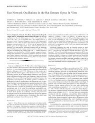
744 A. BAuDE et al.antiserum suggested by immunochemical characterization.Furthermore, the localization of immunoreactionend-product <strong>to</strong> the intracellular face of theplasma membrane confirms the <strong>to</strong>pographical organizationof the subunit in the lipid bilayer as predictedfrom the amino acid sequence. 15.30The granule cell layer is labelled strongly with[3Hjmuscimol,24 showing high-affinity GABA bindingsites, but few benzodiazepine agonist binding siteshave been found (for review, see Ref. 21). However,the partial inverse agonist [3HjRo 15-4513. in thepresence of diazepam, shows binding restricted <strong>to</strong> thegranular layer of the cerebellar cortex. 34 This apparentparadox has been explained by results from theco-expression of the 1X6, f32 and y2 sub units incultured cells. This subunit composition produced aGABA A recep<strong>to</strong>r which binds with high affinity muscimol,as well as RoI5-4513 and the structurallyrelated antagonist RoI5-l788, but not other benzodiazepineagonists or f3-carbolinesY Taken <strong>to</strong>getherwith the restricted localization of the 1X6 subunit, thissuggests that the IX 6 subunit is part of the high-affinityGABA binding site on granule cells.Different subcellular distribution for the 1X6 versus IX Iand f32/3 subunits of the GABA A recep<strong>to</strong>rFrom the distribution of the peroxidase endproductit is concluded that the IX 6 subunit is mainlylocated at synapses, although the presence of loweramounts at non-synaptic recep<strong>to</strong>r in the dendriticmembrane cannot be excluded with the present technique.In any case, the 1X6 subunit was not detectedin the somatic membrane under our conditions,showing the first cvidence of a differential subcellulardistribution of subunits of thc GABA Arecep<strong>to</strong>r.In contrast, the IXI and /32/3 subunits are presentboth at synaptic and extrasynaptic sites. 38 The lattertwo subunits have been found at synaptic and nonsynapticsites in the thalamus and cortex as well,l6,37indicating that the granule cclls are not unique in thisrespect. The possibility that the apparent widespreadlocalization of the IX I and /32/3 subunits is due <strong>to</strong>technical fac<strong>to</strong>rs connected <strong>to</strong> the extracellular epi<strong>to</strong>pes,e.g, the spread of the peroxidase end-productin the extracellular space from synaptic <strong>to</strong> extrasynapticsites, has been discussed earlier, and is veryunlikely.3H The granule cell was chosen for this studybecause it does not receive synapses on its somaticmembrane which is in direct membrane appositioneither <strong>to</strong> astrocytes or <strong>to</strong> other granule cells. Since allthe synapses are concentrated in the glomeruli wellseparated from the somata it is not easy <strong>to</strong> envisagediffusion of reaction product <strong>to</strong> the somatic membrane<strong>to</strong> sites where granule cell bodies are contactingeach other over tens of square micrometres. Thepresence of extrasynaptic recep<strong>to</strong>r/channcl complexeswhich are able <strong>to</strong> bind GABA and pass chloride ions,is also confirmed by whole-cell patch-clamp recordingsfrom cerebellar slices, which show chloride currentswith fast rise-time in response <strong>to</strong> brief (5-10 ms)ionophoretic pulses of GABA applied <strong>to</strong> the soma(Farrant M. and Cull-Cundy S. G., personal communication).It remains <strong>to</strong> be seen whether the channelbehaviour is in line with the predicted subunitcomposition.Composition of GABA A recep<strong>to</strong>r complex in granulecellsLigand binding studies in the cerebellum showedhigh-affinity GABA recep<strong>to</strong>rs in the granule celllayer, but few benzodiazepine agonist bindingsites. 21 ,24,44 However, the expression of the IX I subunitI7 ,31,33 (which is thought <strong>to</strong> be responsible for typeI benzodiazepine recep<strong>to</strong>r pharmacology25) and itslocalization in the granule cell plasma membrane,38suggest the presence of at least some sites for benzodiazepines.Immunoprecipitation experiments indicatethat a native recep<strong>to</strong>r in the cerebellum includesthe 1X6 subunit,15 but the {;(6 subunit probably occursin a separate channel complex from the one containingthe IX 1 subunit (Quirk K., Gillard N., Ragan C. I.,Whiting P., McKernan R. M., unpublished observations).Granule cells also abundantly express theband y2 subunits. Immunoreactivity for the (j subunitseems <strong>to</strong> be concentrated in the glomerulj,2suggesting colocalization with the 1X6 sub unit. Immunoprecipitationresults indeed revcal that both :'(6and b subunits exist in a recep<strong>to</strong>r complex which---- - -------Fig. 3. Electron micrographs showing immunoreactivity for the 0(6 subunit in granule cells of the cerebellarcortex in rat. Immunoreactivity is only present in the dendrites (asterisks in A) of the granule cells (gc).The immunoperoxidase end-product is most dense at the synaptic junctions (thin arrows in B) formedwith Golgi cell terminals (Gt). The weaker diffuse reaction end-product in the dendrites is interpreted asoriginating mainly from synaptic plasma membranes. One of the dendrites (asterisk in B) receives animmunoposilive synapse from a Golgi terminal (thin arrow) and an immunonegative synapsc from amossy fibre terminal (m!). Immunoreactivity for the rx6 subunit is absent from a granule cell soma (gc)and a glial cell (Gl). Scale bars = 0.5 I,m (A); O.21'm (B). Immunocy<strong>to</strong>chemistry was performed asdescribed in Fig. 2 with the difference that no Tri<strong>to</strong>n X-lOO was used. After 3,3'-diaminobenzidincreaction, the sections were treated with 1 % OS04 in 0.1 M phosphate buffer for 1 h, washed in distilledwater and placed for 1 h in uranyl acetate (1 % in distilled water). After dehydration in graded ethanoland propylene oxide, the sections were infiltrated with epoxy resin (DURCUPAN ACM, Fluka) overnightand flat-embedded on glass slides for light microscopic assessment. Areas of interest were re-embeddedfor electron microscopic sectioning. A <strong>to</strong>tal of 13 blocks, including all layers of the cerebellar cortex werestudied for the rx 6 subunit. The ultrathin sections were not stained. Three blocks for the f32/3 subunit andtwo blocks for the rx I subunit were also studied according <strong>to</strong> the same procedure.