Embryonic and larval development of the striped mullet Mugil ...
Embryonic and larval development of the striped mullet Mugil ...
Embryonic and larval development of the striped mullet Mugil ...
- No tags were found...

You also want an ePaper? Increase the reach of your titles
YUMPU automatically turns print PDFs into web optimized ePapers that Google loves.
<strong>Embryonic</strong> <strong>and</strong> <strong>larval</strong> <strong>development</strong> <strong>of</strong> <strong>the</strong> <strong>striped</strong><strong>mullet</strong> <strong>Mugil</strong> eephalus CL)MATHEW ABRAHAM, P. SHIRANEE, P. KISHORE CHANDRA, M. KAILASAMAND V.K. CHARLESCentral Institute <strong>of</strong> Brackishwater Aquaculture, Madras-600 008, IndiaABSTRACT<strong>Mugil</strong> cephalus in ripe condition collected from inshore catches at Muttilkadunear Madras were induced to breed using HCG <strong>and</strong> ovaprim to study <strong>the</strong>embryonic <strong>and</strong> <strong>larval</strong> <strong>development</strong>. Samples <strong>of</strong> unfertilised eggs <strong>and</strong> various<strong>development</strong>al stages from fertilization through embryonic <strong>and</strong> <strong>larval</strong> stageswere preserved for fur<strong>the</strong>r micrographic studies. Rate <strong>of</strong> fertilization wasdetermined at <strong>the</strong> time <strong>of</strong> blastodisc formation <strong>and</strong> was 90 %. Of <strong>the</strong> totalnumber <strong>of</strong> 1.46 million eggs, 1.26 million were fertilised. The pre-hatchedembryo was deeply pigmented, exhibited twitching movements <strong>and</strong> lay curvedover <strong>the</strong> yolk mass. After an incubation period <strong>of</strong> 30-32 hrs, <strong>the</strong> embryo hatchedout. The newly hatched out larvae measuring 2.29 mm, were transparent witha large oval head, a well defined yolk sac <strong>and</strong> short tail encircled by acontinuous finfold. hlouth formation was complete <strong>and</strong> <strong>the</strong> larvae startedfeeding on 3rd day <strong>of</strong> hatching. The paper presents <strong>the</strong> results <strong>of</strong> detailedstudies <strong>of</strong> embryonic <strong>and</strong> larva1 <strong>development</strong> <strong>and</strong> <strong>the</strong> time scale establishedfor critical stages <strong>of</strong> <strong>development</strong>.Introduction<strong>Embryonic</strong> <strong>and</strong> <strong>larval</strong> <strong>development</strong>studies besides providing interestinginformation in itself, are imperative <strong>and</strong>consequential to <strong>the</strong> successful rearing<strong>of</strong> larvae for seed production. Despite<strong>the</strong> successes in artificial propagation <strong>of</strong><strong>the</strong> <strong>mullet</strong>, by induced spawning, <strong>the</strong>reis still a need to refine fur<strong>the</strong>r <strong>the</strong>techniques <strong>of</strong> <strong>larval</strong> rearing particularlyfor practical <strong>and</strong> commercial applications(Liao, 1993). It is an undisputedfact that <strong>larval</strong> rearing remains <strong>the</strong>most critical <strong>and</strong> crucial phase inbrackishwater fish seed production.Development <strong>of</strong> suitable protocols for<strong>the</strong> mass rearing <strong>of</strong> <strong>larval</strong> fish repre-sents one <strong>of</strong> <strong>the</strong> last bamers for <strong>the</strong>successful propagation <strong>of</strong> a variety <strong>of</strong>marine species (Tamaru et al., 1993).Most problems arise from relativelysmaller size <strong>of</strong> <strong>the</strong> mouth <strong>and</strong> limitedyolk reserves (Shirota, 1970) <strong>of</strong> <strong>the</strong>larvae.The present study elaborates withphotographic evidence <strong>the</strong> embryonic<strong>and</strong> <strong>larval</strong> structure <strong>and</strong> <strong>development</strong>with <strong>the</strong> corresponding time scale <strong>of</strong> M.cephalus. A photographic presentation<strong>of</strong> <strong>the</strong> embryonic <strong>development</strong> <strong>of</strong> M.cephalus with time scale has beenelaborated by Tamaru et al. (1993).Vivid photographs, however, with notextual reference have been made by
Ma<strong>the</strong>w Abraham et al.Liao (1993). Earlier studies on <strong>the</strong><strong>development</strong> <strong>of</strong> <strong>the</strong> <strong>mullet</strong>s, conductedon various geographic <strong>and</strong> environrnen-/tal pr<strong>of</strong>iles include that <strong>of</strong> Nair (1957),:Anderson (1958), Kuo et al. (1973), Ling;(1970) <strong>and</strong> Chaudhuri et al. (1977).iMaterials <strong>and</strong> methodsi<strong>Mugil</strong> cephalus breeders were ob- \ The total number <strong>of</strong> eggs was estimated-&-?&1.4 m <strong>and</strong> <strong>the</strong> rate <strong>of</strong> fertiliz 'ontained from commercial sea catches at 90 %. Incubation period spann / over aMuttukadu, near Madras during Febru- period <strong>of</strong> 30-32 hrs <strong>and</strong> <strong>the</strong> hatchingary 1997. Females with an average rate was 41.6 %. Immediately aReroocyte diameter <strong>of</strong> >525pm <strong>and</strong> milting fertilization <strong>the</strong> germplasm migrated tomales were selected. The selected fish <strong>the</strong> animal pole to form a dense cap likewere given a dip treatment in 1 ppm structure called <strong>the</strong> germinal disc (Plateacriflavin <strong>and</strong> 1 hr bath in 10 ppm 1B). Cleavage (Plate 1 C,D,E,F) wasoxytetracyclin. Induced breeding was restricted to <strong>the</strong> germinal disc <strong>and</strong> <strong>the</strong>done following <strong>the</strong> two injection protocol resulting blastoderm assumed a discwith a 24 hr interval. HCG was used as like multi-cellular structure called <strong>the</strong><strong>the</strong> priming dose @ 6,000-10,000 I.U./kg blastodisc, which was more or lessbody weight <strong>and</strong> ovaprim was used as convex in shape <strong>and</strong> enclosed between<strong>the</strong> resolving dose @ 3-5mIJkg body itself <strong>and</strong> <strong>the</strong> uncleaved residue <strong>of</strong> <strong>the</strong>weight. The males were not given any egg - a cavity representing <strong>the</strong> blastohormonetreatment. Dry stripping was coele (Plate 1G) cleacage was followedresorted to <strong>and</strong> fertilization was effected by gastrulation which converted <strong>the</strong>in-vitro. Incubation was done @ 140 embryo into a two layered structureeggsA. Water temperature ranged from (Plate I H). The outer germ layer, <strong>the</strong>26-28°C <strong>and</strong> salinity was 26 ppt. Sam- epiblast, gave rise to <strong>the</strong> ectoderm whileples <strong>of</strong> eggs before fertilization, imme- <strong>the</strong> involuted cell mass, <strong>the</strong> hypoblast,diately after fertilization <strong>and</strong> <strong>the</strong>re- <strong>the</strong> endodermal <strong>and</strong> mesodermal comafterat 30 minute intervals until hatch- ponents.ing <strong>and</strong> including <strong>the</strong> <strong>larval</strong> samples at30 minute intervals were also preservedin a medium made <strong>of</strong> 2 % formalin, 4 %glycerol <strong>and</strong> 94 % water for fur<strong>the</strong>rstudies. The larvae were reared in FRPtanks provided with flow through runningwater system at a density <strong>of</strong> 45-50nosll. The larvae were fed with <strong>the</strong>rotifer Brachionus plicatilis from thirdday onwards.ResultsThe ovulation was complete 22 hrsafter <strong>the</strong> resolving dose. The strippedeggs were translucent <strong>and</strong> non adhesivewith a deep yellow colour. The diameter<strong>of</strong> <strong>the</strong> spawned eggs ranged from 750-760 pm. The yolk was characterised bya single yolk globule <strong>and</strong> <strong>the</strong> eggs weretelolecithal in nature (Plate 1 A). Fertilizationwas effected by stripping miltfrom males <strong>and</strong> mixing it with <strong>the</strong> eggs.Subsequent to gastrulation, <strong>development</strong>continued until a primitive vertebratebody was formed <strong>and</strong> <strong>the</strong> neuralplate established. The embryo was <strong>the</strong>nmore or less cylindrical <strong>and</strong> bilaterallysymmetrical <strong>and</strong> was referred to asneurula (Plate 1. I). The head <strong>and</strong>pharyngeal region projected from <strong>the</strong>yolk mass anteriorly <strong>and</strong> <strong>the</strong> trunkcurved over <strong>the</strong> yolk <strong>and</strong> <strong>the</strong> tailprojected posteriorly (Plate 1. J). Themesoderm on both sides <strong>of</strong> <strong>the</strong> notochordorganised into somites <strong>and</strong>melanophores began to appear (Plate 1K). The pre-hatched embryo showed
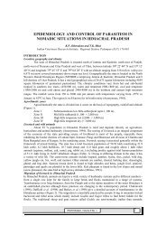
Embryonzc <strong>and</strong> <strong>larval</strong> <strong>development</strong> <strong>of</strong> hlugil cephalus 125Platc: 1. '4. Fertilised egg, B. Germinal disc stage, C. 2-cell stage, U. &cell stage,E. 16-cell stage, F. 32-cell stage, G. Morula stage, H. Gastrula, I. Early neurula,J. Neurula somite stage, K. Late neurula, L. Pre-hatched embryo. (Magnification: x 40).
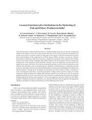
Embryon~c <strong>and</strong> larual <strong>development</strong> <strong>of</strong> Mug1 cephalus 127Plate 2. A. Newly hatched larva, B. 24-hr old larva. (Magnification: x 100).
Ma<strong>the</strong>w Abraham et al.length <strong>of</strong> 2.80 mm <strong>and</strong> 60 % <strong>of</strong> <strong>the</strong> yolkwas utilised. The mouth opening measured135 pm in <strong>the</strong> 96 hrs old larvae(Plate 3 B). The alimentary canal wasdistinct <strong>and</strong> a streaming movement wasobserved from <strong>the</strong> anterior end to <strong>the</strong>anal end. Pigmentation was seen toextend from <strong>the</strong> cephalic to <strong>the</strong> caudalend. The anal aperture was well formed<strong>and</strong> distinct, closely placed with littleinter orbital space. At 144 hrs <strong>the</strong> larvaemeasured an average length <strong>of</strong> 3.92 mm<strong>and</strong> showed deeper pigmentationdorsally <strong>and</strong> laterally. At 160 hrs <strong>the</strong>larvae measured 4.59 mm (Plate 3 E).The alimentary canal was vivid <strong>and</strong>clear, 90 % <strong>of</strong> <strong>the</strong> yolk reserve wasconsumed <strong>and</strong> vestiges <strong>of</strong> <strong>the</strong> pectoralfins appeared as finbuds. At 172 hrs <strong>the</strong>larvae were deeply pigmented. The bodyassumed a fish like shape with a wellformed caudal fin <strong>and</strong> rudimentarypectoral fins (Plate 3 F). The larvaestarted to swim actively <strong>and</strong> wereobserved to feed voraciously. Details <strong>of</strong><strong>larval</strong> <strong>development</strong> with <strong>the</strong> correspondingtime scale is given in Table 2.DiscussionThe diameter <strong>of</strong> <strong>the</strong> fertilised eggs <strong>of</strong>M. cephalus as reported in earlierstudies is seen to vary from 0.48 - 0.80mm, in different environmental pr<strong>of</strong>ileby Chaudhuri et al. (1977), Ling (1970)<strong>and</strong> Kuo et al. (1973). In <strong>the</strong> presentstudy <strong>the</strong> spawned eggs ranged indiameter from 750-760 pm at a temperature<strong>of</strong> 26-28°C <strong>and</strong> salinity <strong>of</strong> 26%0.In a series <strong>of</strong> spawning experimentsduring 1988-'90 Tamaru et al. (1994);Kuo et al. (1973) <strong>and</strong> Nash et al. (1974)observed that <strong>the</strong> diameter <strong>of</strong> spawnedeggs ranged from 863.9 +. 20.7 pm -938.6 * 32.1 pm in M. cephalus.<strong>Embryonic</strong> <strong>development</strong> through <strong>the</strong>critical stages <strong>of</strong> cleavage, blastulation,gastrulation <strong>and</strong> neurula stages with<strong>the</strong> corresponding time scale as observedin <strong>the</strong> present study was seen tobe almost similar to that reported byTamaru et al. (1993). The fertilised eggshave a diameter <strong>of</strong> 770-778 pm after 24hrs <strong>of</strong> fertilization. The embryo showeddeep pigmentation <strong>and</strong> twitching movementsat regular intervals. At 28 hrsafter fertilisation <strong>the</strong> embryonic heartbeat could be observed. Kuo et aL. (1973)reported that in M. cephalus <strong>the</strong> heartbegan to beat 25.10 hrs after fertilisation.Two environmental factors viz., temperature<strong>and</strong> salinity have pr<strong>of</strong>oundinfluence on <strong>the</strong> <strong>development</strong> <strong>and</strong> hatching<strong>of</strong> marine teleost eggs (Blaxter,1998). In M. cephalus it has been demonstratedby Walsh et al. (1991) thatsalinity does not influence <strong>the</strong> time <strong>of</strong>hatching in this species. However, <strong>the</strong>time <strong>of</strong> hatching is inversely proportionalto <strong>the</strong> incubation temperature. Hatchingtime has been observed to vary from 28-54 hrs in different salinity-temperaturecombinations in studies made by Kuo etal. (19731, Liao (1975), Rajyalakshmi etal. (1991) <strong>and</strong> Krishnan et al. (1996). In<strong>the</strong> present study first hatching wasobserved at 30 hrs <strong>and</strong> hatcE-~g wascompleted at 32 hrs at a temperature <strong>of</strong>26-28°C <strong>and</strong> salinity <strong>of</strong> 26 ppt. Tamaruet al. (1993) observed hatching at 28 hrsat a temperature <strong>of</strong> 26°C.Eda et al. (1990) reported that <strong>the</strong>mean total length <strong>of</strong> <strong>the</strong> newly hatchedlarvae was 2.68 + 0.06 mm. In <strong>the</strong>present study <strong>the</strong> newly hatched larvaehad a mean length <strong>of</strong> 2.29 mm. Themouth was observed to open on <strong>the</strong>second day <strong>and</strong> was functional on <strong>the</strong>third day post-hatch. Rajyalakshmi etal. (1991) reported that <strong>the</strong> larvae haddeveloped mouth on <strong>the</strong> third day <strong>and</strong>was hctional on <strong>the</strong> fourth day. Eda
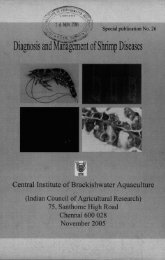
<strong>Embryonic</strong> <strong>and</strong> <strong>larval</strong> <strong>development</strong> <strong>of</strong> <strong>Mugil</strong> cephalus 129Plate 3. A. 72-hr old Iarve, B. 96-hr old larva, C. 108-hr old larva, D. 144-hr old larva,E. 160-hr old larva, F. 172-hr old larva. (Magnification: x 10).et al. (19901, Kuo et al. (19731, Nash etal. (1974) <strong>and</strong> Liao et al. (1971) observedthat <strong>the</strong> mouth <strong>of</strong> <strong>the</strong> <strong>mullet</strong> larvaeopen <strong>and</strong> yolk absorption is completedby <strong>the</strong> second <strong>and</strong> fourth day post-hatchrespectively. Hence, as in <strong>the</strong> presentstudy <strong>the</strong> most critical period in <strong>the</strong>rearing <strong>of</strong> M. cephalus larvae coincideswith <strong>the</strong> opening <strong>of</strong> <strong>the</strong> mouth i.e. <strong>the</strong>secondfthird day post-hatch. Apparentlyactive feeding on rotifers by <strong>the</strong> larvaebegin before <strong>the</strong> completion <strong>of</strong> yolk sacabsorption <strong>and</strong> it is vital <strong>and</strong> imperativethat food organisms should be presentedto <strong>the</strong> <strong>mullet</strong> larvae by <strong>the</strong> second day,36 hrs post-hatch. At 96 hrs <strong>the</strong> mouthopening measured 135 pm <strong>and</strong> has adirect bearing on <strong>the</strong> kind <strong>of</strong> rotifer fedto <strong>the</strong> larvae with reference to size.Brachionus plicatilis, <strong>the</strong> most commonlyused <strong>larval</strong> feed for M. cephalusmay be <strong>of</strong> <strong>the</strong> L or S type <strong>and</strong> is observedto have a lorica length that varies from110-340 pm. The small size <strong>of</strong> <strong>the</strong> larvaerepresents an even smaller mouth sizeat <strong>the</strong> time <strong>of</strong> first feeding. Fresh waterfish larvae, in general not only hatch outat a larger size (ie. possess a largec
Ma<strong>the</strong>w Abraham et al. 130TABLE 2. Larual <strong>development</strong> <strong>of</strong> Mug11 cephalusTime after Description Platehatching0 hr. Just hatched embryo, transparent well defined yolk 2 Asac, with a transparent fin fold encircling <strong>the</strong> body.Total average length 2.29 mm, head length 0.22 mmmaximum width 0.81 mm.24 hr. Average length 2.65 mm; dark prominent eye spot on 2 B<strong>the</strong> anterior part <strong>of</strong> head. There appears a breakin <strong>the</strong> finfold ventrally with <strong>the</strong> anal aperturebeginning to form.72 hr. Well defined mouth opening <strong>and</strong> jaw movement 3 Aobserved. Larva has a long tapering tail <strong>and</strong>exhibits free movements.96 hr. Larvae having an average length <strong>of</strong> 2.80 mm; mouth 3 Bopening 135 pm, 60% <strong>of</strong> yolk utilised. Alimentarycanal distinct.108 hr. Larvae free swimming <strong>and</strong> have an average length <strong>of</strong> 3 C2.84 mm. Eye balls large <strong>and</strong> distinct.144 hr. Larvae measure an average length <strong>of</strong> 3.92 mm <strong>and</strong> 3 Dshow deep pigmentation.160 hr. Rudimentary pectoral fins <strong>and</strong> well defined 3 Emyomeres appear.172 hr. Larvae deeply pigmented, body assumes a fish like 3 Fshape with a well formed caudal fin <strong>and</strong>rudimentary pectoral fms visible.mouth) but are morphologically moreadvanced than <strong>the</strong> marine teleosts atfirst feeding. This allows for <strong>the</strong> exclusiveuse <strong>of</strong> artificial diets in <strong>the</strong>irrearing. In contrast, marine fish larvaehave to rely on live food organisms as<strong>the</strong>ir initial foodstuffs (Tamaru et al.,1993). This fact poses to be <strong>the</strong> mostdifficult <strong>and</strong> critical in sucessful <strong>larval</strong>rearing <strong>of</strong> <strong>mullet</strong> larvae. The presentstudy, with special mention on itsfeeding on <strong>the</strong> embryonic <strong>and</strong> <strong>larval</strong><strong>development</strong> <strong>of</strong> M. cephalus for <strong>the</strong> firsttime in India, will help refine <strong>the</strong>present level <strong>of</strong> knourledge on inducedbreeding <strong>and</strong> <strong>larval</strong> rearing.AcknowledgmentThe authors are grateful to Dr. K.Alagarswami former Director <strong>and</strong> toDr. G. R. M. Rao, Director for <strong>the</strong>irencouragement <strong>and</strong> help during <strong>the</strong>course <strong>of</strong> this study.ReferencesAnderson, W. W. 1958. Larval <strong>development</strong>,growth <strong>and</strong> spawning <strong>of</strong> <strong>striped</strong> <strong>mullet</strong>(<strong>Mugil</strong> cephalus) along <strong>the</strong> SouthAtlantic coast <strong>of</strong> <strong>the</strong> United States.Fish. Bull. US. Fish. Wild. Serv., 58(144) : 510-519.Blaxter, J.H.S. 1988. Pattern <strong>and</strong> variety in<strong>development</strong>. In : W.S. Hoar <strong>and</strong> D.J.R<strong>and</strong>all (Eds.), Fish Physiology, Vol. 11,Part A. Academic Press. San Diego,p. 1-58.Chaudhuri, H., R.M. Bhowmic, G.V. Kowtal.M.M. Bagchi, R.K. Jana <strong>and</strong> S.D.Guptha 1977. Experiments in artificialpropagation <strong>and</strong> <strong>larval</strong> <strong>development</strong> <strong>of</strong>M. cephalus (Linnaeus) in India. J.Inl<strong>and</strong> Fish. Sac. India., 9 : 30-41.Eda, H., R. RIurashige, Y. Oozeki, A.Hagiwara, B. Eastham, P. Bass, C.S.Tamaru <strong>and</strong> C.S. Lee 1990. Factorsaffecting intensive <strong>larval</strong> rearing in<strong>striped</strong> <strong>mullet</strong>, <strong>Mugil</strong> cephalus.Aquaculture, 91 : 281-294.
<strong>Embryonic</strong> <strong>and</strong> larual deuelopment <strong>of</strong> <strong>Mugil</strong> ceph alus 131Krishnan, L., K.V. Ramakrishna, P.K Ghosh,R.D. Prasadam <strong>and</strong> D. Raja Babu 1996.Experiments on induced breeding <strong>of</strong> <strong>the</strong>grey <strong>mullet</strong> <strong>Mugil</strong> cephalus (L) inChilka Lake. J. mar. biol. Ass. India.,38 (18~2): 150-152.Kuo, C.hf., Z.H. Shehadeh <strong>and</strong> K.K Milisen1973. A preliminary report on <strong>the</strong><strong>development</strong>, growth <strong>and</strong> survival <strong>of</strong>laboratory reared larvae <strong>of</strong> <strong>the</strong> grey<strong>mullet</strong>, <strong>Mugil</strong> cephalus (L). J. Fish.Biol., 5 : 459-470.Liao, I.C. 1975. Experiments on <strong>the</strong> inducedbreeding <strong>of</strong> <strong>the</strong> grey <strong>mullet</strong>, in Taiwanfrom 1963-'73. Aquaculture, 6 (1) :31-58.Liao, I.C. 1993. Finfish hatcheries in Taiwan: Recent advances. In : Finfish hatcheryin Asia : Proc. Finfish Hatchery in Asia'91. TML Conference Proc. No. 3. C. S.Lee, hl.S. Su <strong>and</strong> I.C. Liao (Eds.), p.1-25.Liao, I.C., Y.J. Lu, T.L. Huang <strong>and</strong> M.C. Lin1971. Experiments on induced breeding<strong>of</strong> <strong>the</strong> grey <strong>mullet</strong> <strong>Mugil</strong> cephalus (L).Aquaculture, 1 : 15-34.Ling, S.N. 1970. A brief review on <strong>the</strong> workdone on <strong>the</strong> induced breeding <strong>of</strong> <strong>Mugil</strong>cephalus in Taiwan. J. Inl<strong>and</strong>. Fish.Soc. India, 1 : 1-12.Nair, G.S. 1957. Notes on <strong>the</strong> early <strong>development</strong><strong>of</strong> <strong>Mugil</strong> cephalus (L). Bull. Res.Inst. Univ. Trauancore, 15(1) : 77-84.Nash, C.E., C.M. Kuo <strong>and</strong> S.C. McConnell1974. Operational procedures for rearinglarvae <strong>of</strong> <strong>the</strong> grey <strong>mullet</strong> <strong>Mugil</strong>cephalus (L). Aquaculture, 3 : 15-24.Rajyalakshmi, T., S.M. Pillai <strong>and</strong> P.Ravich<strong>and</strong>ran 1991. Experiments oninduced breeding <strong>and</strong> <strong>larval</strong> rearing <strong>of</strong>grey <strong>mullet</strong>s <strong>and</strong> sea bream at ChilkaLake. J. Inl<strong>and</strong> Fish. Soc. India, 23 (1): 16-26.Shirota, A. 1970. Studies on <strong>the</strong> mouth size<strong>of</strong> fish larvae. Bull. Jap. Soc. Sci. Fish,36 : 353-368.Tamaru, C.S., J. William Fitzgerald Jr. <strong>and</strong>S. Vernon 1993. In. : Hatchery manualfor <strong>the</strong> artificial propagation <strong>of</strong> <strong>the</strong><strong>striped</strong> <strong>mullet</strong> <strong>Mugil</strong> cephalus (L).Published by Dept. <strong>of</strong> Commerce. Guam96911, p. 1-167.Tamaru, C.S., C. Sh. Lee, C.D. Kelly, G.Miyamotu <strong>and</strong> A. Moriwake 1994.Oocyte growth in <strong>the</strong> <strong>striped</strong> <strong>mullet</strong><strong>Mugil</strong> cephalus (L) maturing at differentsalinities. J. World AquacultureSociety., 25(1) :109-115.Walsh, W.A., C. Swanson <strong>and</strong> C. S. Lee1991. Combined effects <strong>of</strong> temperature<strong>and</strong> salinity on <strong>development</strong> <strong>and</strong> hatching<strong>of</strong> <strong>striped</strong> <strong>mullet</strong> <strong>Mugil</strong> cephalus.Aquaculture 37 : 281-289.