Estradiol
Estradiol
Estradiol
- No tags were found...

You also want an ePaper? Increase the reach of your titles
YUMPU automatically turns print PDFs into web optimized ePapers that Google loves.
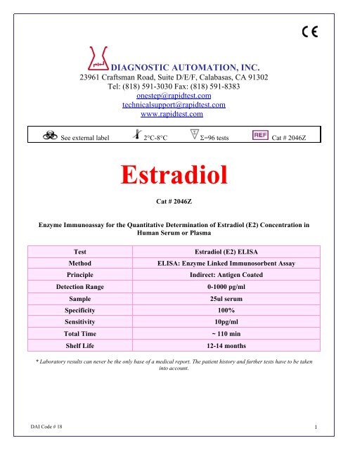
DIAGNOSTIC AUTOMATION, INC.23961 Craftsman Road, Suite D/E/F, Calabasas, CA 91302Tel: (818) 591-3030 Fax: (818) 591-8383onestep@rapidtest.comtechnicalsupport@rapidtest.comwww.rapidtest.comSee external label 2°C-8°C Σ=96 tests Cat # 2046Z<strong>Estradiol</strong>Cat # 2046ZEnzyme Immunoassay for the Quantitative Determination of <strong>Estradiol</strong> (E2) Concentration inHuman Serum or PlasmaTestMethodPrincipleDetection RangeSample<strong>Estradiol</strong> (E2) ELISAELISA: Enzyme Linked Immunosorbent AssayIndirect: Antigen Coated0-1000 pg/ml25ul serumSpecificity 100%SensitivityTotal TimeShelf Life10pg/ml~ 110 min12-14 months* Laboratory results can never be the only base of a medical report. The patient history and further tests have to be takeninto account.DAI Code # 18 1
INTENDED USEFor the quantitative determination of <strong>Estradiol</strong> (E2) concentration in human serumINTRODUCTION<strong>Estradiol</strong> (E2) is a C18 steroid hormone with a phenolic A ring. This steroid hormone has a molecularweight of 272.4. It is the most potent natural Estrogen, produced mainly by the ovary, placenta, and insmaller amounts by the adrenal cortex, and the male testes (1,2,3).<strong>Estradiol</strong> (E2) is secreted into the blood stream where 98% of it circulates bound to sex hormonebinding globulin (SHBG). To a lesser extent it is bound to other serum proteins such as albumin. Onlya tiny fraction circulates as free hormone or in the conjugated form (4,5). Estrogenic activity is effectedvia estradiol-receptor complexes which trigger the appropriate response at the nuclear level in thetarget sites. These sites include the follicles, uterus, breast, vagina, urethra, hypothalamus, pituitaryand to a lesser extent the liver and skin.In non-pregnant women with normal menstrual cycles, estradiol secretion follows a cyclic, biphasicpattern with the highest concentration found immediately prior to ovulation (6,7). The rising estradiolconcentration is understood to exert a positive feedback influence at the level of the pituitary where itinfluences the secretion of the gonadotropins, follicle stimulating hormone (FSH), and luteinizinghormone (LH), which are essential for follicular maturation and ovulation, respectively (8,9). Followingovulation, estradiol levels fall rapidly until the luteal cells become active resulting in a secondary gentlerise and plateau of estradiol in the luteal phase. During pregnancy, maternal serum <strong>Estradiol</strong> levelsincrease considerably, to well above the pre-ovulatory peak levels and high levels are sustainedthroughout pregnancy (10).Serum <strong>Estradiol</strong> measurements are a valuable index in evaluating a variety of menstrual dysfunctionssuch as precocious or delayed puberty in girls (11) and primary and secondary amenorrhea andmenopause (12). <strong>Estradiol</strong> levels have been reported to be increased in patients with feminizingsyndromes (14), gynaecomastia (15) and testicular tumors (16).In cases of infertility, serum <strong>Estradiol</strong> measurements are useful for monitoring induction of ovulationfollowing treatment with, for example, clomiphene citrate, LH-releasing hormone (LH-RH), orexogenous gonadotropins (17, 18). During ovarian hyperstimulation for in vitro fertilization (IVF), serumestradiol concentrations are usually monitored daily for optimal timing of human chorionic gonadotropin(hCG) administration and oocyte collection (19).PRINCIPLE OF THE TESTThe E2 EIA is based on the principle of competitive binding between E2 in the test specimen and E2-HRP conjugate for a constant amount of rabbit anti-<strong>Estradiol</strong>. In the incubation, goat anti-rabbit IgGcoatedwells are incubated with 25 µl E2 standards, controls, patient samples, 100 µl <strong>Estradiol</strong>-HRPConjugate Reagent and 50 µl rabbit anti-<strong>Estradiol</strong> reagent at room temperature (18-25°C) for 90minutes. During the incubation, a fixed amount of HRP-labeled E2 competes with the endogenous E2in the standard, sample, or quality control serum for a fixed number of binding sites of the specific E2antibody. Thus, the amount of E2 peroxidase conjugate immunologically bound to the wellprogressively decreases as the concentration of E2 in the specimen increases. Unbound E2 peroxidaseconjugate is then removed and the wells washed. Next, a solution of TMB Reagent is added andincubated at room temperature for 20 minutes, resulting in the development of blue color. The colordevelopment is stopped with the addition of 1N HCl, and the absorbance is measuredDAI Code # 18 2
spectrophotometrically at 450 nm. The intensity of the color formed is proportional to the amount ofenzyme present and is inversely related to the amount of unlabeled E2 in the sample. A standardcurve is obtained by plotting the concentration of the standard versus the absorbance. The E2concentration of the specimens and controls run concurrently with the standards can be calculated fromthe standard curve.REAGENTSMaterials provided with the kit:• Goat Anti-Rabbit IgG-coated microtiter wells, 96 wells• <strong>Estradiol</strong> Reference Standards: 0, 10, 30, 100, 300, and 1000 pg/ml. Liquid, 0.5 ml each, ready touse.• Rabbit Anti-<strong>Estradiol</strong> Reagent (pink color), 7 ml• <strong>Estradiol</strong>-HRP Conjugate Reagent (blue color), 12 ml• <strong>Estradiol</strong> Control 1, Liquid, 0.5 ml, Ready to use.• <strong>Estradiol</strong> Control 2, Liquid, 0.5 ml, Ready to use.• TMB Reagent (One-Step), 11 ml.• Stop Solution (1N HCl), 11 ml.Materials required but not provided:• Precision pipettes: 25 µl , 50 µl, 100 µl, 200 µl, and 1.0 ml.• Disposable pipette tips.• Distilled and deionized water.• Vortex mixer or equivalent.• Absorbent paper or paper towel.• Linear-linear graph paper.• Microtiter plate reader.WARNINGS AND PRECAUTIONS FOR USERSTest methods are not available which can offer complete assurance that Hepatitis B virus, HumanImmunodeficiency Virus (HIV/HTLV-III/LAV), or other infectious agents are absent from the reagents inthis kit. Therefore, all human blood products, including patient samples, should be consideredpotentially infectious. Handling and disposal should be in accordance with the procedures defined byan appropriate national biohazard safety guideline or regulation, where it exists (e.g., USA Center forDisease Control/National Institute of Health Manual, “Biosafety in Microbiological and BiomedicalLaboratories,” 1984) (22).SPECIMEN COLLECTION AND PREPARATIONS1. Only human serum should be used.2. No special pretreatment of sample is necessary.3. Serum samples may be stored at 2-8°C for up to 24 hours, and should be frozen at −10°C or lowerfor longer periods. Do not use grossly hemolyzed or grossly lipemic specimens.4. Please note: Samples containing sodium azide should not be used in the assay.DAI Code # 18 3
STORAGE OF TEST KIT AND INSTRUMENTATIONUnopened test kits should be stored at 2-8°C upon receipt and the microtiter plate should be kept in asealed bag with desiccants to minimize exposure to damp air. Opened test kits will remain stable untilthe expiration date shown, provided it is stored as described above. A microtiter plate reader with abandwidth of 10 nm or less and an optical density range of 0-3 O.D. at 450 nm wavelength isacceptable for use in absorbance measurement.REAGENT PREPARATION1. All reagents should be brought to room temperature (18-25°C) before use.2. Samples with expected <strong>Estradiol</strong> concentrations over 1000 pg/ml may be quantitated by dilutionwith diluent available from vendor.ASSAY PROCEDURE1. Secure the desired number of coated wells in the holder.2. Dispense 25 µl of standards, specimens and controls into appropriate wells.3. Dispense 100 µl of <strong>Estradiol</strong>-HRP Conjugate Reagent into each well.4. Dispense 50 µl of rabbit anti-<strong>Estradiol</strong> (E2) reagent to each well.5. Thoroughly mix for 30 seconds. It is very important to mix them completely.6. Incubate at room temperature (18-25°C) for 90 minutes.7. Rinse and flick the microwells 5 times with distilled or deionized water. (Please do not use tapwater.)8. Dispense 100 µl of TMB Reagent into each well. Gently mix for 10 seconds.9. Incubate at room temperature (18-25°C) for 20 minutes.10. Stop the reaction by adding 100 µl of Stop Solution to each well.11. Gently mix 30 seconds. It is important to make sure that all the blue color changes to yellowcolor completely.12. Read absorbance at 450 nm with a microtiter well reader within 15 minutes.CALCULATION OF RESULTS1. Calculate the mean absorbance value (A 450 ) for each set of reference standards, controls andsamples.2. Construct a standard curve by plotting the mean absorbance obtained for each reference standardagainst its concentration in pg/ml on a linear-linear graph paper, with absorbance values on thevertical or Y axis, and concentrations on the horizontal or X axis.3. Use the mean absorbance values for each specimen to determine the corresponding concentrationof <strong>Estradiol</strong> in pg/ml from the standard curve.4. Any values obtained for diluted samples must be further converted by applying the appropriatedilution factor in the calculations.DAI Code # 18 4
EXAMPLE OF STANDARD CURVEResults of a typical standard run with optical density readings at 450 nm shown in the Y axis against<strong>Estradiol</strong> concentrations shown in the X axis. Note: This standard curve is for the purpose of illustrationonly, and should not be used to calculate unknowns. Each laboratory must provide its own data andstandard curve in each experiment.<strong>Estradiol</strong> (pg/ml) Absorbance (450 nm)0 2.94310 2.55130 2.055100 1.624300 0.9251000 0.571Absorbance (450 nm)32.521.510.500 200 400 600 800 1000<strong>Estradiol</strong> Conc. (pg/ml)EXPECTED VALUES AND SENSITIVITYEach laboratory should establish its own normal range based on the patient population. The <strong>Estradiol</strong>EIA was performed on randomly selected outpatient clinical laboratory samples. The results of thesedeterminations are as follows:Males:< 60 pg/mlFemales: postmenopausal phase < 18 pg/mlovulating, early follicular 30-100 pg/mllate follicular100-400 pg/mlluteal phase60-150 pg/mlpregnant, normalup to 35,000 pg/mlprepubertal children, normal < 10 pg/mlDAI Code # 18 5
PERFORMANCE CHARACTERISTICSSensitivityThe minimum detectable concentration of the <strong>Estradiol</strong> ELISA assay as measured by 2 SD fromthe mean of a zero standard is estimated to be 10 pg/ml.PrecisionWithin-run precision was determined by replicate determinations of four different serum samples in oneassay. Within-assay variability is shown below:Samples 1 2 3 4# Replicates 24 24 24 24Mean <strong>Estradiol</strong> (pg/ml)13 73 247 633S.D. 3 7 10 31C.V. (%) 24.1 10.3 4.1 4.9Inter-Assay PrecisionBetween-run precision was determined by replicate measurements of six different serum samples overa series of individually calibrated assays. Between-assay variability is shown below:Recovery StudiesSamples 1 2 3 4# Replicates 20 20 20 20Mean <strong>Estradiol</strong> (pg/ml) 14 82 264 691S.D. 4 8 17 45C.V. (%) 26.7 10.3 6.4 6.6Various patient serum samples of known <strong>Estradiol</strong> levels were combined and assayed in duplicate.The mean recovery was 101.3%.PAIR EXPECTED [<strong>Estradiol</strong>] OBSERVED [<strong>Estradiol</strong>] % RECOVERYNO.(pg/ml)(pg/ml)1 580 599 103.32 810 733 90.43 280 249 89.24 265 285 107.55 81 91 112.66 62 67 107.37 18 18 98.7DAI Code # 18 6
SpecificityThe following materials have been checked for cross reactivity. The percentage indicates crossreactivity at 50% displacement compared to <strong>Estradiol</strong>.Data on the cross-reactivity for several endogenous and pharmaceutical steroids are summarized in thefollowing table:Cross-reactivity (%) = Observed <strong>Estradiol</strong> Concentration × 100Steroid ConcentrationSteroidCross-Reactivity<strong>Estradiol</strong> 100%Estrone 2.10%Estriol 1.50%17a <strong>Estradiol</strong> 0.30%Cortisol
LIMITATIONS OF THE PROCEDURE1. Reliable and reproducible results will be obtained when the assay procedure is carried out with acomplete understanding of the package insert instructions and with adherence to good laboratorypractice.2. The wash procedure is critical. Insufficient washing will result in poor precision and falsely elevatedabsorbance readings.3. Serum samples demonstrating gross lipemia, gross hemolysis, or turbidity should not be used withthis test.4. The results obtained from the use of this kit should be used only as an adjunct to other diagnosticprocedures and information available to the physician.QUALITY CONTROLGood laboratory practice requires that controls are run with each calibration curve. A statisticallysignificant number of controls should be assayed to establish mean values and acceptable ranges toassure proper performance.We recommend using Bio-Rad Lyphochek Immunoassay Control Sera as controls. The <strong>Estradiol</strong> EIAkit also provides with internal controls, Level 1 and 2.REFERENCES1. Tsang, B.K., Armstrong, D.T. and Whitfield, J.F., Steroid biosynthesis by isolated human ovarianfollicular cells in vitro, J. Clin. Endocrinol. Metab., 1980; 51: 1407-1411.2. Gore-Langton, R.E. and Armstrong, D.T., Follicular steroidogenesis and its control. In: Knobil, E.,and Neill, J. et al., ed. The Physiology of Reproduction. Raven Press, New York; 1988: 331-385.3. Hall, P.F., Testicular steroid synthesis: Organization and regulation. In: Knobil, E., and Neill, J. etal., ed. The Physiology of Reproduction. Raven Press, New York; 1988: 975-998.4. Siiteri, P.K., Murai, J.T., Hammond, G.L., Nisker, J.A., Raymoure, W.J. and Kuhn, R.W., The serumtransport of steroid hormones, Rec. Prog. Horm. Res., 1982; 38: 457-510.5. Baird, D.T., Ovarian steroid secretion and metabolism in women. In: James, V.H.T., Serio, M. andGiusti, G., eds. TheEndocrine Function of the Human Ovary. Academic Press, New York; 1976: 125-133.6. McNatty, K.P., Baird, D.T., Bolton, A., Chambers, P., Corker, C.S. and McLean, H., Concentrationof oestrogens and androgens in human ovarian venous plasma and follicular fluid throughout themenstrual cycle, J. Endocrinol., 1976; 71: 77-85.7. Abraham, G.E., Odell, W.D., Swerdloff, R.S., and Hopper, K., Simultaneous radioimmunoassay ofplasma FSH, LH, progesterone, 17-hydroxyprogesterone and estradiol-17β during the menstrualcycle, J. Clin. Endocrinol. Metab., 1972; 34: 312-318.8. March, C.M., Goebelsmann, U., Nakumara, R.M., and Mishell, D.R. Jr., Roles of estradiol andprogesterone in eliciting the midcycle luteinizing hormone and follicle-stimulating hormone surges.J. Clin. Endocrinol. Metab., 1979; 49: 507-513.9. Simpson, E.R., and MacDonald, P.C., Endocrinology of pregnancy. In: Williams, R.H., ed.,Textbook of Endocrinology. Saunders Company, Philadelphia; 1981: 412-422.10. Jenner, M.R., Kelch, R.P., Kaplan, S.L. and Grumbach, M.M., Hormonal changes in puberty: IV.Plasma estradiol, LH, and FSH in prepubertal children, pubertal females and in precocious puberty,DAI Code # 18 8
premature thelarche, hypogonadism and in a child with feminizing ovarian tumor. J. Clin.Endocrinol. Metab., 197 2; 34: 521-530.11. Goldstein, D., Zuckerman, H., Harpaz, S., et al., Correlation between estradiol and progesterone incycles with luteal phase deficiency. Fertil. Steril., 1982; 37: 348-354.12. Kirschner, M.A., The role of hormones in the etiology of human breast cancer. Cancer, 1977; 39:2716-2726.13. Odell, W.D. and Swerdloff, R.S., Abnormalities of gonadal function in men. Clin. Endocr., 1978; 8:149-180.14. MacDonald, P.C., Madden, J.D., Brenner, P.F., Wilson, J.D. and Siiteri, P.K., Origin of estrogen innormal men and in women with testicular feminization, J.Clin. Endocrinol. Metabl., 1979; 49: 905-916.15. Fishel, S.B., Edwards, R.G., Purdy, J.M., Steptoe, P.C., Webster, J., Walters, E., Cohen, J., Fehilly,C. Hewitt, J., and Rowland, G., Implantation, abortion and birth after in vitro fertilization using thenatural menstrual cycle or follicular stimulation with clomiphene citrate and human menopausalgonadotropin, J. In Vitro Fertil. Embryo Transfer, 1985; 2: 123-131.16. Ratcliffe, W.A., Carter, G.D., Dowsett, M., et al., Oestradiol assays: applications and guidelines forthe provision of a clinical biochemistry service, Ann. Clin. Biochem., 1988; 25:466-483.17. Tietz, N.W. ed., Clinical Guide to Laboratory Tests, 3 rd Edition, W.B. Saunders, Co., Philadelphia,1995: 216-217.18. USA Center for Disease Control/National Institute of Health Manual, “Biosafety in Microbiologicaland Biomedical Laboratories”, 1984.19. ICN Guide to Endocrine Testing. Diagnostic Division, ICN Biomedicals, Inc. pp. 2:15-19.Date Adopted Reference No.2006-28-05 DA-<strong>Estradiol</strong> -2009DIAGNOSTIC AUTOMATION, INC.23961 Craftsman Road, Suite D/E/F, Calabasas, CA 91302Tel: (818) 591-3030 Fax: (818) 591-8383ISO 13485-2003Revision Date: 10/10/2009DAI Code # 18 9