Scientific Report 2003-2004 - Cleveland Clinic Lerner Research ...
Scientific Report 2003-2004 - Cleveland Clinic Lerner Research ...
Scientific Report 2003-2004 - Cleveland Clinic Lerner Research ...
- No tags were found...
You also want an ePaper? Increase the reach of your titles
YUMPU automatically turns print PDFs into web optimized ePapers that Google loves.
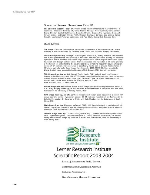
Continued from Page 199SCIENTIFIC SUPPORT SERVICES— PAGE 181LRI <strong>Scientific</strong> Support: People-empowered Cores provide infrastructure support for CCF researchers.Clockwise from top left, Cathy Stanko, Manager Flow Cytometry Core; CarmelBurns, Director, Central Cell Services Core; Earl Poptik, Director, the Hybridoma Core; JeffHowell, smiling, and Eldon Walker, Ph.D., Director, Computer Services, also smiling; JamesProudfit, Mechanical Prototype Laboratory; and Pam Clark, Central Cell Services Core.BACK COVERTop Image: Full color 3-dimensional tomographic assessment of the human coronary arteryanatomy in vivo in real time. By Geoffrey Vince, Ph.D., the Whitaker Imaging Laboratory.Second image from top, on right: Human cystic fibrosis (CF) airway epithelial cells infectedwith human parainfluenza virus (HPIV3) 24 hr earlier. Immunofluorescence staining for ribonucleoproteinof HPIV3 identifies virus within single infected cells and in large multinucleated syncytia,which form through cell-cell fusion. There is increased viral replication in CF cells, providinga mechanism for understanding the severe respiratory symptoms frequently requiring hospitalizationof CF children. Increased virus is specifically due to lack of antiviral host defense inCF airway epithelial cells. Cover image from Immunity, (<strong>2003</strong>) 18:619-630. From an article byZheng, S. et al. Image produced in the laboratory of S.C. Erzurum, M.D. Used with permission.Third image from top, on left: Normal T cells (nuclei DAPI stained, small blue) becometrapped in the Hyaluronic Acid (HA) (FITC stained, green) cables formed on a renal cell carcinomacell line (nuclei DAPI stained, large blue), SK-RC-45. The HA ligand, CD44 (Alexa 568stained, red), can be seen on both the RCC line and the T cells.Mark Thornton, from Dr. Jim Finke’s lab.Fourth image from top: Normal human femur. Image generated with high-resolution micro-CT,a 3D x-ray imaging technology, to evaluate bone microarchitecture in early bone loss and boneformation in the laboratory of Kimerly Powell, Ph.D.Fifth image from top, on left: Confocal micrograph of human colon tissue from a patient withactive ulcerative colitis. Hyaluronan (green), CD-44 (red) and nuclei (blue) are fluorescently labeledin this section. By Carol de la Motte, with Judy Drazba, from the Laboratory of ScottStrong M.D.Sixth image from top: Molecular surface of PINCH LIM domain involved in mediating cell adhesion.The regions colored in blue are involved in protein-protein recognition in focal adhesionassembly. From the laboratory of Jun Qin, Ph.D.Seventh image from top: Confocal micrograph of poly I:C-treated mouse colon parenchymalcells. Hyaluronan (green), TNF-stimulated gene 6 (TSG-6) (red) and nuclei (blue) are fluorescentlylabeled in this image. By Carol de la Motte, with Judy Drazba, from the Laboratory ofScott Strong M.D.<strong>Lerner</strong> <strong>Research</strong> Institute<strong>Scientific</strong> <strong>Report</strong> <strong>2003</strong>-<strong>2004</strong>RUSSELL J.VANDERBOOM, PH.D., EDITORCHRISTINE KASSUBA, EDITORIAL ASSISTANTJIM LANG, PHOTOGRAPHYDAVID SCHUMICK, MEDICAL ILLUSTRATOR200


