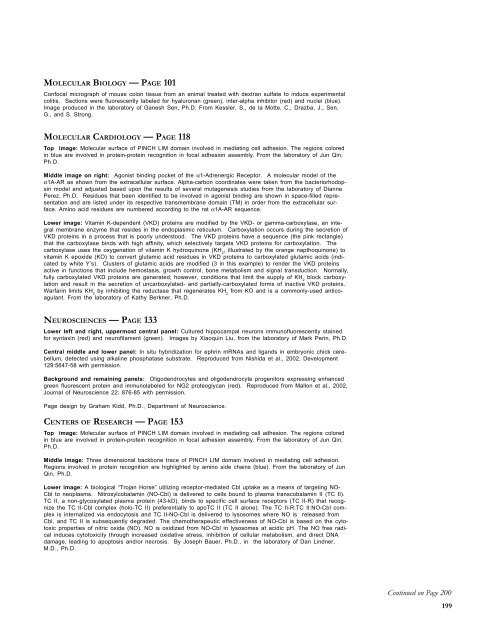
Illustrations LegendsBIOMEDICAL ENGINEERING—PAGE 11Top Left Triad: Stem Cell technology, the laboratory of George Muschler, M.D. Left Image: In Situ hybridizationexpression of Collagen Type 1 on day 6 Human CTPs in vitro. Control hybridized with sense to bothCbfa 1 and BMP6. Center Image: Proliferation of Human CTPs and Expression of Alkaline Phosphatase onLoaded Coralline HA disks, day 9 culture. Graphic Image on right: Schematic diagram of the osteoblasticstem cell system. This conceptual drawing illustrates the primary candidate populations of stem cells and transitcells thought to be associated with bone formation and remodeling: Vascular pericytes (green), Westen-Baintoncells (orange), type I or pre-osteoblasts (pink), secretory osteoblasts (maroon), osteocytes (brown), liningcells (purple), and adipocytes (yellow). Vascular pericytes may give rise to the Westen-Bainton cells. Pericytesand Westen-Bainton cells may contribute to the formation of pre-osteoblasts and also adipocytes. New osteoblastare added in the region immediately behind the advancing front of osteoclastic resorption. Secretory osteoblastsproduce new bone matrix until they become quiescent on the surface of bone as a lining cells (purple)or become embedded in the matrix as osteocytes (brown), or die via apoptosis. Osteoclast formation is also illustrated.A fraction of the monocytes population in systemic circulation (blue) will become resident in thebone marrow space. Osteoclasts are formed by fusion of monocytes resident in bone marrow to form multinucleatedfunctional units. The nuclei in active osteoclasts continue to be turned over as a result of nuclear lossand ongoing fusion events with new marrow derived monocytes. The black arrow indicates the direction of boneresorption by the osteoclastic front, followed by bone formation.Double panel, top right: Normal human femur contrasted with Human femur degenerated by osteoporosis(Top right twin panels). Images generated with high-resolution micro-CT, a 3D x-ray imaging technology, to evaluatebone microarchitecture in early bone loss and bone formation in the laboratory of Kimerly Powell, Ph.D.Central image: An array of microneedles (center image) 30 mm wide by 300 mm high in development in theBiological Micro Electrical Mechanisms Systems laboratory of Shuvo Roy, Ph.D. and Aaron Fleischman, Ph.D.Lower panel: “Biomechanics of a walk.” Graphic (lower panel) provided by Ton van den Bogert, Ph.D., BiomechanicsLaboratory.CANCER BIOLOGY—PAGE 44Top figure: Diagram illustrating the progression of prostate cancer. Human prostate cancer involves stagesthat correlate with loss of tumor suppressor genes. From Robert Silverman, Ph.D.Middle figure:Model hereditary prostate cancer family. From Graham Casey, Ph.D.Bottom figure: Immunohistochemical analysis of the expression of RNase L protein in a prostate tumor specimenfrom a mutation carrier. The cytoplasm of normal prostate epithelium stains positively (arrow on right),whereas the tumor cells are negative (arrow on left). From Robert Silverman, Ph.D.CELL BIOLOGY—PAGE 65Top image: Distribution of EGFP-fascin in a syndecan-1-activated cell, (top image). Activation of syndecan-1,by cell attachment to surfaces coated with either syndecan-1 antibody or thrombospondin-1, results in lamellipodialcell spreading and recruitment of fascin into the core F-actin bundles of microspikes and filopodia. Illustrationfrom the laboratory of Jo Adams.Lower Panels: Confocal micrographs showing surface binding (lower left) and internalization (lower right) ofHDL 2(red) and HDL 3(green) mediated by the scavenger receptor Bl in cultured adrenal cells. Areas of HDL 2and HDL 3colocalization are yellow. Images by Diane Green, B.S0., from the laboratory of Rick Morton, Ph.D.,with Judy Drazba, Ph.D., Imaging Core.IMMUNOLOGY — PAGE 85Top image: Normal T cells (nuclei DAPI stained, small blue) become trapped in the Hyaluronic Acid (HA) (FITCstained, green) cables formed on a renal cell carcinoma cell line (nuclei DAPI stained, large blue), SK-RC-45.The HA ligand, CD44 (Alexa 568 stained, red), can be seen on both the RCC line and the T cells.Mark Thornton, from Dr. Jim Finke’s lab.Lower image: Confocal micrograph of poly I:C-treated mouse colon parenchymal cells. Hyaluronan (green),TNF-stimulated gene 6 (TSG-6) (red) and nuclei (blue) are fluorescently labeled in this image. From de laMotte, C., Drazba, J., Hascall, V., Day, A., and S. Strong.198
MOLECULAR BIOLOGY — PAGE 101Confocal micrograph of mouse colon tissue from an animal treated with dextran sulfate to induce experimentalcolitis. Sections were fluorescently labeled for hyaluronan (green), inter-alpha inhibitor (red) and nuclei (blue).Image produced in the laboratory of Ganesh Sen, Ph.D. From Kessler, S., de la Motte, C., Drazba, J., Sen,G., and S. Strong.MOLECULAR CARDIOLOGY — PAGE 118Top image: Molecular surface of PINCH LIM domain involved in mediating cell adhesion. The regions coloredin blue are involved in protein-protein recognition in focal adhesion assembly. From the laboratory of Jun Qin,Ph.D.Middle image on right: Agonist binding pocket of the α1-Adrenergic Receptor. A molecular model of theα1A-AR as shown from the extracellular surface. Alpha-carbon coordinates were taken from the bacteriorhodopsinmodel and adjusted based upon the results of several mutagenesis studies from the laboratory of DiannePerez, Ph.D. Residues that been identified to be involved in agonist binding are shown in space-filled representationand are listed under its respective transmembrane domain (TM) in order from the extracellular surface.Amino acid residues are numbered according to the rat α1A-AR sequence.Lower image: Vitamin K-dependent (VKD) proteins are modified by the VKD- or gamma-carboxylase, an integralmembrane enzyme that resides in the endoplasmic reticulum. Carboxylation occurs during the secretion ofVKD proteins in a process that is poorly understood. The VKD proteins have a sequence (the pink rectangle)that the carboxylase binds with high affinity, which selectively targets VKD proteins for carboxylation. Thecarboxylase uses the oxygenation of vitamin K hydroquinone (KH 2, illustrated by the orange napthoquinone) tovitamin K epoxide (KO) to convert glutamic acid residues in VKD proteins to carboxylated glutamic acids (indicatedby white Y’s). Clusters of glutamic acids are modified (3 in this example) to render the VKD proteinsactive in functions that include hemostasis, growth control, bone metabolism and signal transduction. Normally,fully carboxylated VKD proteins are generated; however, conditions that limit the supply of KH 2block carboxylationand result in the secretion of uncarboxylated- and partially-carboxylated forms of inactive VKD proteins.Warfarin limits KH 2by inhibiting the reductase that regenerates KH 2from KO and is a commonly-used anticoagulant.From the laboratory of Kathy Berkner, Ph.D.NEUROSCIENCES — PAGE 133Lower left and right, uppermost central panel: Cultured hippocampal neurons immunofluorescently stainedfor syntaxin (red) and neurofilament (green). Images by Xiaoquin Liu, from the laboratory of Mark Perin, Ph.D.Central middle and lower panel: In situ hybridization for ephrin mRNAs and ligands in embryonic chick cerebellum,detected using alkaline phosphatase substrate. Reproduced from Nishida et al., 2002, Development129:5647-58 with permission.Background and remaining panels: Oligodendrocytes and oligodendrocyte progenitors expressing enhancedgreen fluorescent protein and immunolabeled for NG2 proteoglycan (red). Reproduced from Mallon et al., 2002,Journal of Neuroscience 22: 876-85 with permission.Page design by Graham Kidd, Ph.D., Department of Neuroscience.CENTERS OF RESEARCH — PAGE 153Top image: Molecular surface of PINCH LIM domain involved in mediating cell adhesion. The regions coloredin blue are involved in protein-protein recognition in focal adhesion assembly. From the laboratory of Jun Qin,Ph.D.Middle image: Three dimensional backbone trace of PINCH LIM domain involved in mediating cell adhesion.Regions involved in protein recognition are highlighted by amino side chains (blue). From the laboratory of JunQin, Ph.D.Lower image: A biological “Trojan Horse” utilizing receptor-mediated Cbl uptake as a means of targeting NO-Cbl to neoplasms. Nitrosylcobalamin (NO-Cbl) is delivered to cells bound to plasma transcobalamin II (TC II).TC II, a non-glycosylated plasma protein (43-kD), binds to specific cell surface receptors (TC II-R) that recognizethe TC II-Cbl complex (holo-TC II) preferentially to apoTC II (TC II alone). The TC II-R:TC II:NO-Cbl complexis internalized via endocytosis and TC II-NO-Cbl is delivered to lysosomes where NO is released fromCbl, and TC II is subsequently degraded. The chemotherapeutic effectiveness of NO-Cbl is based on the cytotoxicproperties of nitric oxide (NO). NO is oxidized from NO-Cbl in lysosomes at acidic pH. The NO free radicalinduces cytotoxicity through increased oxidative stress, inhibition of cellular metabolism, and direct DNAdamage, leading to apoptosis and/or necrosis. By Joseph Bauer, Ph.D., in the laboratory of Dan Lindner,M.D., Ph.D.Continued on Page 200199
- Page 7 and 8:
From the Chairman, Board of Governo
- Page 9 and 10:
Continued from Page 6sources. The p
- Page 11 and 12:
Continued from Page 8initiative rec
- Page 13 and 14:
BiomedicalEngineering
- Page 15 and 16:
The Department of Biomedical Engine
- Page 17 and 18:
The Department of Biomedical Engine
- Page 19 and 20:
The Department of Biomedical Engine
- Page 21 and 22:
Examples of devices that are being
- Page 23 and 24:
The Department of Biomedical Engine
- Page 25 and 26:
The Department of Biomedical Engine
- Page 27 and 28:
The Department of Biomedical Engine
- Page 29 and 30:
The Department of Biomedical Engine
- Page 31 and 32:
The Department of Biomedical Engine
- Page 33 and 34:
Deep brain stimulation (DBS) of the
- Page 36 and 37:
ORTHOPAEDICBIOLOGY ANDBIOENGINEERIN
- Page 38 and 39:
ORTHOPAEDICBIOLOGY ANDBIOENGINEERIN
- Page 40 and 41:
ORTHOPAEDICBIOLOGY ANDBIOENGINEERIN
- Page 42 and 43:
CONNECTIVETISSUEBIOLOGYTHE MIDURALA
- Page 44 and 45:
The Department of Biomedical Engine
- Page 46 and 47:
DEPARTMENT OFCANCER BIOLOGYCHAIRMAN
- Page 48 and 49:
The Department of Cancer Biology46D
- Page 50 and 51:
THE ALMASANLABORATORYVISITING SCHOL
- Page 52 and 53:
Graham Casey, Ph.D.THE CASEYLABORAT
- Page 54 and 55:
THE DANESHGARILABORATORYPOSTDOCTORA
- Page 56 and 57:
THE ERZURUMLABORATORYRESEARCH ASSOC
- Page 58 and 59:
THE HESTONLABORATORYSTAFF SCIENTIST
- Page 60 and 61:
The Department of Cancer BiologyIde
- Page 62 and 63:
THE SIZEMORELABORATORYRESEARCH FELL
- Page 64 and 65:
THE WILLIAMSLABORATORYPROJECT SCIEN
- Page 66 and 67:
THE YI LABORATORYPOSTDOCTORAL FELLO
- Page 68 and 69:
DEPARTMENT OFCELL BIOLOGYINTERIM CH
- Page 70 and 71:
The Department of Cell BiologyRole
- Page 72 and 73:
THE CHISOLMLABORATORYRESEARCH ASSOC
- Page 74 and 75:
THE DRISCOLLLABORATORYPOSTDOCTORAL
- Page 76 and 77:
THE HAZENLABORATORYRESEARCH ASSOCIA
- Page 78 and 79:
The Department of Cell BiologyHyper
- Page 80 and 81:
The Department of Cell BiologyRole
- Page 82 and 83:
THE J. SMITHLABORATORYTECHNOLOGISTG
- Page 84 and 85:
THE WEIMBSLABORATORYPROJECT SCIENTI
- Page 86 and 87:
The Department of Cell BiologyCell
- Page 88 and 89:
DEPARTMENTOF IMMUNOLOGYCHAIRMANThom
- Page 90 and 91:
THE ARONICALABORATORYSENIOR RESEARC
- Page 92 and 93:
THE FAIRCHILDLABORATORYPOSTDOCTORAL
- Page 94 and 95:
THE HAMILTONLABORATORYPROJECT SCIEN
- Page 96 and 97:
THE LARNERLABORATORYPOSTDOCTORAL FE
- Page 98 and 99:
THE M. SIEMIONOWLABORATORYRESEARCH
- Page 100 and 101:
The Department of ImmunologyHyaluro
- Page 102 and 103:
The Department of ImmunologyImmunol
- Page 104 and 105:
DEPARTMENT OFMOLECULAR BIOLOGYCHAIR
- Page 106 and 107:
THE GUDKOVLABORATORYPROJECT SCIENTI
- Page 108 and 109:
THE HARTERLABORATORYPOSTDOCTORAL FE
- Page 110 and 111:
THE PADGETTLABORATORYPROJECT SCIENT
- Page 112 and 113:
THE G. SENLABORATORYPROJECT SCIENTI
- Page 114 and 115:
THE STARKLABORATORYPROJECT SCIENTIS
- Page 116 and 117:
THE PELLETTLABORATORYRESEARCH ASSOC
- Page 118 and 119:
The Department of Molecular Biology
- Page 120 and 121:
DEPARTMENT OFMOLECULARCARDIOLOGYThe
- Page 122 and 123:
THE BERKNERLABORATORYPOSTDOCTORAL F
- Page 124 and 125:
THE J. FOXLABORATORYPROJECT SCIENTI
- Page 126 and 127:
THE KARNIKLABORATORYRESEARCH ASSOCI
- Page 128 and 129:
THE MORAVECLABORATORYRESEARCH SCHOL
- Page 130 and 131:
THE PLOWLABORATORYCOLLABORATORSTati
- Page 132 and 133:
THE S. SENLABORATORYPOSTDOCTORAL FE
- Page 134 and 135:
The Department of Molecular Cardiol
- Page 136 and 137:
DEPARTMENT OFNEUROSCIENCESCHAIRMANB
- Page 138 and 139:
THE BOULISLABORATORYPOSTDOCTORAL FE
- Page 140 and 141:
THE KOMUROLABORATORYCOLLABORATORSAr
- Page 142 and 143:
THE MACKLINLABORATORYPROJECT STAFFT
- Page 144 and 145:
THE NAJMLABORATORYCOLLABORATORSWill
- Page 146 and 147:
THE PERINLABORATORYPOSTDOCTORAL FEL
- Page 148 and 149:
THE RANSOHOFFLABORATORYThe Departme
- Page 150 and 151: THE STAUGAITISLABORATORYCOLLABORATO
- Page 152 and 153: THE TRAPPLABORATORYINVESTIGATORSAns
- Page 154 and 155: CENTER FORANESTHESIOLOGYRESEARCHDIR
- Page 156 and 157: Role of Receptors and Ion Channels
- Page 158 and 159: CENTER FORCEREBROVASCULASRESEARCHCe
- Page 160 and 161: THE JANIGROLABORATORYPROJECT SCIENT
- Page 162 and 163: THE HOLLYFIELDLABORATORYPROJECT SCI
- Page 164 and 165: THE ANAND-APTELABORATORYPOSTDOCTORA
- Page 166 and 167: THE HAGSTROMLABORATORYSENIOR TECHNO
- Page 168 and 169: THE V. PEREZLABORATORYTECHNOLOGISTA
- Page 170 and 171: Continued from Page 167active train
- Page 172 and 173: J.J. JACOBS CENTERFOR THROMBOSIS AN
- Page 174 and 175: THE BORDENLABORATORYRESEARCH ASSOCI
- Page 176 and 177: CLEVELAND CENTERFOR STRUCTURALBIOLO
- Page 178 and 179: THE QIN LABORATORYPOSTDOCTORAL FELL
- Page 180 and 181: The view from the skyway between th
- Page 182 and 183: OFFICE OFSPONSORED RESEARCHEXECUTIV
- Page 184 and 185: BIOLOGICAL RESOURCES UNITThe Clevel
- Page 186 and 187: IMAGING COREIMAGING COREDIRECTORJud
- Page 188 and 189: HYBRIDOMA COREHYBRIDOMA COREDIRECTO
- Page 190 and 191: LERNER RESEARCHINSTITUTECOMPUTING S
- Page 192 and 193: ELECTRONICS COREELECTRONICS COREMAN
- Page 194 and 195: MEDICAL SCIENTIFIC COMMUNICATIONSME
- Page 196 and 197: Keyword IndexACESen, I. 129Acoustic
- Page 198 and 199: Keyword IndexMacrophagesHamilton 92
- Page 202: Continued from Page 199SCIENTIFIC S


