Pleomorphic adenoma arising from the tail of the parotid gland ...
Pleomorphic adenoma arising from the tail of the parotid gland ...
Pleomorphic adenoma arising from the tail of the parotid gland ...

You also want an ePaper? Increase the reach of your titles
YUMPU automatically turns print PDFs into web optimized ePapers that Google loves.
Cases Journal<br />
BioMed Central<br />
Case Report<br />
<strong>Pleomorphic</strong> <strong>adenoma</strong> <strong>arising</strong> <strong>from</strong> <strong>the</strong> <strong>tail</strong> <strong>of</strong> <strong>the</strong> <strong>parotid</strong> <strong>gland</strong> –<br />
value <strong>of</strong> preoperative multi planar imaging: a case report<br />
R Vaidhyanath* 1 , S Harieaswar1 , C Kendall2 and P Conboy3 Open Access<br />
Address: 1Department <strong>of</strong> Radiology, Leicester Royal Infirmary, University Hospitals <strong>of</strong> Leicester, Leicester, LE1 5WW, UK, 2Department <strong>of</strong><br />
Histopathology, Leicester Royal Infirmary, University Hospitals <strong>of</strong> Leicester, Leicester, LE1 5WW, UK and 3Department <strong>of</strong> ENT, Leicester Royal<br />
Infirmary, University Hospitals <strong>of</strong> Leicester, Leicester, LE1 5WW, UK<br />
Email: R Vaidhyanath* - ram.vaidhyanath@uhl-tr.nhs.uk; S Harieaswar - sreemathi.harieaswar@uhl-tr.nhs.uk; C Kendall - charles.kendall@uhltr.nhs.uk;<br />
P Conboy - peter.conboy@uhl-tr.nhs.uk<br />
* Corresponding author<br />
Published: 3 July 2008<br />
Cases Journal 2008, 1:23 doi:10.1186/1757-1626-1-23<br />
This article is available <strong>from</strong>: http://www.casesjournal.com/content/1/1/23<br />
Abstract<br />
Background: Lesions <strong>of</strong> <strong>the</strong> '<strong>tail</strong>' <strong>of</strong> <strong>the</strong> <strong>parotid</strong> <strong>gland</strong> are difficult to assess clinically and provide<br />
a diagnostic dilemma on imaging, especially in <strong>the</strong> axial plane. Pedunculated lesions <strong>of</strong> <strong>the</strong> '<strong>tail</strong>' <strong>of</strong><br />
<strong>parotid</strong> can be mistaken for an extra <strong>parotid</strong> lesion. Accurate localisation <strong>of</strong> <strong>the</strong>se lesions on<br />
imaging is essential to assist <strong>the</strong> clinical diagnosis, to prevent inadequate/incomplete excision and<br />
complications, especially damage to facial nerve.<br />
Case Report: In this case report, we present a case <strong>of</strong> a pleomorphic <strong>adenoma</strong> <strong>arising</strong> <strong>from</strong> <strong>the</strong><br />
'<strong>tail</strong>' <strong>of</strong> <strong>the</strong> <strong>parotid</strong> <strong>gland</strong>, which on imaging, appears to be extra <strong>parotid</strong> in location. We also review<br />
<strong>the</strong> anatomy <strong>of</strong> <strong>the</strong> <strong>parotid</strong> '<strong>tail</strong>' and relevant literature.<br />
Conclusion: Lesions <strong>of</strong> <strong>the</strong> <strong>parotid</strong> '<strong>tail</strong>' are a diagnostic challenge to clinicians and Radiologists.<br />
Pedunculated lesions <strong>arising</strong> <strong>from</strong> <strong>the</strong> '<strong>tail</strong>' <strong>of</strong> <strong>the</strong> <strong>parotid</strong> <strong>gland</strong> can appear extra <strong>parotid</strong> in location.<br />
Knowledge <strong>of</strong> <strong>parotid</strong> <strong>gland</strong> anatomy and use <strong>of</strong> multi planar imaging is essential in <strong>the</strong> accurate<br />
localisation <strong>of</strong> <strong>the</strong>se lesions. This will also prevent inadequate or incomplete excision.<br />
Background<br />
Lesions <strong>of</strong> <strong>the</strong> '<strong>tail</strong>' <strong>of</strong> <strong>the</strong> <strong>parotid</strong> <strong>gland</strong> are difficult to<br />
assess clinically and provide a diagnostic dilemma on<br />
imaging, especially in <strong>the</strong> axial plane. Pedunculated<br />
lesions <strong>of</strong> <strong>the</strong> '<strong>tail</strong>' <strong>of</strong> <strong>parotid</strong> can be mistaken for an extra<br />
<strong>parotid</strong> lesion. Accurate localisation <strong>of</strong> <strong>the</strong>se lesions on<br />
imaging is essential to assist <strong>the</strong> clinical diagnosis, to prevent<br />
inadequate/incomplete excision and complications,<br />
especially damage to facial nerve.<br />
In this case report, we present a case <strong>of</strong> a pleomorphic <strong>adenoma</strong><br />
<strong>arising</strong> <strong>from</strong> <strong>the</strong> '<strong>tail</strong>' <strong>of</strong> <strong>the</strong> <strong>parotid</strong> <strong>gland</strong>, which on<br />
imaging, appears to be extra <strong>parotid</strong> in location. We also<br />
Received: 16 May 2008<br />
Accepted: 3 July 2008<br />
© 2008 Vaidhyanath et al; licensee BioMed Central Ltd.<br />
This is an Open Access article distributed under <strong>the</strong> terms <strong>of</strong> <strong>the</strong> Creative Commons Attribution License (http://creativecommons.org/licenses/by/2.0),<br />
which permits unrestricted use, distribution, and reproduction in any medium, provided <strong>the</strong> original work is properly cited.<br />
review <strong>the</strong> anatomy <strong>of</strong> <strong>the</strong> <strong>parotid</strong> '<strong>tail</strong>' and relevant literature.<br />
Case report<br />
A 65 yr old female presented to <strong>the</strong> Ear Nose and Throat<br />
[ENT] clinic <strong>of</strong> our institution with a one-month history<br />
<strong>of</strong> a lump in <strong>the</strong> right neck. On examination, a mobile<br />
lump was palpable in <strong>the</strong> angle <strong>of</strong> <strong>the</strong> mandible/upper<br />
cervical region. A flexible endoscopy did not reveal any<br />
mucosal lesion in <strong>the</strong> posterior nasal space, oropharynx,<br />
hypo pharynx or in <strong>the</strong> larynx. Clinically a lymph node<br />
mass was considered as a possible diagnosis. Fine needle<br />
Page 1 <strong>of</strong> 3<br />
(page number not for citation purposes)
Cases Journal 2008, 1:23 http://www.casesjournal.com/content/1/1/23<br />
aspiration [FNA] and Magnetic Resonance Imaging [MRI]<br />
were <strong>the</strong>n performed.<br />
MRI showed a well-defined oval shaped mass measuring<br />
3 × 2 cm just inferior to <strong>the</strong> right <strong>parotid</strong> <strong>gland</strong>. The epicenter<br />
<strong>of</strong> <strong>the</strong> lesion was in <strong>the</strong> inter muscular plane,<br />
medial to <strong>the</strong> sternocleidomastoid muscle but separate<br />
<strong>from</strong> it. It was heterogeneous on both T1 & T2 W images<br />
with a well defined margin (Fig 1 & 2). Following contrast<br />
administration, <strong>the</strong>re was moderate heterogeneous<br />
enhancement (Fig 3) In view <strong>of</strong> <strong>the</strong> location <strong>of</strong> <strong>the</strong> lesion,<br />
a differential diagnosis <strong>of</strong> lymph nodal mass & Spinal<br />
Accessory nerve Schwannoma were considered.<br />
FNA <strong>of</strong> <strong>the</strong> lesion showed a mix <strong>of</strong> bland epi<strong>the</strong>lial cell,<br />
clusters <strong>of</strong> spindle cells and myxoid matrix, which was<br />
typical <strong>of</strong> a pleomorphic <strong>adenoma</strong>.<br />
A decision to perform an excision biopsy was subsequently<br />
made. At surgery, <strong>the</strong> tumor was found to be a<br />
pedunculated mass <strong>arising</strong> <strong>from</strong> <strong>the</strong> inferior aspect <strong>of</strong> <strong>the</strong><br />
<strong>tail</strong> <strong>of</strong> <strong>the</strong> <strong>parotid</strong> <strong>gland</strong>. It was closely related and superficial<br />
to <strong>the</strong> spinal accessory nerve but separate <strong>from</strong> it.<br />
The mass was excised completely incorporating a limited<br />
cuff <strong>of</strong> macroscopically normal <strong>parotid</strong> tissue, taking care<br />
not to injure <strong>the</strong> inferior branches <strong>of</strong> <strong>the</strong> facial nerve.<br />
Histopathology <strong>of</strong> <strong>the</strong> excised specimen showed uniform<br />
proliferation <strong>of</strong> epi<strong>the</strong>lial elements within a patially<br />
T1 arrow) Figure & medial Coronal inferior 1to<br />
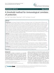
<strong>the</strong> image: to sternocleidomastoid <strong>the</strong> There right is <strong>parotid</strong> a well defined <strong>gland</strong> muscle (short mass (arrowheads) white (long arrow) white<br />
T1 Coronal image: There is a well defined mass (long white<br />
arrow) inferior to <strong>the</strong> right <strong>parotid</strong> <strong>gland</strong> (short white arrow)<br />
& medial to <strong>the</strong> sternocleidomastoid muscle (arrowheads).<br />
T2 Figure Axial 2image:<br />
The lesion has mixed signal intensity<br />
T2 Axial image: The lesion has mixed signal intensity. The<br />
lesion appears to be separate <strong>from</strong> <strong>the</strong> <strong>parotid</strong> <strong>gland</strong>.<br />
myxo-chondroid stroma, again, consistent with a diagnosis<br />
<strong>of</strong> a pleomorphic <strong>adenoma</strong>. There was no definite salivary<br />
<strong>gland</strong> tissue within <strong>the</strong> specimen.<br />
Post ment Figure contrast following 3 T1 contrast coronal administration<br />
image: There is moderate enhance-<br />
Post contrast T1 coronal image: There is moderate enhancement<br />
following contrast administration.<br />
Page 2 <strong>of</strong> 3<br />
(page number not for citation purposes)
Cases Journal 2008, 1:23 http://www.casesjournal.com/content/1/1/23<br />
Discussion<br />
The anatomy and description <strong>of</strong> <strong>the</strong> <strong>parotid</strong> '<strong>tail</strong>' is confusing<br />
as it is not a distinct anatomical entity. It is a triangular<br />
shaped <strong>parotid</strong> tissue seen anterolateral to <strong>the</strong><br />
sternocleidomastoid muscle, posterolateral to <strong>the</strong> posterior<br />
belly <strong>of</strong> diagastric muscle & deep to <strong>the</strong> platysma [1].<br />
Hamilton et al [2] define <strong>the</strong> '<strong>tail</strong>' <strong>of</strong> <strong>the</strong> <strong>parotid</strong> <strong>gland</strong> as<br />
inferior 2 cms <strong>of</strong> <strong>the</strong> superficial lobe <strong>of</strong> <strong>the</strong> <strong>gland</strong>. Many<br />
surgeons, however, consider <strong>the</strong> retromandibular portion<br />
<strong>of</strong> <strong>the</strong> superficial <strong>gland</strong> as <strong>the</strong> '<strong>tail</strong>' <strong>of</strong> <strong>the</strong> <strong>parotid</strong> <strong>gland</strong>.<br />
Lesions <strong>arising</strong> <strong>from</strong> <strong>the</strong> '<strong>tail</strong>' <strong>of</strong> <strong>parotid</strong> <strong>gland</strong> can be challenging<br />
to <strong>the</strong> clinicians and is <strong>of</strong>ten mistaken as a nodal<br />
mass or a posterior submandibular space lesion. This is<br />
particularly true <strong>of</strong> a pedunculated mass <strong>arising</strong> <strong>from</strong> <strong>the</strong><br />
inferior aspect <strong>of</strong> <strong>the</strong> <strong>parotid</strong> <strong>gland</strong>.<br />
Imaging plays an important role, not in <strong>the</strong> diagnosis <strong>of</strong><br />
<strong>the</strong> mass, but, to accurately locate <strong>the</strong> intra or extra<br />
<strong>parotid</strong> location <strong>of</strong> <strong>the</strong>se lesions. This has important<br />
implications on <strong>the</strong> surgical management <strong>of</strong> <strong>the</strong> patient.<br />
Parotidectomy with facial nerve preservation is <strong>the</strong><br />
method <strong>of</strong> choice in treating benign lesions <strong>of</strong> <strong>the</strong> <strong>parotid</strong><br />
<strong>gland</strong> [3]. This technique is not only diagnostic but minimises<br />
<strong>the</strong> risk <strong>of</strong> recurrence in most cases, with a very low<br />
risk <strong>of</strong> permanent facial nerve injury. There is a significant<br />
risk <strong>of</strong> tumour recurrence, <strong>of</strong>ten multi focal, with incomplete<br />
excision [4].<br />
On CT or MRI lesions <strong>arising</strong> <strong>from</strong> <strong>the</strong> <strong>parotid</strong> '<strong>tail</strong>' could<br />
appear to be extra <strong>parotid</strong> in location especially in <strong>the</strong><br />
axial plane. Lack <strong>of</strong> surrounding <strong>parotid</strong> tissue can only<br />
add to <strong>the</strong> confusion. Coronal images are helpful in<br />
assessing <strong>the</strong> location <strong>of</strong> <strong>the</strong> lesion. In general, lesions<br />
<strong>arising</strong> <strong>from</strong> <strong>the</strong> <strong>parotid</strong> '<strong>tail</strong>' (intra <strong>parotid</strong> mass) are<br />
anterolateral to <strong>the</strong> sternocleidomastoid muscle while<br />
extra <strong>parotid</strong> masses are anteromedial to sternocleidomastoid<br />
muscle [2]. In our patient, <strong>the</strong> mass was seen<br />
anteromedial to <strong>the</strong> sternocleidomastoid muscle in <strong>the</strong><br />
axial plane.<br />
The standard approach to a neck mass is clinical examination<br />
followed by fine needle aspiration. An ultrasound<br />
examination combined with fine needle aspiration is <strong>the</strong><br />
preferred choice. This allows <strong>the</strong> correct location <strong>of</strong> <strong>the</strong><br />
lesion and ensures adequate material is obtained for cytological<br />
examination.<br />
Lesions <strong>of</strong> <strong>the</strong> <strong>parotid</strong> '<strong>tail</strong>' are a diagnostic challenge to<br />
<strong>the</strong> imager. Knowledge <strong>of</strong> <strong>the</strong> <strong>parotid</strong> <strong>gland</strong> anatomy &<br />
use <strong>of</strong> multi planar imaging is essential in <strong>the</strong> accurate<br />
localisation <strong>of</strong> <strong>the</strong>se lesions.<br />
Conclusion<br />
Lesions <strong>of</strong> <strong>the</strong> <strong>parotid</strong> '<strong>tail</strong>' are a diagnostic challenge to<br />
clinicians and radiologists. Pedunculated lesions <strong>arising</strong><br />
<strong>from</strong> <strong>the</strong> '<strong>tail</strong>' <strong>of</strong> <strong>the</strong> <strong>parotid</strong> <strong>gland</strong> can appear extra<br />
<strong>parotid</strong> in location. Knowledge <strong>of</strong> <strong>parotid</strong> <strong>gland</strong> anatomy<br />
and use <strong>of</strong> multi planar imaging is essential in <strong>the</strong> accurate<br />
localisation <strong>of</strong> <strong>the</strong>se lesions. This will also prevent inadequate<br />
or incomplete excision.<br />
Competing interests<br />
The authors declare that <strong>the</strong>y have no competing interests.<br />
Authors' contributions<br />
RV, PC conceived <strong>the</strong> case report. SH prepared <strong>the</strong> manuscript.<br />
RV, PC, CK finalised <strong>the</strong> manuscript. All authors<br />
read and approved <strong>the</strong> final manuscript.<br />
Consent<br />
Written informed consent was obtained <strong>from</strong> <strong>the</strong> patient<br />
for publication <strong>of</strong> this case report and any accompanying<br />
images. A copy <strong>of</strong> <strong>the</strong> written consent is available for<br />
review by <strong>the</strong> Editor-in-Chief <strong>of</strong> this journal.<br />
References<br />
1. Som PM, Brandwein M: Salivary <strong>gland</strong>: Anatomy & Pathology.<br />
In Head and Neck Imaging Volume 2. 4th edition. Edited by: Som P,<br />
Curtin H. St. Louis: Mosby; 2003:2005-2133.<br />
2. Hamilton , Bronwyn E, Salzman , Karen L, Wiggins , Richard H III,<br />
Harnsberger HR: Earring Lesions <strong>of</strong> <strong>the</strong> Parotid Tail. Am J Neuroradiol<br />
2003, 24:1757-1764.<br />
3. H<strong>of</strong>fman H, Funk G, Endres D: Evaluation and surgical treatment<br />
<strong>of</strong> tumors <strong>of</strong> <strong>the</strong> salivary <strong>gland</strong>s. In Comprehensive Management<br />
<strong>of</strong> Head and Neck Tumors Volume 2. 2nd edition. Edited by:<br />
Thawley SE, Panje W, Batsakis L, Robert J. Philadelphia: W. B. Saunders<br />
Company; 1999:1147-1177.<br />
4. Eisele DW, Johns ME: Complications <strong>of</strong> surgery <strong>of</strong> <strong>the</strong> salivary<br />
<strong>gland</strong>s. In Complications in Head and Neck Surgery Edited by: David E.<br />
St. Louis: Mosby Year Book, Inc; 1993:183-200.<br />
Publish with BioMed Central and every<br />
scientist can read your work free <strong>of</strong> charge<br />
"BioMed Central will be <strong>the</strong> most significant development for<br />
disseminating <strong>the</strong> results <strong>of</strong> biomedical research in our lifetime."<br />
Sir Paul Nurse, Cancer Research UK<br />
Your research papers will be:<br />
available free <strong>of</strong> charge to <strong>the</strong> entire biomedical community<br />
peer reviewed and published immediately upon acceptance<br />
cited in PubMed and archived on PubMed Central<br />
yours — you keep <strong>the</strong> copyright<br />
BioMedcentral<br />
Submit your manuscript here:<br />
http://www.biomedcentral.com/info/publishing_adv.asp<br />
Page 3 <strong>of</strong> 3<br />
(page number not for citation purposes)