metabolism of diallyl disulfide by human liver microsomal ...
metabolism of diallyl disulfide by human liver microsomal ...
metabolism of diallyl disulfide by human liver microsomal ...

You also want an ePaper? Increase the reach of your titles
YUMPU automatically turns print PDFs into web optimized ePapers that Google loves.
0090-9556/99/2707-0835–841$02.00/0<br />
DRUG METABOLISM AND DISPOSITION Vol. 27, No. 7<br />
Copyright © 1999 <strong>by</strong> The American Society for Pharmacology and Experimental Therapeutics Printed in U.S.A.<br />
ABSTRACT:<br />
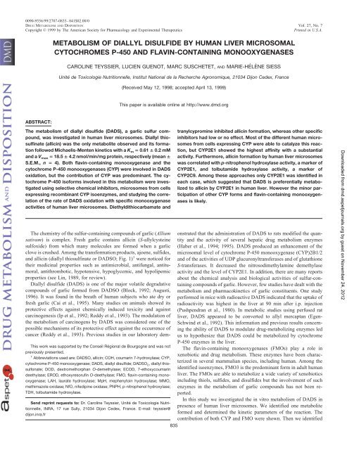
METABOLISM OF DIALLYL DISULFIDE BY HUMAN LIVER MICROSOMAL<br />
CYTOCHROMES P-450 AND FLAVIN-CONTAINING MONOOXYGENASES<br />
CAROLINE TEYSSIER, LUCIEN GUENOT, MARC SUSCHETET, AND MARIE-HÉLÈNE SIESS<br />
Unité de Toxicologie Nutritionnelle, Institut National de la Recherche Agronomique, 21034 Dijon Cedex, France<br />
The <strong>metabolism</strong> <strong>of</strong> <strong>diallyl</strong> <strong>disulfide</strong> (DADS), a garlic sulfur compound,<br />
was investigated in <strong>human</strong> <strong>liver</strong> microsomes. Diallyl thiosulfinate<br />
(allicin) was the only metabolite observed and its formation<br />
followed Michaelis-Menten kinetics with a K m � 0.61 � 0.2 mM<br />
and a V max � 18.5 � 4.2 nmol/min/mg protein, respectively (mean �<br />
S.E.M., n � 4). Both flavin-containing monooxygenase and the<br />
cytochrome P-450 monooxygenases (CYP) were involved in DADS<br />
oxidation, but the contribution <strong>of</strong> CYP was predominant. The cytochrome<br />
P-450 is<strong>of</strong>orms involved in this <strong>metabolism</strong> were investigated<br />
using selective chemical inhibitors, microsomes from cells<br />
expressing recombinant CYP isoenzymes, and studying the correlation<br />
<strong>of</strong> the rate <strong>of</strong> DADS oxidation with specific monooxygenase<br />
activities <strong>of</strong> <strong>human</strong> <strong>liver</strong> microsomes. Diethyldithiocarbamate and<br />
The chemistry <strong>of</strong> the sulfur-containing compounds <strong>of</strong> garlic (Allium<br />
sativum) is complex. Fresh garlic contains allicin (S-allylcysteine<br />
sulfoxide) from which many molecules are formed when a garlic<br />
clove is crushed. Among the transformation products, ajoene, sulfides,<br />
and allicin (<strong>diallyl</strong> thiosulfinate or DADSO; Fig. 1) 1 were noticed for<br />
their medicinal properties such as antimicrobial, antifungal, antitumoral,<br />
antithrombotic, hypotensive, hypoglycemic, and hypolipemic<br />
properties (see Lin, 1989, for review).<br />
Diallyl <strong>disulfide</strong> (DADS) is one <strong>of</strong> the major volatile degradative<br />
compounds <strong>of</strong> garlic formed from DADSO (Block, 1992; Augusti,<br />
1996). It was found in the breath <strong>of</strong> <strong>human</strong> subjects who ate dry or<br />
fresh garlic (Cai et al., 1995). Many studies on animals showed its<br />
protective effects against chemically induced toxicity and against<br />
carcinogenesis (Ip et al., 1992; Reddy et al., 1993). The modulation <strong>of</strong><br />
the <strong>metabolism</strong> <strong>of</strong> carcinogens <strong>by</strong> DADS was considered one <strong>of</strong> the<br />
possible mechanisms <strong>of</strong> its protective effect against the occurrence <strong>of</strong><br />
cancer (Reddy et al., 1993). Previous studies in our laboratory dem-<br />
This work was supported <strong>by</strong> the Conseil Régional de Bourgogne and was not<br />
previously presented.<br />
1 Abbreviations used are: DADSO, allicin; COH, coumarin 7-hydroxylase; CYP,<br />
cytochrome P-450 monooxygenase; DADS, <strong>diallyl</strong> <strong>disulfide</strong>; DADSO 2, <strong>diallyl</strong> thiosulfonate;<br />
DOD, dextromethorphan O-demethylase; ECOD, 7-ethoxycoumarin<br />
deethylase; EROD, ethoxyresorufin O-deethylase; FMO, flavin-containing monooxygenase;<br />
LAH, laurate hydroxylase; MpH, mephenytoin hydroxylase; MMO,<br />
methimazole oxidase; NfO, nifedipine oxidase; PNPH, p-nitrophenol hydroxylase;<br />
TDH, tolbutamide hydroxylase.<br />
Send reprint requests to: Dr. Caroline Teyssier, Unité de Toxicologie Nutritionnelle,<br />
INRA, 17 rue Sully, 21034 Dijon Cedex, France. E-mail: teyssier@<br />
dijon.inra.fr<br />
(Received May 12, 1998; accepted April 13, 1999)<br />
This paper is available online at http://www.dmd.org<br />
835<br />
tranylcypromine inhibited allicin formation, whereas other specific<br />
inhibitors had low or no effect. Most <strong>of</strong> the different <strong>human</strong> microsomes<br />
from cells expressing CYP were able to catalyze this reaction,<br />
but CYP2E1 showed the highest affinity with a substantial<br />
activity. Furthermore, allicin formation <strong>by</strong> <strong>human</strong> <strong>liver</strong> microsomes<br />
was correlated with p-nitrophenol hydroxylase activity, a marker <strong>of</strong><br />
CYP2E1, and tolbutamide hydroxylase activity, a marker <strong>of</strong><br />
CYP2C9. Among these approaches only CYP2E1 was identified in<br />
each case, which suggested that DADS is preferentially metabolized<br />
to allicin <strong>by</strong> CYP2E1 in <strong>human</strong> <strong>liver</strong>. However the minor participation<br />
<strong>of</strong> other CYP forms and flavin-containing monooxygenases<br />
is likely.<br />
onstrated that the administration <strong>of</strong> DADS to rats modified the quantity<br />
and the activity <strong>of</strong> several hepatic drug <strong>metabolism</strong> enzymes<br />
(Haber et al., 1994; 1995). DADS produced an enhancement <strong>of</strong> the<br />
<strong>microsomal</strong> level <strong>of</strong> cytochrome P-450 monooxygenase (CYP)2B1/2<br />
and <strong>of</strong> the activities <strong>of</strong> UDP glucuronyltransferases and <strong>of</strong> glutathione<br />
S-transferases. It decreased the nitrosodimethylamine demethylase<br />
activity and the level <strong>of</strong> CYP2E1. In addition, there are many reports<br />
about the chemical analysis and biological activities <strong>of</strong> sulfur-containing<br />
compounds <strong>of</strong> garlic. However, few studies have dealt with the<br />
<strong>metabolism</strong> and pharmacokinetics <strong>of</strong> garlic constituents. One study<br />
performed in mice with radioactive DADS indicated that the uptake <strong>of</strong><br />
radioactivity was highest in the <strong>liver</strong> at 90 min after i.p. injection<br />
(Pushpendran et al., 1980). In metabolic studies using perfused rat<br />
<strong>liver</strong>, DADS appeared to be converted to allyl mercaptan (Egen-<br />
Schwind et al., 1992). This information and previous results concerning<br />
the ability <strong>of</strong> DADS to modulate drug-metabolizing enzymes led<br />
us to hypothesize that DADS could be metabolized <strong>by</strong> cytochrome<br />
P-450 enzymes in the <strong>liver</strong>.<br />
The flavin-containing monooxygenases (FMOs) play a role in<br />
xenobiotic and drug <strong>metabolism</strong>. These enzymes have been characterized<br />
in several mammalian species, including <strong>human</strong>. Among the<br />
identified isoenzymes, FMO3 is the predominant form in adult <strong>human</strong><br />
<strong>liver</strong>. The FMOs are able to metabolize a wide variety <strong>of</strong> xenobiotics<br />
including thiols, sulfides, and <strong>disulfide</strong>s but the involvement <strong>of</strong> such<br />
enzymes in the <strong>metabolism</strong> <strong>of</strong> garlic compounds has not been reported.<br />
In this study we investigated the in vitro <strong>metabolism</strong> <strong>of</strong> DADS in<br />
presence <strong>of</strong> <strong>human</strong> <strong>liver</strong> microsomes. We identified one metabolite<br />
formed and determined the kinetic parameters <strong>of</strong> the reaction. The<br />
contribution <strong>of</strong> both CYP and FMO were shown. Then we identified<br />
Downloaded from<br />
dmd.aspetjournals.org <strong>by</strong> guest on November 24, 2012
836 TEYSSIER ET AL.<br />
FIG. 1.Structures <strong>of</strong> DADS and DADSO.<br />
the CYP isoenzymes mainly involved in the reaction using several<br />
approaches: 1) effect <strong>of</strong> selective chemical inhibitors on DADS oxidation,<br />
2) determination <strong>of</strong> DADS oxidation <strong>by</strong> microsomes from<br />
cells expressing recombinant CYP isoenzymes, and 3) study <strong>of</strong> the<br />
correlation <strong>of</strong> DADS oxidation with marker activities <strong>of</strong> CYP isoenzymes<br />
in different <strong>human</strong> <strong>liver</strong> microsomes.<br />
Experimental Procedures<br />
Materials. DADS (purity <strong>of</strong> 80%) was obtained from Aldrich Chemical Co.<br />
(Strasbourg, France). The other 20% were identified <strong>by</strong> HPLC as <strong>diallyl</strong><br />
sulfide and <strong>diallyl</strong> trisulfide. DADS was used without purification for the<br />
inhibition assays. DADS was further purified <strong>by</strong> vacuum distillation to a level<br />
<strong>of</strong> 95% for the determination <strong>of</strong> kinetic parameters and the study <strong>of</strong> the<br />
involvement <strong>of</strong> FMOs. DADSO was donated <strong>by</strong> Pr<strong>of</strong>essor Auger, Institut de<br />
Recherche sur la Biologie de l’Insecte, University F. Rabelais <strong>of</strong> Tours<br />
CYP<br />
EROD a<br />
1A2<br />
COH b<br />
2A6<br />
TDH b<br />
2C9<br />
TABLE 1<br />
Biochemical characteristics <strong>of</strong> <strong>human</strong> <strong>liver</strong> samples<br />
MpH a<br />
2C19<br />
(France). It was synthesized from the corresponding <strong>disulfide</strong> according to the<br />
method <strong>of</strong> Ferary (1996). 5,5-Dithiobis(2-nitrobenzoic acid), �-naphth<strong>of</strong>lavone,<br />
chlorzoxazone, coumarin, dextromethorphan, diethyldithiocarbamate,<br />
NADPH, nifedipine, orphenadrine, quinidine, sulfaphenazole, tolbutamide,<br />
tranylcypromine, and troleandomycin were purchased from Sigma-Aldrich<br />
Chimie (Saint Quentin Fallavier, France). Dextrorphan and S-(�)-mephenytoin<br />
were purchased from Ultrafine Chemicals (Salford, England). 1-Aminobenzotriazole<br />
was donated <strong>by</strong> Dr. Cabanne, Laboratoire de Phytopharmacie,<br />
Institut National de la Recherche Agronomique (Dijon, France). [1- 14 C]Lauric<br />
acid and [ 14 C] S-(�)-mephenytoin were purchased from Amersham Pharmacia<br />
Biotech (Les Ulis, France). Microsomes from B-lymphoblastoid cell lines<br />
expressing individual <strong>human</strong> CYPs or baculovirus-infected insect cells expressing<br />
individual <strong>human</strong> FMOs were purchased from Gentest (Woburn,<br />
MA). All other chemicals and reagents used were <strong>of</strong> the highest commercial<br />
quality available.<br />
Human Liver Samples. Human <strong>liver</strong> samples were provided <strong>by</strong> Pr<strong>of</strong>essor<br />
Favre, Département de Chirurgie Digestive Thoracique et Cancérologique <strong>of</strong><br />
the General Hospital <strong>of</strong> Dijon, France. The protocol was approved <strong>by</strong> a local<br />
ethics committee. The used tissues came from patients undergoing <strong>liver</strong> resection<br />
for various clinical reasons. The samples were frozen in liquid nitrogen<br />
and stored at �80°C until use for microsome preparation as described below.<br />
Biochemical characteristics <strong>of</strong> the microsomes are mentioned in Table 1.<br />
Preparation <strong>of</strong> Microsomal Fractions and Determination <strong>of</strong> Protein and<br />
Total CYP Contents. The microsomes were prepared as described previously<br />
(Haber et al., 1994). Microsomal proteins were quantified <strong>by</strong> the method <strong>of</strong><br />
Bradford (1976) using bovine serum albumin fraction V as standard. Cytochrome<br />
P-450 was assayed according to Omura and Sato (1964).<br />
Microsomal Metabolism <strong>of</strong> DADS. The reaction medium described below<br />
was the same for each experiment. This reaction medium consisted <strong>of</strong> <strong>human</strong><br />
<strong>liver</strong> microsomes corresponding to 300 pmol <strong>of</strong> CYP, 1 mM DADS, 1 mM<br />
DOD b<br />
2D6<br />
PNPH b<br />
2E1<br />
(�-1)LAH b<br />
2E1<br />
KS1 100 4.3 0.09 66.1 0.58 1.3 3.95 2.2 17.0<br />
K12 18 2.9 0.15 nd nd 3.9 6.05 2.9 5.8<br />
K17 36 2.7 0.43 0 2.72 2.1 6.41 1.0 24.3<br />
KS21 33 3.6 0.33 249 0.14 1.4 5.44 2.1 13.0<br />
K25 75 7.3 0.28 nd nd 1.8 7.04 1.8 19.8<br />
K26 45 4.5 0.37 nd 0.15 2.2 8.75 2.4 19.1<br />
KS27 22 2.1 0.08 162 0.14 0.6 2.47 0.6 7.7<br />
KS28 32 8.0 0.27 nd 0.03 1.9 5.49 2.7 15.4<br />
KT28 95 6.9 0.35 nd 0.06 2.2 6.18 4.5 15.7<br />
KS29 67 6.3 0.21 23.9 0.21 1.1 3.42 1.8 10.0<br />
KS30 81 2.6 0.28 95.1 0.35 1.4 5.90 2.0 12.8<br />
KT30 62 1.8 0.46 21.5 0.34 1.3 4.69 2.5 11.2<br />
K32 138 4.0 0.31 328 0.06 2.3 7.97 3.4 18.0<br />
K33 147 5.2 0.22 nd 0.35 1.7 4.54 3.7 15.7<br />
KT34 244 5.1 0.61 197 1.11 2.4 2.68 4.6 14.8<br />
KS38 45 6.8 0.03 nd 0.48 2.4 1.77 1.7 14.3<br />
K40 146 3.2 0.15 15.6 0.46 nd 3.89 1.8 12.2<br />
KS41 69 4.6 0.25 nd nd 1.8 3.73 2.2 17.9<br />
K44 100 4.1 0.17 149 0.02 2.4 2.28 2.4 13.7<br />
KS47 85 3.4 0.58 0.00 0.64 2.3 4.94 0.8 10.4<br />
KS48 100 3.7 0.33 175 0.67 3.3 6.21 0.9 19.8<br />
K49 20 5.0 0.49 17.1 0.89 2.7 4.39 0.8 13.5<br />
K50 36 6.0 0.36 294 0.18 1.5 3.14 0.5 11.1<br />
K51 99 4.7 0.78 91.4 1.00 3.0 12.85 1.7 22.8<br />
K52 114 4.7 0.79 354 nd 2.0 6.00 0.9 15.3<br />
K53 43 2.3 0.63 135 0.45 1.2 5.30 0.9 17.7<br />
K55 nd nd 0.68 4.3 0.42 nd nd nd nd<br />
K56 nd nd 0.69 54.5 1.61 nd nd nd nd<br />
KS57 nd nd 0.52 0.0 0.79 nd nd nd nd<br />
K59 nd nd 0.82 24.6 0.71 nd nd nd nd<br />
K61 nd nd 0.21 93.6 0.50 nd nd nd nd<br />
KS63 nd nd 0.94 18.9 nd nd nd nd nd<br />
KT63 nd nd 0.76 39.3 0.24 nd nd nd nd<br />
a pmol/min/nmol CYP<br />
b nmol/min/nmol CYP<br />
nd, not determined<br />
NfO b<br />
3A4<br />
(�)LAH b<br />
4A<br />
Downloaded from<br />
dmd.aspetjournals.org <strong>by</strong> guest on November 24, 2012
DIALLYL DISULFIDE METABOLISM BY HUMAN LIVER MONOOXYGENASES<br />
NADPH, 50 mM Tris-HCl pH 7 in a total volume <strong>of</strong> 500 �l. After 30 min at<br />
37°C, the reaction was stopped <strong>by</strong> adding 320 �l <strong>of</strong> acetonitrile. After 15 min<br />
<strong>of</strong> protein precipitation, the mixture was centrifuged at 10,500g for 10 min and<br />
40 �l <strong>of</strong> the supernatant was analyzed <strong>by</strong> HPLC. The same protocol was<br />
applied with 25 pmol for each cDNA-expressed <strong>human</strong> CYP microsome. In<br />
case <strong>of</strong> cDNA-expressed <strong>human</strong> FMO3 microsomes, 100 �g <strong>of</strong> protein was<br />
added in a total volume <strong>of</strong> 125 �l. Many DADS concentrations were used to<br />
determine the kinetic parameters.<br />
Thermal inactivation <strong>of</strong> microsomes was performed <strong>by</strong> a preincubation <strong>of</strong><br />
microsomes in Tris-HCl buffer for 10 min at 37°C in the absence <strong>of</strong> NADPH,<br />
or in the presence <strong>of</strong> NADPH for control microsomes. Then DADS and<br />
NAPDH were added subsequently to start the incubation for 30 min.<br />
Inhibition <strong>of</strong> DADS Metabolism. Inhibitors were added to the incubation<br />
mixtures before initiation <strong>of</strong> the reaction. Only with the mechanism-based<br />
inhibitors such as diethyldithiocarbamate or aminobenzotriazole, microsomes<br />
were preincubated for 10 min at 37°C before the addition <strong>of</strong> DADS. Nonhydrosoluble<br />
inhibitors were dissolved in 100% ethanol and the following volumes<br />
<strong>of</strong> ethanol were added to the incubation medium (total volume, 500 �l):<br />
0.2 �l for sulfaphenazole and orphenadrine, 0.3 �l for coumarin, 0.4 �l for<br />
diethyldithiocarbamate, 0.5 �l for aminobenzotriazole and quinidine, 0.8 �l<br />
for �-naphth<strong>of</strong>lavone, and 2.5 �l for nifedipine. The <strong>human</strong> samples KS1,<br />
K12, K25, KS28, K33, K40, KS41, and KS63 were used for the inhibitory<br />
experiments.<br />
HPLC Analysis. HPLC analysis was carried out using a Waters (Saint<br />
Quentin-en-Yrelines, France) system equipped with a model 600 pump, a<br />
model 717 auto sampler, a model 996 photodiode array UV detector, and a GL<br />
Sciences Inc. (Tokyo, Japan). Interstil ODS-3 column (4.6 � 150 mm). The<br />
flow rate was 0.6 ml/min and the solvent-isocratic program was 30:70 (v/v,<br />
acetonitrile/water) for 20 min. The spectrum from 190 to 300 nm was used to<br />
detect DADS and its metabolites. The quantification was made at 254 nm. Data<br />
were processed <strong>by</strong> Waters Millenium s<strong>of</strong>tware.<br />
Determination <strong>of</strong> Kinetic Constants. K m and V max were determined with<br />
a range <strong>of</strong> substrate concentrations <strong>of</strong> 0 to 7.5 mM with <strong>human</strong> <strong>liver</strong> microsomes<br />
and FMO cDNA-expressed microsomes, and 0 to 5 mM with CYP<br />
cDNA-expressed microsomes. The values were estimated <strong>by</strong> fitting the<br />
Michaelis-Menten equation using a nonlinear regression program <strong>of</strong> SAS<br />
S<strong>of</strong>tware (Cary, NC). The <strong>human</strong> samples KS1, KS30, K33, and K40 were<br />
used in this experiment. The apparent V max <strong>of</strong> FMO3 cDNA-expressed microsomes<br />
was determined considering that 100 �g <strong>of</strong> protein corresponded to<br />
approximately 100 pmol <strong>of</strong> enzyme. This consideration was based on the<br />
specific activity <strong>of</strong> recent lots <strong>of</strong> FMO3 from Gentest.<br />
Enzyme Assays. Detailed information for these assays are given Table 2.<br />
The coumarin 7-hydroxylase (COH) activity was determined according<br />
to Maurice et al. (1991); dextromethorphan O-demethylase (DOD) according<br />
to Bourrié et al. (1996); 7-ethoxycoumarin deethylase (ECOD) according to<br />
Ullrich and Weber (1972); ethoxyresorufin O-deethylase (EROD) according to<br />
Burke et al. (1985); laurate hydroxylase (LAH) according to Parker and Orton<br />
(1980) with few modifications; mephenytoin hydroxylase (MpH) according to<br />
Meier et al. (1985); methimazole oxidase (MMO) according to Dixit and<br />
Roche (1984); nifedipine oxidase (NfO) according to Guengerich et al. (1986);<br />
p-nitrophenol hydroxylase (PNPH) according to Tassaneeyakul et al. (1993);<br />
and tolbutamide hydroxylase (TDH) according to Bourrié et al. (1996).<br />
Correlation Analysis. To investigate the involvement <strong>of</strong> different CYP<br />
isoenzymes in DADS oxidation, marker activities <strong>of</strong> several isoenzymes<br />
(CYP1A2, CYP2A6, CYP2C9, CYP2C19, CYP2D6, CYP2E1, CYP3A4, and<br />
CYP4A) were measured for at least 24 samples <strong>of</strong> <strong>human</strong> <strong>liver</strong> microsomes.<br />
The enzyme assays were performed as described above. Calculations <strong>of</strong> the<br />
correlation coefficient between the rate <strong>of</strong> transformation <strong>of</strong> DADS and the<br />
marker CYP activities were made with the SAS system (Cary, NC) with the<br />
following equation: r � covariance (x,y) / (variance (x) � variance (y)) 1/2 .<br />
Results<br />
Identification <strong>of</strong> Metabolite. When DADS was incubated with<br />
<strong>human</strong> <strong>liver</strong> microsomes and NADPH, only one peak was detected <strong>by</strong><br />
HPLC (Fig. 2). This peak was identified as DADSO <strong>by</strong> comparing its<br />
retention time in different HPLC gradients and its absorption spectrum<br />
with that <strong>of</strong> synthesized DADSO. No other metabolite was<br />
detected for various incubation times <strong>of</strong> DADS. When either NADPH<br />
or microsomes were omitted, no DADSO was detected.<br />
Kinetics <strong>of</strong> Reaction. The formation <strong>of</strong> DADSO was linear over a<br />
period <strong>of</strong> 45 min. An incubation time <strong>of</strong> 30 min was therefore<br />
routinely used. A concentration <strong>of</strong> 1 mM NADPH was optimal for the<br />
reaction. The kinetic <strong>of</strong> formation <strong>of</strong> DADSO <strong>by</strong> <strong>liver</strong> microsomes<br />
was consistent with the Michaelis-Menten equation. The apparent Km was 0.61 � 0.2 mM and the apparent Vmax was 18.5 � 4.2 nmol/<br />
min/mg protein. Values are means � S.E.M. for four samples.<br />
Contribution <strong>of</strong> FMOs to DADS Oxidation. To evaluate the<br />
respective roles <strong>of</strong> FMOs and CYP in the oxidation <strong>of</strong> DADS, we<br />
initiated incubations <strong>of</strong> DADS in the presence <strong>of</strong> : 1) 1-aminobenzotriazole,<br />
a suicide inhibitor <strong>of</strong> CYPs (De Montellano and Mathews,<br />
1981; Fig. 3 shows the inhibition <strong>of</strong> DADS oxidation <strong>by</strong> this inhibi-<br />
TABLE 2<br />
Incubation conditions for the determination <strong>of</strong> marker enzyme activities in <strong>human</strong> <strong>liver</strong> microsomes<br />
COH DOD ECOD EROD LAH MpH MMO NfO PNPH TDH<br />
Buffer A B C D E B F G H B<br />
NADPH (mM) 1 1 0.05 0.25 1.5 1 0.1 1 1 1<br />
Microsomal proteins<br />
(mg/ml)<br />
0.28 1 0.13 1 0.125 2 0.3 2 1 2<br />
Substrate (mM) 0.1 0.05 1 0.001 0.13 0.2 1 0.2 0.1 2<br />
Total volume (ml) 0.36 0.5 2 0.4 0.5 0.5 1 0.5 0.5 1<br />
Temperature (°C) 37 37 30 30 37 37 37 37 37 37<br />
Incubation time (min) 3 20 4 3 15 30 5 10 20 30<br />
Stop reagent (�l) HCl 1N HCl 1N HCl 1N AcN/TCA<br />
20%, 50/50<br />
Perchloric acid 0.15 M H3PO4 HPLC separation Yes Yes Yes Yes Yes Yes<br />
UV detection (nm) 412 254 340 230<br />
Radiometric detection 14C 14C Fluorometric<br />
detection<br />
Excitation (nm)<br />
380 280 380 522<br />
Emission (nm) 450 312 450 586<br />
A: 50 mM sodium phosphate buffer (pH 7.4)<br />
B: 100 mM potassium phosphate buffer (pH 7.4)<br />
C: 100 mM Tris-HCl buffer (pH 7.7), 1 mM EDTA<br />
D: 100 mM Tris-HCl buffer (pH 7.7), 25 mM MgCl 2<br />
E: 66 mM Tris-HCl buffer (pH 7.4)<br />
F: 0.1 M tricine (pH 8.4), 1 mM EDTA, 0.06 mM 5,5�-dithiobis(2-nitrobenzoate), 6 mM potassium phosphate (pH 8.4), 0.02 mM dithiothreitol<br />
G: 50 mM potassium phosphate buffer (pH 7.4), 3 mM MgCl 2<br />
H: 100 mM sodium phosphate buffer (pH 6.8)<br />
837<br />
Downloaded from<br />
dmd.aspetjournals.org <strong>by</strong> guest on November 24, 2012
838 TEYSSIER ET AL.<br />
FIG. 2.HPLC pr<strong>of</strong>ile <strong>of</strong> an incubation <strong>of</strong> DADS with microsomes and NADPH,<br />
obtained with UV detection at 254 nm.<br />
If DADSO2 has been formed, its elution would be between the peak <strong>of</strong> NADPH<br />
(AU � 0.03) and the peak <strong>of</strong> DADSO.<br />
FIG. 3.Effects <strong>of</strong> 1-aminobenzotriazole on DADS oxidase activity.<br />
Values are mean <strong>of</strong> four samples � S.E.M. and are expressed relative to control<br />
(without inhibitor).<br />
TABLE 3<br />
Effect <strong>of</strong> thermal treatment <strong>of</strong> microsomes on few activities<br />
Values are the means <strong>of</strong> five determinations � S.E.M.<br />
CYP DADS a Oxidase MMO b<br />
ECOD b<br />
10 min 37°C 20.0 � 6.3 40.2 � 9.6 0.25 � 0.0<br />
Control 23.0 � 7.4 194 � 50.1 0.25 � 0.0<br />
a pmol/min/pmol CYP<br />
b nmol/min/mg protein<br />
tor), 2) microsomes containing irreversibly inactivated FMOs (this FMO<br />
inactivation is produced <strong>by</strong> heating the microsomes, Grothusen et al.,<br />
1996); the results indicated that FMO inactivation <strong>by</strong> heat treatment<br />
caused a 13% decrease <strong>of</strong> DADS oxidation (Table 3); it was verified that<br />
this thermal treatment inactivated the FMOs <strong>by</strong> measure <strong>of</strong> MMO activity<br />
although residual MMO activity was observed after 10 min heating;<br />
this was due to the CYP ability to metabolize the methimazole (results not<br />
shown and Poulsen et al., 1974); we checked also that thermal inactivation<br />
did not modify the ECOD activity; this reaction is <strong>of</strong>ten used as a<br />
standard marker for CYP-mediated reactions as several forms <strong>of</strong> CYP are<br />
involved in this activity), and 3) microsomes prepared from baculovirusinfected<br />
insect cell lines expressing the <strong>human</strong> FMO3 (in this medium,<br />
the same product DADSO was obtained as with <strong>human</strong> <strong>liver</strong> microsomes;<br />
the kinetic parameters <strong>of</strong> the reaction were calculated: K m �<br />
10.15 mM and V max � 41.9 pmol/min/pmol FMO3).<br />
Effect <strong>of</strong> Human CYP Inhibitors on DADS Oxidation. To further<br />
assess which CYP isoenzymes are involved in DADS oxidation,<br />
the effects <strong>of</strong> the following chemical inhibitors were determined:<br />
�-naphth<strong>of</strong>lavone (specific <strong>of</strong> CYP1A), chlorzoxazone (CYP2E1;<br />
Amet et al., 1995), coumarin (CYP2A6; Harris et al., 1994), diethyldithiocarbamate<br />
(CYP2E1; Guengerich et al., 1991), mephenytoin<br />
(CYP2C19; Harris et al., 1994), nifedipine (CYP3A4; Harris et al.,<br />
1994), orphenadrine (CYP2B6; Chang et al., 1993), quinidine<br />
(CYP2D6; Harris et al., 1994), sulfaphenazole (CYP2C9; Baldwin et<br />
al., 1995), tolbutamide (CYP2C9; Harris et al., 1994), tranylcypromine<br />
(CYP2C19; Postlind et al., 1998), and troleandomycin<br />
(CYP3A4; Rodrigues, 1994).<br />
The results are presented in Fig. 4. The strongest inhibitions were<br />
observed in the presence <strong>of</strong> diethyldithiocarbamate, followed <strong>by</strong> tranylcypromine.<br />
A slighter inhibition rate appeared with coumarin,<br />
chlorzoxazone, mephenytoin, nifedipine, and sulfaphenazole. Orphenadrine,<br />
tolbutamide, and troleandomycin had almost no effect on<br />
the DADSO formation, and �-naphth<strong>of</strong>lavone and quinidine had no<br />
effect at all. To verify the selectivity <strong>of</strong> some <strong>of</strong> the inhibitors used,<br />
we measured PNPH activity, a marker <strong>of</strong> CYP2E1, in the presence <strong>of</strong><br />
30 �M tranylcypromine, or <strong>of</strong> 22 or 200 �M coumarin. The result is<br />
shown in Table 4. Tranylcypromine and coumarin, described as<br />
selective inhibitors <strong>of</strong> CYP2C19 and CYP2A6 respectively, were also<br />
inhibitors <strong>of</strong> PNPH activity. These results suggest that tranylcypromine<br />
and coumarin can inhibit other CYPs, at least CYP2E1.<br />
Metabolism <strong>by</strong> Microsomes Containing cDNA-Expressed<br />
CYPs. The abilities <strong>of</strong> different <strong>human</strong> CYP enzymes to catalyze the<br />
S-oxidation <strong>of</strong> DADS to DADSO were investigated in many microsomes<br />
derived from cells expressing CYP DNA. CYP2A6, CYP2C8,<br />
CYP2C9, CYP2C19, CYP2D6, and CYP2E1 catalyzed the S-oxidation<br />
<strong>of</strong> DADS. CYP2D6, CYP2C19, and CYP2E1 were the most<br />
active enzymes. We can observe in Fig. 5, that every CYP is<strong>of</strong>orm<br />
followed a Michaelis-Menten kinetic except CYP2E1. For this latter<br />
enzyme, the DADS oxidase activity decreased quickly as soon as a<br />
certain amount <strong>of</strong> DADSO was produced. This could be explained <strong>by</strong><br />
the CYP2E1 mechanism-based inhibitor property <strong>of</strong> DADSO. No<br />
detectable amounts <strong>of</strong> DADSO were found when using microsomes<br />
expressing CYP1A2, CYP3A4, or CYP4A11, or when using microsomes<br />
from B-lymphoblastoid cells transfected with an empty vector.<br />
The kinetic parameters were determined for CYP2A6, CYP2C8,<br />
CYP2C9, CYP2C19, CYP2D6, and CYP2E1 (Table 5). CYP2E1<br />
exhibited the smallest K m whereas CYP2D6, CYP2C19, and CYP2E1<br />
had close V max values. The intrinsic clearance (V max/K m ratio) was<br />
2010 for CYP2E1 whereas it reached the maximum value <strong>of</strong> 108 for<br />
the other CYPs (Table 5).<br />
Correlation Study between DADS Oxidation and Specific CYP<br />
Activities. The correlation between DADS oxidation and CYP marker<br />
activities was analyzed with a panel <strong>of</strong> at least 24 individual samples<br />
<strong>of</strong> <strong>human</strong> <strong>liver</strong> microsomes. The characteristic transformations <strong>by</strong><br />
individual CYPs were EROD for CYP1A2, COH for CYP2A6, TDH<br />
for CYP2C9, MpH for CYP2C19, DOD for CYP2D6, (�-1)LAH and<br />
PNPH for CYP2E1, NfO for CYP3A4, and (�)LAH for CYP4A.<br />
These results are shown in Table 6. The best correlations (r) were<br />
found with PNPH, TDH, and (�-1)LAH activities, whereas the correlations<br />
with EROD, COH, DOD, NfO, MpH, and (�)LAH were<br />
very low.<br />
Downloaded from<br />
dmd.aspetjournals.org <strong>by</strong> guest on November 24, 2012
DIALLYL DISULFIDE METABOLISM BY HUMAN LIVER MONOOXYGENASES<br />
FIG. 4.Inhibition <strong>of</strong> DADS oxidation <strong>by</strong> chemical CYP inhibitors in <strong>human</strong> <strong>liver</strong> microsomes.<br />
Values are the percentage <strong>of</strong> control (without inhibitor). They are expressed as means � S.E.M. <strong>of</strong> four <strong>human</strong> samples.<br />
TABLE 4<br />
Inhibitory effects <strong>of</strong> various compounds on PNPH activity<br />
Values are the percentages <strong>of</strong> control activity (without inhibitor) and are means � S.E.M.<br />
<strong>of</strong> four <strong>human</strong> samples (KS27, KS30, KS44, and KS52).<br />
Compound PNPH Activity<br />
Tranylcypromine, 30 �M 21.3 � 1.0<br />
Coumarin, 22 �M 39.0 � 1.5<br />
Coumarin, 200 �M 34.6 � 1.8<br />
FIG. 5.Michaelis-Menten plot for the oxidation <strong>of</strong> DADS <strong>by</strong> <strong>human</strong><br />
cDNA-expressed CYP microsomes.<br />
Values are means <strong>of</strong> duplicate.<br />
Discussion<br />
We have studied the <strong>metabolism</strong> <strong>of</strong> DADS in the presence <strong>of</strong><br />
<strong>human</strong> <strong>liver</strong> microsomes and we have observed the appearance <strong>of</strong><br />
DADSO formed <strong>by</strong> the oxidation <strong>of</strong> one sulfur atom. Both DADS and<br />
TABLE 5<br />
Kinetic parameters <strong>of</strong> DADS oxidation <strong>by</strong> cDNA-expressed <strong>human</strong> CYP or<br />
FMO microsomes<br />
Values are the means <strong>of</strong> two determinations.<br />
Enzyme K m V max V max/K m<br />
mM pmol/min/pmol enzyme<br />
CYP1A2 nd nd<br />
CYP2A6 0.33 20.9 63.3<br />
CYP2C8 2.03 3.8 1.87<br />
CYP2C9 0.50 5.2 10.4<br />
CYP2C19 1.71 96.3 56.3<br />
CYP2D6 1.12 121 108.0<br />
CYP2E1 0.03 60.3 2010<br />
CYP3A4 nd nd<br />
CYP4A1 nd nd<br />
Control nd nd<br />
FMO3 10.15 41.9 4.13<br />
nd, not determined in absence <strong>of</strong> any activity.<br />
839<br />
DADSO are natural compounds found in crushed garlic but the latter<br />
has been considered as the most important biologically active compound<br />
<strong>of</strong> garlic (Reuter, 1995; Augusti, 1996). Among several sulfides<br />
from Allium studied, DADS exhibited in vivo the strongest<br />
anticarcinogenic properties (Ip et al., 1992; Haber-Mignard et al.,<br />
1996) and protected against carcinogen-induced DNA strand breaks<br />
(Le Bon et al., 1997). These effects were attributed to in vivo modulation<br />
<strong>of</strong> hepatic drug-metabolizing enzymes <strong>by</strong> DADS (Haber et al.,<br />
1994). Knowing the in vitro DADS <strong>metabolism</strong>, we suppose that the<br />
in vivo effects <strong>of</strong> DADS are in fact due to DADSO.<br />
In this study, we have observed the oxidation <strong>of</strong> DADS to sulfoxide.<br />
The in vivo <strong>metabolism</strong>s <strong>of</strong> <strong>diallyl</strong> sulfide (DAS; Brady et al.,<br />
1991, Chen et al., 1994) and <strong>of</strong> dipropyl sulfide (Nickson and Mitchell,<br />
1994), two sulfur compounds from Allium, implicate a first step <strong>of</strong><br />
oxidation to form a sulfoxide and a second one to form a sulfone. With<br />
DADS two steps <strong>of</strong> oxidation were not excluded, even if formation <strong>of</strong><br />
<strong>diallyl</strong> thiosulfonate (DADSO 2) was not observed in the incubation<br />
medium. In fact we have characterized DADSO as a mechanismbased<br />
inactivator <strong>of</strong> CYP2E1 (results not published). This should<br />
mean that DADSO could be oxidized to a reactive species (possibly<br />
Downloaded from<br />
dmd.aspetjournals.org <strong>by</strong> guest on November 24, 2012
840 TEYSSIER ET AL.<br />
TABLE 6<br />
Correlation between DADS oxidation and CYP is<strong>of</strong>orm specific activities in a<br />
panel <strong>of</strong> <strong>human</strong> <strong>liver</strong> microsomes<br />
Enzyme Activity CYP<br />
Correlation<br />
Coefficient (r)<br />
EROD 1A2 0.07<br />
COH 2A6 0.06<br />
TDH 2C9 0.52 a<br />
MpH 2C19 0.05<br />
DOD 2D6 0.05<br />
PNPH 2E1 0.63 b<br />
(�-1)LAH 2E1 0.40 c<br />
NfO 3A4 0.19<br />
(�)LAH 4A 0.07<br />
a p � .001, n � 33<br />
b p � .001, n � 25<br />
c p � .05, n � 26<br />
DADSO 2) that should interact with the enzyme and should not be<br />
released from the active site <strong>of</strong> the enzyme.<br />
Egen-Schwind et al. (1992) have studied the <strong>metabolism</strong> <strong>of</strong><br />
DADSO in a perfused rat <strong>liver</strong>. They observed that while passing<br />
through the <strong>liver</strong>, DADSO was metabolized to DADS. The discrepancy<br />
could be explained <strong>by</strong> the models that were used in both studies.<br />
They did not add NADPH during the <strong>liver</strong> perfusion. Due to the<br />
sensitivity <strong>of</strong> DADSO to temperature, a dismutation <strong>of</strong> DADSO in<br />
DADS could be possible. We observed the formation <strong>of</strong> DADS when<br />
DADSO was incubated with microsomes at 37°C without NADPH. In<br />
addition, Jin and Baillie (1997) studied the <strong>metabolism</strong> <strong>of</strong> DAS in rat.<br />
They proposed that the reduction <strong>of</strong> allyl sulfoxide to DAS was<br />
impossible with respect to the glutathione conjugates observed with<br />
rat fed with DAS or allyl sulfoxide.<br />
Flavin or CYPs are the only enzymes present in microsomes that<br />
can catalyze NADPH- and oxygen-dependent oxidation <strong>of</strong> xenobiotics.<br />
The inhibition <strong>of</strong> CYP <strong>by</strong> 1-aminobenzotriazole, a suicide inhibitor,<br />
as well as the irreversible inactivation <strong>of</strong> FMO <strong>by</strong> heating<br />
induced a decrease <strong>of</strong> the rate <strong>of</strong> DADS oxidation. Moreover, the<br />
DADS oxidation was observed with microsomes prepared from cells<br />
expressing <strong>human</strong> FMO3. These results suggest a contribution <strong>of</strong> both<br />
CYP and FMO-containing monooxygenases. Some results allow the<br />
evaluation <strong>of</strong> the FMO implication in this oxidation: its K m is much<br />
higher than the one obtained for FMOs associated with CYP in <strong>human</strong><br />
microsomes (10.15 versus 0.61 mM). The similar comparison made<br />
with cDNA-expressed isoenzymes gave 10.15 mM for FMO3 and<br />
0.03 mM for CYP2E1. When the incubation <strong>of</strong> microsomes and<br />
DADS was made in the presence <strong>of</strong> inhibited CYP, the DADS oxidase<br />
activity is very low, whereas when the FMOs were inactivated this<br />
activity was slightly decreased. Each <strong>of</strong> these results suggests that<br />
FMOs are less active than CYP in the <strong>metabolism</strong> <strong>of</strong> DADS. Nevertheless,<br />
to our knowledge, this is the first study that describes the<br />
implication <strong>of</strong> FMOs in the oxidation <strong>of</strong> sulfur compounds issued<br />
from garlic.<br />
Three approaches have been developed to identify the CYP isoenzymes<br />
involved in DADS oxidation. They were based on: 1) the use<br />
<strong>of</strong> cDNA-expressed CYP isoenzymes, 2) the use <strong>of</strong> selective chemical<br />
inhibitors, and 3) the study <strong>of</strong> the correlation <strong>of</strong> DADS oxidation<br />
activity with marker activities <strong>of</strong> CYP isoenzymes. Several pieces <strong>of</strong><br />
evidence indicate that CYP2E1 is the major cytochrome P-450 responsible<br />
for the metabolic process: 1) the rate <strong>of</strong> formation <strong>of</strong><br />
DADSO was substantially inhibited <strong>by</strong> the CYP2E1 inhibitors diethyldithiocarbamate<br />
and chlorzoxazone, 2) PNPH and (�-1)LAH activities<br />
(two marker activities <strong>of</strong> CYP2E1) in 25 individual <strong>human</strong> <strong>liver</strong><br />
microsomes exhibited the best correlation with the formation <strong>of</strong><br />
DADSO, and 3) with a K m <strong>of</strong> 0.03 mM and a V max/K m ratio <strong>of</strong> 2010<br />
calculated with cDNA-expressed CYP2E1, this isoenzyme exhibited<br />
the highest affinity and the highest intrinsic clearance among the<br />
is<strong>of</strong>orms tested. Even if the quantity <strong>of</strong> CYP2E1 in <strong>human</strong> microsomes<br />
was evaluated to 7% <strong>of</strong> whole CYP versus 18% for CYP2C and<br />
29% for CYP3A (Shimada et al., 1994), the relative involvement <strong>of</strong><br />
CYP2E1 is predominant.<br />
Nevertheless, our results suggest the involvement <strong>of</strong> other CYPs.<br />
Many isoenzymes, such as CYP2A6, CYP2C9, CYP2C19, CYP2D6,<br />
and CYP2E1 were able to oxidize DADS to DADSO. Their K m values<br />
showed that in a competitive context like in <strong>human</strong> microsomes, only<br />
CYP2E1 would be involved. The apparent velocity <strong>of</strong> CYP2E1 was<br />
not the highest one. We demonstrated that DADSO was a mechanismbased<br />
inhibitor <strong>of</strong> CYP2E1 (Martin, Teyssier and Siess, publication in<br />
preparation). This means that the more DADSO is produced, more<br />
CYP2E1 is inhibited. This observation may serve as an explanation<br />
for the low V max <strong>of</strong> CYP2E1.<br />
In the inhibitory experiments, tranylcypromine, an inhibitor described<br />
as being specific <strong>of</strong> CYP2C19 (Postlind et al., 1998), strongly<br />
inhibited DADS oxidase activity, suggesting the participation <strong>of</strong> this<br />
isoenzyme. However, the specificity <strong>of</strong> this inhibitor should be reconsidered<br />
because Draper et al. (1997) recently demonstrated that this<br />
molecule is among the strongest inhibitors <strong>of</strong> the coumarin hydroxylase<br />
activity (CYP2A6 activity marker), and we showed the PNPH<br />
activity inhibition <strong>by</strong> tranylcypromine. Therefore, CYP2C19 does not<br />
seem to be involved in DADS oxidation.<br />
DADS has a chemical structure similar to DAS; they differ only <strong>by</strong><br />
one sulfur atom. These two molecules are metabolized <strong>by</strong> CYP2E1<br />
and produce a mechanism-based inhibitor <strong>of</strong> CYP2E1. The <strong>metabolism</strong><br />
<strong>of</strong> DADS and its pathway is one more similarity between these<br />
two molecules issued <strong>of</strong> garlic.<br />
In conclusion, the oxidation <strong>of</strong> DADS to DADSO in <strong>human</strong> <strong>liver</strong><br />
microsomes is mainly mediated <strong>by</strong> CYPs but also <strong>by</strong> FMOs. Among<br />
the CYP isoenzymes, CYP2E1 seems to be the most involved isoenzyme<br />
even if a few other isoenzymes are also able to catalyze this<br />
reaction.<br />
Acknowledgments. We thank Jacques Auger for providing<br />
DADSO and Marie-France Vernevaut for her technical assistance.<br />
References<br />
Amet Y, Berthou F, Baird S, Dreano Y, Bail JP and Menez JF (1995) Validation <strong>of</strong> the<br />
(omega-1)-hydroxylation <strong>of</strong> lauric acid as an in vitro substrate probe for <strong>human</strong> <strong>liver</strong> CYP2E1.<br />
Biochem Pharmacol 50:1775–1782.<br />
Augusti KT (1996) Therapeutic values <strong>of</strong> onion (Allium cepa L) and garlic (Allium sativum L).<br />
Indian J Exp Biol 34:634–640.<br />
Baldwin SJ, Bloomer JC, Smith GJ, Ayrton AD, Clarke SE and Chenery RJ (1995) Ketoconazole<br />
and sulphaphenazole as the respective selective inhibitors <strong>of</strong> P4503A and 2C9. Xenobiotica<br />
25:261–270.<br />
Block E (1992) The organosulfur chemistry <strong>of</strong> the genus Allium. Implications for the organic<br />
chemistry <strong>of</strong> sulfur. Angew Chem Int Ed Eng 31:1135–1178.<br />
Bourrié M, Meunier V, Berger Y and Fabre G (1996) Cytochrome P450 is<strong>of</strong>orm inhibitors as a<br />
tool for the investigation <strong>of</strong> metabolic reactions catalyzed <strong>by</strong> <strong>human</strong> <strong>liver</strong> microsomes.<br />
J Pharmacol Exp Ther 277:321–332.<br />
Brady JF, Ishizaki H, Fukuto JM, Lin MC, Fadel A, Gapac JM and Yang CS (1991) Inhibition<br />
<strong>of</strong> cytochrome P-450 2E1 <strong>by</strong> <strong>diallyl</strong> sulfide and its metabolites. Chem Res Toxicol 4:642–647.<br />
Burke MD, Thompson S, Elcombe CR, Halpert J, Haaparanta T and Mayer RT (1985) Ethoxy-,<br />
pentoxy- and benzyloxyphenoxazones and homologues: A series <strong>of</strong> substrates to distinguish<br />
between different induced cytochromes P-450. Biochem Pharmacol 34:3337–3345.<br />
Cai Y, Baer-Dubowska W, Ashwood-Smith MJ, Ceska D, Tachibana S and Digiovanni J (1995)<br />
Mechanism based inactivation <strong>of</strong> hepatic ethoxyresorufin O-dealkylation activity <strong>by</strong> naturally<br />
occurring coumarin. Chem Res Toxicol 9:729–736.<br />
Chang T, Weber C, Crespi C and Waxman D (1993) Differential activation <strong>of</strong> cyclophosphamide<br />
and ifosphamide <strong>by</strong> cytochromes P450 2B and 3A in <strong>human</strong> <strong>liver</strong> microsomes. Cancer Res<br />
53:5629–5637.<br />
Chen L, Lee M, Hong JY, Huang W, Wang E and Yang CS (1994) Relationship between<br />
cytochrome P450 2E1 and acetone catabolism in rats as studied with <strong>diallyl</strong> sulfide as an<br />
inhibitor. Biochem Pharmacol 48:2199–2205.<br />
De Montellano O and Mathews JM (1981) Autocatalytic alkylation <strong>of</strong> the cytochrome P-450<br />
prosthetic haem group <strong>by</strong> 1-aminobenzotriazole. Isolation <strong>of</strong> an NN-bridged benzyneprotoporphyrin<br />
IX adduct. Biochem J 195:761–764.<br />
Dixit A and Roche TE (1984) Spectrophotometric assay <strong>of</strong> the flavin-containing monooxygenase<br />
Downloaded from<br />
dmd.aspetjournals.org <strong>by</strong> guest on November 24, 2012
DIALLYL DISULFIDE METABOLISM BY HUMAN LIVER MONOOXYGENASES<br />
and changes in its activity in female mouse <strong>liver</strong> with nutritional and diurnal conditions. Arch<br />
Biochem Biophys 233:50–63.<br />
Draper AJ, Madan A and Parkinson A (1997) Inhibition <strong>of</strong> coumarin 7-hydroxylase activity in<br />
<strong>human</strong> <strong>liver</strong> microsomes. Arch Biochem Biophys 341:47–61.<br />
Egen-Schwind C, Eckard R, Jekat FW and Winterh<strong>of</strong>f H (1992) Pharmacokinetics <strong>of</strong> vinyldithiins,<br />
transformation products <strong>of</strong> allicin. Planta Med 58:8–13.<br />
Ferary S (1996) Analyse des composés soufrés émis par quelques espèces végétales dans les<br />
relations plantes-insectes. P.h.D. thesis l’Université de Tours, France.<br />
Grothusen A, Hardt J, Brautigam L, Lang D and Bocker R (1996) A convenient method to<br />
discriminate between cytochrome P450 enzymes and flavin-containing monooxygenases in<br />
<strong>human</strong> <strong>liver</strong> microsomes. Arch Toxicol 71:64–71.<br />
Guengerich FP, Kim DH and Iwasaki M (1991) Role <strong>of</strong> <strong>human</strong> cytochrome P-450 IIE1 in the<br />
oxidation <strong>of</strong> many low molecular weight cancer suspects. Chem Res Toxicol 4:168–179.<br />
Guengerich FP, Martin MV, Beaune PH, Kremers P, Wolff T and Waxman DJ (1986) Characterization<br />
<strong>of</strong> rat and <strong>human</strong> <strong>liver</strong> <strong>microsomal</strong> cytochrome P-450 forms involved in nifedipine<br />
oxidation, a prototype for genetic polymorphism in oxidative drug <strong>metabolism</strong>. J Biol Chem<br />
261:5051–5060.<br />
Haber D, Siess MH, Canivenc-Lavier MC, Le Bon AM and Suschetet M (1995) Differential<br />
effects <strong>of</strong> dietary <strong>diallyl</strong> sulfide and <strong>diallyl</strong> <strong>disulfide</strong> on rat intestinal and hepatic drugmetabolizing<br />
enzymes. J Toxicol Environ Health 44:423–434.<br />
Haber D, Siess MH, De Waziers I, Beaune P and Suschetet M (1994) Modification <strong>of</strong> hepatic<br />
drug-metabolizing enzymes in rat fed naturally occurring allyl sulphides. Xenobiotica 24:169–<br />
182.<br />
Haber-Mignard D, Suschetet M, Berges R, Astorg P and Siess MH (1996) Inhibition <strong>of</strong> aflatoxin<br />
B1- and N-nitrosodiethylamine-induced <strong>liver</strong> preneoplastic foci in rats fed naturally occurring<br />
allyl sulfides. Nutr Cancer 25:61–70.<br />
Harris JW, Rahman A, Kim BR, Guengerich FP and Collins JM (1994) Metabolism <strong>of</strong> Taxol <strong>by</strong><br />
<strong>human</strong> hepatic microsomes and <strong>liver</strong> slices: Participation <strong>of</strong> cytochrome P450 3A4 and an<br />
unknown P450 enzyme. Cancer Res 54:4026–4035.<br />
Ip C, Lisk DJ and Stoewsand GS (1992) Mammary cancer prevention <strong>by</strong> regular garlic and<br />
selenium-enriched garlic. Nutr Cancer 17:279–286.<br />
Jin L and Baillie TA (1997) Metabolism <strong>of</strong> the chemoprotective agent <strong>diallyl</strong> sulfide to<br />
glutathione conjugates in rats. Chem Res Toxicol 10:318–327.<br />
Le Bon AM, Roy C, Dupont C and Suschetet M (1997) In vivo antigenotoxic effects <strong>of</strong> dietary<br />
allyl sulfides in the rat. Cancer Lett 114:131–134.<br />
841<br />
Lin RI (1989) Garlic in Nutrition and Medicine. Nutrition International Co., Irvine, CA.<br />
Maurice M, Emiliani S, Dalet BI, Derancourt J and Lange R (1991) Isolation and characterization<br />
<strong>of</strong> a cytochrome P450 <strong>of</strong> the IIA subfamily from <strong>human</strong> <strong>liver</strong> microsomes. Eur J Biochem<br />
200:511–517.<br />
Meier UT, Kronbach T and Meyer UA (1985) Assay <strong>of</strong> mephenytoin <strong>metabolism</strong> in <strong>human</strong> <strong>liver</strong><br />
microsomes <strong>by</strong> high-performance liquid chromatography. Anal Biochem 151:286–291.<br />
Nickson RM and Mitchell SC (1994) Fate <strong>of</strong> dipropyl sulphide and dipropyl sulphoxide in rat.<br />
Xenobiotica 24:157–168.<br />
Parker GL and Orton TC (1980) Induction <strong>by</strong> oxyisobutyrates <strong>of</strong> hepatic and kidney <strong>microsomal</strong><br />
cytochrome P-450 with specificity towards hydroxylation <strong>of</strong> fatty acids, in Biochem Biophys<br />
and Regulation <strong>of</strong> Cytochrome P-450 (Gustafsson JA, Carlstedt-Duke J, Mode A and Rafter<br />
J eds) p 373–377, Elsevier, Amsterdam.<br />
Postlind H, Danielson, Lindgren A and Andersson SHG (1998) Tolterodine, a new muscarinic<br />
receptor antagonist, is metabolized <strong>by</strong> cytochromes P450 2D6 and 3A in <strong>human</strong> <strong>liver</strong><br />
microsomes. Drug Metab Dispos 26:289–293.<br />
Poulsen LL, Hyslop RM and Ziegler DM (1974) S-oxygenation <strong>of</strong> N-substituted thioureas<br />
catalyzed <strong>by</strong> the pig <strong>liver</strong> <strong>microsomal</strong> FAD-containing monooxygenases. Arch Biochem<br />
Biophys 198:78–88.<br />
Pushpendran CK, Devasagayam TP, Chintalwar GJ, Banerji A and Eapen J (1980) The metabolic<br />
fate <strong>of</strong> [35S]-<strong>diallyl</strong> disulphide in mice. Experientia 36:1000–1001.<br />
Reddy BS, Rao CV, Rivenson A and Kell<strong>of</strong>f G (1993) Chemoprevention <strong>of</strong> colon carcinogenesis<br />
<strong>by</strong> organosulfur compounds. Cancer Res 53:3493–3498.<br />
Reuter HD (1995) Allium sativum and allium ursinum: Part 2. Pharmacology and medicinal<br />
application. Phytomedicine 2:73–91.<br />
Rodrigues AD (1994) Use <strong>of</strong> in vitro <strong>human</strong> <strong>metabolism</strong> studies in drug development. An<br />
industrial perspective. Biochem Pharmacol 48:2147–2156.<br />
Shimada T, Yamazaki H, Mimura M, Inui Y and Guengerich FP (1994) Interindividual variations<br />
in <strong>human</strong> <strong>liver</strong> cytochrome P-450 enzymes involved in the oxidation <strong>of</strong> drugs, carcinogens and<br />
toxic chemicals: Studies with <strong>liver</strong> microsomes <strong>of</strong> 30 Japanese and 30 Caucasians. J Pharmacol<br />
Exp Ther 270:414–423.<br />
Tassaneeyakul W, Veronese ME, Birkett DJ, Gonzalez FJ and Miners JO (1993) Validation <strong>of</strong><br />
4-nitrophenol as an in vitro substrate probe for <strong>human</strong> <strong>liver</strong> CYP2E1 using cDNA expression<br />
and <strong>microsomal</strong> kinetic techniques. Biochem Pharmacol 46:1975–1981.<br />
Ullrich V and Weber P (1972) The O-dealkylation <strong>of</strong> 7-ethoxycoumarin <strong>by</strong> <strong>liver</strong> microsomes. A<br />
direct fluorometric test. Hoppe-Seyler’s Z Physiol Chem 353:1171–1177.<br />
Downloaded from<br />
dmd.aspetjournals.org <strong>by</strong> guest on November 24, 2012