Mar - International Buffalo Information Centre
Mar - International Buffalo Information Centre
Mar - International Buffalo Information Centre

Create successful ePaper yourself
Turn your PDF publications into a flip-book with our unique Google optimized e-Paper software.
<strong>International</strong> <strong>Buffalo</strong> <strong>Information</strong> <strong>Centre</strong>Aims(IBIC)IBIC is a specialized information center onwater buffalo. Established in 1981 by KasetsartUniversity (Thailand) with an initial financialsupport from the <strong>International</strong> DevelopmentResearch Center (IDRC) of Canada. IBIC aims atbeing the buffalo information center of buffaloresearch community through out the world.Main Objectives1. To be world source on buffaloinformation2. To provide literature search andphotocopy services3. To disseminate information innewsletter4. To publish occasional publicationssuch as an inventory of ongoingresearch projectsBUFFALO BULLETINISSN : 0125-6726<strong>Buffalo</strong> Bulletin is published quarterly in <strong>Mar</strong>ch,June, September and December. Contributions onany aspect of research or development, progressreports of projects and news on buffalo will beconsidered for publication in the bulletin. Manuscriptsmust be written in English and follow theinstruction for authors which describe at inside ofthe back cover.EditorS. SophonPublisher<strong>International</strong> <strong>Buffalo</strong> <strong>Information</strong> <strong>Centre</strong>,Office of University Library,Kasetsart UniversityOnline availible:http://ibic.lib.ku.ac.th/e-BulletinBUFFALO BULLEITNIBIC, KASETSART UNIVERSITY, P.O. BOX 1084BANGKOK 10903, THAILANDURL : http://ibic.lib.ku.ac.thE-mail : libibic@ku.ac.thTel : 66-2-9428616 ext. 344Fax : 66-2-9406688
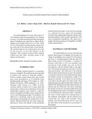
<strong>Buffalo</strong> Bulletin (<strong>Mar</strong>ch 2011) Vol.30 No.1lignocaine hydrochloride was given encircling thelesion. An elliptical skin incision was then takenat the level of fistulous opening. It became clearat surgery that the fistula involved the abomasum.Haemorrhage was controlled and abomasum wasfreed of adhesions, freshened and sutured by doublelayer of inversion sutures using chromic catgutno.1. The muscle wall and skin was freshened andsutured routinely.Post operatively dicrysticine sulfate 1 gmi/m for 5 days and meloxicam 0.5 mg/kg b.wt for 3days were administered. Antiseptic dressing of thesurgical wound was carried out and skin sutureswere removed on the 12 th post operative day.Animal was kept on fluid therapy for three postoperative days and thereafter a restricted soft dietwas allowed. The calf recovered uneventfully andno recurrence of fistulation was noticed during atwo-month observation period.DISCUSSIONFistulas of compound stomach have beenfrequently observed and usually associated withtrauma. Due to the infrequency of abomasalTable 1. Haemato-biochemical parameters observed on presentation.Haematological parameters:Hb (g%) PCV (%) TEC (x106/l) TLC (x103/l)8.0 30 4.62 6.31Biochemical parameters:SerumGlucose (mg%)SerumCalcium (mg%)SerumSodium (mmol/L)SerumPotassium (mmol/L)52.35 7.96 145.63 5.83Figure 1. Fistula communicating throughthe umbilical opening.Figure 2. Milk passing through the abomasalfistula2
<strong>Buffalo</strong> Bulletin (<strong>Mar</strong>ch 2011) Vol.30 No.1disorders in calves, they need to be recognized andtreated promptly to obtain a successful outcome(Nowrouzian, 1994). A case of reticular fistula ina buffalo (Singh, 2004) and omasal hernia withfistulation in a bullock (Bhardwaj et al., 2000)have been reported. The present case reportsthe successful surgical treatment of congenitalabomasal fistula in a buffalo calf. Thoughabomasum has been usually involved in fistula inheifer calves (Rijkenhuizen and Sickmann, 1994),it is also reported in association with hernia in acow (Balagopalan et al., 1993) and buffalo (Sobti etal., 1998), however associated with trauma. But thecause of the abnormality in the present case couldnot be ascertained.REFERENCESBalagopalan, T.P., K. Rajankutty and S.R. Nayar.1993. Ventral hernia associated withabomasal fistula in a cow - A clinical report.J. Vet. Anim. Sci., 24(2): 203-204.Bhardwaj, H.R., M.S. Bhadwal and M.M.S. Zama.2000. Surgical management of omasal herniawith fistulation in a cross-bred bullock.Indian J. Vet. Surg., 21(1): 60.Newman, S.J., T.L. Bailey, J.C. Jones, W.A.DiGrassie and W.D. Whittier. 1999. Multiplecongenital anomalies in a calf. J. Vet. Diagn.Invest., 11: 368-371.Nowrouzian, I., F. Adibhashemi, F. Shokuhi SabetJalali and M.R. Ahmadi. 1994. Clinicosurgicalstudy of abomasal disorders in Calf.J. Fac. Vet. Med. Univ. Tehran., 49(1-2): 31-40.Rijkenhuizen, A.B. and H.G. Sickmann. 1994.Incarcerated umbilical hernia withenterocutaneous fistula in a calf. TijdschrDiergeneeskd., 120(1): 8-10.Singh, R. 2004. Unusual case of reticular fistulain a buffalo treated under field condition.Abstracts from 28 th ISVS Congress, IndianJ. Vet. Surg., 25(2): 131.Sobti, V.K., N.S. Saini, K.I. Singh, P.S. Bansal andP.S. Simran. 1998. Surgical management ofabmomasal herniation and its fistulation in abuffalo. Indian Vet. J., 75: 1046-1047.3
Case Report<strong>Buffalo</strong> Bulletin (<strong>Mar</strong>ch 2011) Vol.30 No.1AN UNUSUAL CASE OF OESOPHAGEAL OBSTRUCTIONIN A FEMALE BUFFALON.V.V. Hari Krishna 1 , Makkena Sreenu 2 and V.S.C. Bose 3ABSTRACTThis paper reports a rare case ofoesophageal obstruction anterior to hiatusoesophagi along with ruminal tympany in a femalegraded Murrah buffalo caused by a palm kerneland its successful surgical treatment throughrumenotomy.Keywords: female buffaloes, Murrah, oesophagealINTRODUCTIONObstruction of the oesophagus is a rareoccurrence in ruminants. Apart from cattle,oesophageal obstruction has been occasionallyreported in buffaloes (Tyagi and Jit Singh, 1999).This paper reports a rare case of oesophagealobstruction at cardia, caused by a regurgitatedpalm kernel.HISTORY AND CLINICAL SIGNSA nine-year-old female graded Murrahbuffalo weighing about 420 kg was presented to theclinic with a history of not taking feed and water andsudden development of bloat since the morning. Ithad calved three months before and had been sentout for grazing daily. The bloat was relieved using16 G needle at a local hospital but the conditionrecurred. Clinical examination revealed distendedleft flank. Pulse, temperature and respiration werewithin normal physiological limits. On passageof a stomach tube, an obstruction was felt at thethoracic region anterior to the diaphragm. It wastentatively diagnosed as oesophageal obstruction,and it was decided to relieve the obstructionthrough rumenotomy, as thoracotomy in ruminantsrequires special equipment, and the procedure is adifficult one.RESULTS AND DISCUSSIONRumenotomy was performed followingstandard surgical procedure under inverted L -block using 2% Lignocaine hydrochloride. Onopening the rumen, surprisingly, it was full of palmkernels. The obstructing palm kernel anterior todiaphragm was taken out by hand with force afterclearing the entangled fibrous food material and allthe remaining palm kernels found in rumen werealso removed. Probiotics were placed in the rumenbefore closing it. The laparotomy wound was closed1TVCSC, NTR College of Veterinary Science, Sri Venkateswara Veterinary University, Gannavaram - 521102, A.P., India2Department of Surgery and Radiology3(PTT)4
<strong>Buffalo</strong> Bulletin (<strong>Mar</strong>ch 2011) Vol.30 No.1routinely. The animal was given streptopenicillin5.0 g i/m for seven days, meloxicam 20 ml i/mfor three days and DNS 2 L i/v for three days aspostoperative care. The cutaneous sutures wereremoved on 10 th postoperative day.The intraluminal oesophageal obstruction,commonly known as choke, may occur in buffaloesdue to vegetables, fruits and phytobezoars (Tyagiand Jit Singh, 1999) or to pieces of leather orrubber (Salunke et al., 2003). In ruminants,obstruction occurs mostly in the cervical region,and obstruction in the thoracic oesophagus is rare.In buffaloes, obstruction of the oesophagus hasmostly been recorded in the distal cervical regionas the lumen of the oesophagus narrows down atthe junction of middle and distal third (Tyagi and JitSingh, 1999). Madhava Rao et al. (2009) reporteda case of cervical oesophageal obstruction due tococonut, and thoracic oesophageal obstruction hasbeen reported by few workers (Ojha and Mohanthy,1970; Yadav et al., 2008). Shivprakash (2003)found a higher incidence in pregnant buffaloes andyoung calves and attributed the same to nutritionaldeficiencies, pica and difficulty in adaptationduring transition from milk to fodder, respectively.The prognosis of oesophageal obstruction is notalways favourable as the oesophageal surgeryis associated with various complications suchas suture dehiscence, perforation or fistula, andstenosis due to scar.In the present case, 92 palm kernels wererecovered from the rumen (Figure 1). It may be dueto the greediness of the animal during early rainyseason, during which time ripened palm kernels areabundant in the fields and swallowed by animalssent out for grazing. The probable reason for theunusual site of the obstruction was that when theanimal is ruminating one of the palm kernel hasmigrated and occluded at the hiatus oesophagi ofoesophagus anterior to diaphragm, and this resultedin its present condition. Attempts to push the objectwith a probang were not successful, perhaps dueto a change in position of kernel at the site, alongwith entangled food material which was removedFigure 1. Photographs showing the palm kernels recovered from the rumen.*Continued to page 95
Case Report<strong>Buffalo</strong> Bulletin (<strong>Mar</strong>ch 2011) Vol.30 No.1DIAGNOSIS AND MANAGEMENT OF POLIOENCEPHALOMALACIA IN INDIANBUFFALOES UNDER FARM CONDITIONSSumit Mahajan, Rajesh Agrawal and S.M. RashidABSTRACTIn this study, thiamine-responsivepolioencephalomalacia (PEM) was detected inseven non-descript buffaloes of a dairy farm inJammu and Kashmir State of India. Clinical casesshowed variable signs including disorientation,aimless walking with a high stepping gait dueto blindness, anorexia, opisthotonus or headretraction (star gazing), muscle tremor, convulsionand recumbency. The result of the study of clinicalcases treated by parenteral injection of thiaminepreparation showed a good response within 12 hafter initial treatment. The immediate responseto the specific treatment coupled with the reportsof general hematology and other diagnostic testused routinely in field was diagnostic for PEM anddifferentiated it from other neurological conditionsbased on the animals’ response to injection ofthiamine beside specific clinical findings.Keywords: polioencephalomalacia, thiamine,buffaloesINTRODUCTIONPolioencephalomalacia (PEM) is a noninfectiousthiamine responsive neurologicaldisease of buffaloes characterized by neurologicalmanifestations (Debasis et al., 2009). The diseaseis seen sporadically or as a herd outbreak, andgenerally young animals on high concentratediet are at high risk (Boyad et al., 1977). Theprobable mechanisms that may cause thiaminedeficiency PEM in ruminants are disorders ofabsorption, synthesize and destruction of thiamineby thiaminase (Bakker et al., 1980; Debasis et al.,2009; Jeffrey et al., 1994; Radostit et al., 2007). Thethiaminase produced by bacteria lead to declinein thiamine concentration in the digesta (Brent etal., 1984). Thiaminase-1 produced by the Bacillusthiaminalyticus and Clostridium sporogenes andthe thiaminase-11 by Bacillus aneurinalyticuscatalyze the cleavage of thiamine in the rumen andalso chemicals in drinking water, toxins releasedby nitrate-utilizing bacteria, and some toxic plantsin pastures are considered as the etiological agents(Dickie et al., 1979). Evidence linking thiaminewith the ruminant PEM disorder includes clinicalresponse to thiamine injection in some individuals(Mouli et al., 2004). Under the field conditions,presumptive diagnosis of PEM cases may dependon the pattern of clinical findings mentionedabove which may be suggestive for the disease.However, other diseases such as lead poisoning,focal symmetrical encephalomalacia, hepaticencephalopathy, or head trauma may show similarclinical signs (Milad et al., 2009). Laboratoryconfirmation of thiamine deficiency can bebased on histopathology and blood biochemistrySher-e-Kashmir University of Agricultural Sciences and Technology Jammu (J&K), India6
<strong>Buffalo</strong> Bulletin (<strong>Mar</strong>ch 2011) Vol.30 No.1including erythrocyte transketolase level, bloodpyruvate and lactate levels (Kiupel et al., 2003).The purpose of the present investigationwas to study and evaluate treatment with thiamineas a tool for rapid diagnosis of PEM and todifferentiate it from other neurological diseasesshowing similar nervous signs under conditionsof normal farm management in the absence oflaboratory facilities.MATERIALS AND METHODSThe clinical cases: In November 2009, asporadic naturally occurring outbreak of PEM wasreported from a farm house in Udhampur districtof Jammu and Kashmir State (India). A total ofseven animals were showing major complaintof inappetance, profuse salivation, sudden incoordinationwith occasional aimless movement,loss of vision and falling down. The clinical featuresrevealing congested mucus membrane, swollen andbulging tendency of eye ball, profuse salivation withfacial tremors and periodic convulsions. Althoughthere is ruminal atony in some cases, temperature,pulse and respiration were all in normal rangeswith slight variation along with normal micturationand defecation. Blood and urine samples werecollected for further investigation. Blood sampleswere subjected to routine hematological testslike total leukocyte count, differential leukocytecount, blood glucose level, blood hemoglobin leveland blood smear for blood protista examination,whereas urine samples were screened for presenceof ketone bodies by Rothera’s test.Treatment regimen: Initially thesymptomatic treatment includes triflupromazine,a potent neuroleptic agent used 0.25 mg/kg BWintramuscularly (I/M), Injection of atropinesulphate 0.20 mg/kg BW I/M, antibiotic injection ofdicrystine of 2.5 gm /animal I/M and intra venous(I/V) infusion of dextrose 10% 2000 ml /animalalong with 20 ml of multivitamin injection mixedwith dextrose for slow I/V infusion. However, oncethe hematological investigation revealed that all thevital blood parameters were in normal range withnegative reports of blood smears and Rothera’s test,keeping in mind the blood picture and the other tests,the cases were presumed to be cases of thiamineassociated PEM and accordingly high doses ofneurotropic vitamin-B injection (Inj. Neuroxin-12-v) were given 10 ml I/M twice a day for five days.RESULTS AND DISCUSSIONSThe thiamine associated PEM is aneurological disorder in buffaloes and is reportedoccasionally in India (Debasis et al., 2009) becauseof its difficult diagnosis under field conditions.The present outbreak was initially confused withpoisoning, ketosis, other similar neurologicaldisorders and blood protozoan infection. However,normal hemato-biochemical values coupledwith negative blood smear and Rothera’s test ledto the diagnosis of thiamine associated PEM,which was clinically confirmed by dramatic andinstant response to vitamin-B therapy. All theanimals showing signs of PEM responded to thetreatment within 12 h of injection with normalvision, stoppage of salivation, and normal gait,and this was consistent with the findings of othersin responsiveness toward thiamine treatment(Debasis et al., 2009; Gholami et al., 2003; Miladet al., 2009; Mouli et al., 2004). Laboratory testsused to confirm PEM in ruminants are basedupon blood chemistry to determine the depressedlevel of blood thiamine and RBC’s transketolase7
<strong>Buffalo</strong> Bulletin (<strong>Mar</strong>ch 2011) Vol.30 No.1activity and rising of pyruvate and lactate levels(Horino et al., 1994; Ramos et al., 2005; Ramos etal., 2006). Rumen and fecal samples may be testedfor thiamine activity; however, these tests are notroutinely available (<strong>Mar</strong>go et al., 2002). However,signs of progressive CNS involvement coupledwith the responsiveness to thiamine therapy asin the present study can be used as diagnosticfor thiamine induced PEM (Strain et al., 1990;Milad et al., 2009; Mouli et al., 2004). Successfultreatment of affected buffaloes in the present studywas attributed to early and aggressive therapy withthiamine. The recovery in all animals was steadyas all nervous signs and symptoms disappeared bythe third day post therapy.This study offers a method for earlydiagnosis of the PEM through rapid responseto parenteral treatment with no need to waitfor the results of laboratory tests, which takestime to confirm under field conditions. Earlytreatment is necessary for ruminants withPEM as the prognosis is considered favorableif the treatment is started early in the disease.ACKNOWLEDGEMENTThe authors wish to thank the Dean, Facultyof Veterinary Science and Animal Husbandry,SKUAST-Jammu, for providing necessary facilitiesto carry out this work.REFERENCESBakker, H.J., J. Dickson, P. Steele and M.C. Nottle.1980. Experimental induction of ovinepolioencephalomalacia. Vet. Record., 107:464-466.Bayod, J.W. and J.R. Walton. 1977. Cerebrocorticalnecrosis in ruminant: an attempt to identifythe source of thiaminase in affected animals.J. Comp. Pathol., 87: 930-935.Brent, B.E. and E.E. Bartley. 1984. Thiamin andniacin in the rumen. J. Anim. Sci., 59(3): 813-822.Debasis, J. and M. Ghosh. 2009. Management ofpolioencephalomalacia in a buffalo heifer: acase report. Northeast Vet., 11(2): 11.Dickie, C.W., R.J. Nelson, D.G. Frazee,L.D. Krugman and E. Browns. 1979.Polioenecephalomalacia in range cattle. J.Vet. Med. Assoc., 175; 5:460-462.Gholami, M.R., M.H. Hablolvarid, T. Bazargani andA. Ezzi. 2003. Case report of encephalopathyin goats. Arch. Razi Ins., 55: 111-116.Horino, R., T. Itabisahi and K. Hirano. 1994.Biochemical and pathological observationson sheep and calves dying of experimentalcerebrocortical necrosis. J. Vet. Med. Sci.,56: 481-485.Jeffrey, M.D., J.P. Duff, R.J. Higgins, V.R. Simpson,R. Jakman, T.O. Jons, S.C. Mechie and C.T.Liversey. 1994. Polioencephalomalaciaassociated with ingestion of ammoniumsulfate by sheep and cattle. Vet. Record.,134: 343-348.Kiupel, M., W.V. Alstine and C. Chilcoat.2003. Gross and microscopic lesions ofpolioencephalomalacia in a llama (lamaglama). J. Zoo Wildl Med., 34: 309-313.<strong>Mar</strong>go, R.M., M.W. Bryan, C. Christopher, C.<strong>Mar</strong>garet, B.B Ellen, H.W. Lisa and D.G.Pugh. 2002. Diseases of Neurologic System,Polioencephalomalacia, p. 309-311. InSheep and Goat Medicine, Pugh, D.G. (ed.)Saunders Company, U.S.A.Milad, K.E and G.S. Ridha. 2009. The occurrence of8
<strong>Buffalo</strong> Bulletin (<strong>Mar</strong>ch 2011) Vol.30 No.1thiamine-responsive polioencephalomalaciain dromedary breeding camels in Libya:preliminary investigation of diagnosis. IraqiJ. Vet. Sci., 23(1): 199-122.Mouli, S.P. and S.N. Babu. 2004. Thiamineresponsive polioencephalomalacia in buffalocalves. Indian Vet. J., 81(7): 819-820.Radostits, O.M., C.C. Gay, K.W. Hinchcliff andP.D. Constable. 2007. Veterinary Medicine:A Textbook of Cattle, Sheep, Pigs, Goats andHorses, 10 th ed. Bailliere Tindall Publishers,United Kingdom. 2156p.Ramos, J.J., M.F. Luis, G. Laura, F. Antonio andL. Araceli. 2005. Polioencephalomalacia inadult sheep grazing pastures with prostratepigweed. Can. Vet. J., 46: 59-61.Ramos, J.J., C. <strong>Mar</strong>ca, L.M.M. Ferrer, A. Losteand L.M. Cebrain. 2006. Fecal thiaminase,plasma lactate and pyruvate concentrationand erythrocyte transketolase activitychanges in apparently normal ewes after theinitiation to the pasture. Res. Vet. Sci., 80:11-16.Strain, G.M., M.S. Claxton, B.M. Olctt and S.E.Turnquist. 1990. Visual-evoked potentialsand electroretinograms in ruminants withthiamine responsive polioencephalomalaciaor suspected listeriosis. Am. J. Vet. Res., 51:1513-1517.*Continued from page 5first with fingers before retrieving the palm kernelfrom the oesophagus. The animal made uneventfulrecovery, and no complications were reported up to6 months post surgery.REFERENCESMadhava Rao, T., S. Bharathi and K.B.P.Raghavender. 2009. Oesophageal obstructionin a buffalo : a case report. Intas Polivet, 10:1-3.Ojha, S.C. and J. Mohanthy. 1970. Thoracicoesophgeal obstruction in a calf. Indian Vet.J., 47: 900-903.Salunke, V.M., M.S. Ali, A.P. Bhokre and V.S.Panchbhai. 2003. Oesophagotomy in standingposition : An easy approach to successfultreatment of oesophageal obstruction inbuffalo : A report of 18 cases. Intas Polivet,4: 366-367.Shivprakash, B.V. 2003. Pregnancy and young age:prone factors for oesophageal obstruction inbuffaloes. Intas Polivet, 4: 284-288.Tyagi, R.P.S. and Jit Singh. 1999. Ruminant Surgery,1 st . CBS Publishers and Distributors, NewDelhi, India. 192p.Yadav, G.U., M.G. Thorat, A.G. Somwamshi andM.J. Talekar. 2008. Thoracic oesophagealobstruction in a <strong>Mar</strong>athwadi buffalo. Vet.9
Case Report<strong>Buffalo</strong> Bulletin (<strong>Mar</strong>ch 2011) Vol.30 No.1AN OMPHALOCELE IN A BUFFALO CALF: A CASE REPORTP. Vidya Sagar, K.S.Vadde, K.S. Sai Krishna and S. VenkateswarluABSTRACTAn omphalocele in a buffalo calfimmediately after birth with ruptured amnion andits prognosis was reported.musculature and skin to the extent of 10 cm caudalto the sternum was noticed. No other congenitalabnormalities were found. It was diagnosed as anomphalocele, and it was decided to correct it byhernioraphy.Keywords: buffalo calf, umbilicus, omphaloceleTREATMENT AND DISCUSSIONINTRODUCTIONAn omphalocele is a congenital defectin the body wall in which eviscerated abdominalorgans are covered by amnion rather than skin(Baird, 1993). It should be differentiated fromumbilical hernias, in which skin covers over theherniated organs.CASE REPORT AND CLINICALHISTORYA new born graded Murrah buffalo calfpresented to the veterinary polyclinic within fewhours of birth with a history of prolapsed bowels.On gross examination, it was noticed the herniatedmass included the abomasum, small intestines andpart of the colon through the abdominal defect at theumbilical region (Figure 1). The amnion coveringthe herniated mass was ruptured and congestion ofthe everted organs set in. An absence of abdomenPrior to the surgery, the soiled herniatedmass thoroughly washed with normal salineand the calf was sedated with triflupromazinehydrochloride 0.01 mg/kg bwt.During laporotomy, it was found difficultto maintain the proper approximation of the edgesbecause of the absence of the abdomen musculatureand skin to a larger extent and with severe grosscontamination of the abdominal cavity, it wasdecided to euthanize the calf.An omphalocele is a hernia that occursin the embryo in which the abdominal contentsprotrude through the umbilicus and remain inthe umbilical stalk, therefore covered by theamnion (Noden and Lahunta, 1985). This probablyresults from the failure of normal withdrawal ofdeveloping intestinal loop. During early stages offetal development the intestines rest partly withinthe extra embryonic celome of the umbilical card.Later, the body wall encloses this area and theintestines are internalized. Failure of the intestinesto return or failure of the four body folds to migrateVeterinary Poly Clinic, Gudiwada, Krishna District, 521301, A.P., India10
<strong>Buffalo</strong> Bulletin (<strong>Mar</strong>ch 2011) Vol.30 No.1normally results in an omphalocele (Noden andLahunta, 1985). It is also called as congenitalumbilical hernia, abdominal fissure, umbilicaleventration and examphalocele.The cause of isolated omphalocele is notknown, and while it is a developmental defect, it isnot necessarily a heritable anomaly (Baird, 1993)although it has been suggested that it may be arecessive genetic trait (Ko et al., 1990).The prognosis with omphalocele isoften poor if severe abdominal contaminationand ischemic necrosis of the everted organsoccurres and there is extensive loss of abdominalmusculature and skin, as observed in the presentcase. Compared with the umbilical hernia, risk ofmorality was high. However, successful surgicalrecovery was achieved in some cases in whichthe amnion covering of the herniated organs waspresent (Baird, 1993).REFERENCESBaird, A.N. 1993. Omphalocele in two calves.JAVMA, 202: 1481-1482.Ko, J.C.H., L.E. Evans and J.S. Haynes. 1990.Multiple congenital defects in a female calf:a case report. Theriogenology, 34: 181-187.Noden, D.M., A. De Lahunta. 1985. Digestivesystem. In: The Embryology of DomesticAnimals. Williams and Wilkins, Baltimore,292-311.11
Case Report<strong>Buffalo</strong> Bulletin (<strong>Mar</strong>ch 2011) Vol.30 No.1DYSTOCIA DUE TO A CONJOINED TWIN MONSTER FOETUSIN A FEMALE BUFFALOS.P. Shukla, Qazi Mudasir and S.P. NemaABSTRACTA case of dystocia due to a conjoined twinmonster foetus with thoracopagus in a femalebuffalo is reported. An emergency cesareansection was decided upon to relieve the subsequentdystocia.Keywords: female buffalo, twin monster, dystosiaINTRODUCTIONafter proper lubrication revealed that the foetuswas in anterior longitudinal presentation, with twofore limbs protruding from the vulva and other twoforelimbs in flexed positions. Repulsion and deeperexploration revealed a conjoined twin monster, withthe presence of two foetal heads. The fetuses weredead, and it appeared to be a twin pregnancy astwo foetal heads joined at the thorax were palpable.Hence, it was diagnosed as a dystocia due to aconjoined twin monster foetus and an emergencycesarean section was decided upon. Previous firstcalving of the animal was reported to be normal.Foetal anomalies and monstrosities arecommon cause of dystocia in bovines (Shukla etal., 2007). Twin monsters are characterized byduplication of anterior, posterior or both parts offoetal body and are common in ruminants.CASE HISTORY AND OBSERVATIONSA pluriparous full-term pregnant Murrahbuffalo was presented to the Teaching VeterinaryClinical Services Complex, College of VeterinaryScience and Animal Husbandry, Mhow, withhistory of severe straining for the previous 12-14 h after the rupture of water bag. Two foetallegs were protruding from the vulva without anyprogress in parturition. Per vaginal examinationTREATMENT AND DISCUSSIONThe buffalo was premedicated withxylazine (0.2 mg/kg b.wt. I/M) and caudal epiduralanalgesia and local infiltration anaesthesia wasachieved using lignocaine Hcl. The left paramedianincision between linea alba and left subcutaneousabdominal vein was used for laparohysterotomy,and a dead conjoined twin monster foetus wasdelivered. The foetal membranes were alsoremoved and eight Furea boli (Nitrofurazone 60mg+urea 6 gm) were left in uterus. The uterus,peritoneum, muscles and skin were sutured inthe routine manner. Post operatively the animalwas given parenteral fluid therapy Inj. N.S (4 lts.)Inj. DNS (2 lts) Inj. Mifex (450 ml) and antibioticDepartment of Animal Reproduction Gynaecology and Obstetrics, College of Veterinary Science and AnimalHusbandry (Mhow) 453446, India12
<strong>Buffalo</strong> Bulletin (<strong>Mar</strong>ch 2011) Vol.30 No.1Table 1. Postmortem examination of fetuses.Internal organs Upper foetus Lower foetusHeart Present AbsentLungs Present AbsentLiver Present UnderdevelopedSpleen Present UnderdevelopedKidneys Normal NormalInternal genital organs Normal Normaltherapy using Strepto-Penicillin (2.5 gms) twicedaily with other supportive treatment includinganti-inflammatory and analgesics (Pheniraminemaleate 15 ml I/M and Meloxicam 15 ml I/M)once daily for the next 5 days. Antiseptic dressingof the surgical wound was done on alternate daysusing povidone iodine solution, and sutures wereopened on the 14 th postoperative day. The buffalomade an uneventful recovery.The twin monster had two normal heads,two necks, two pairs of fore and hind limbs withtwo separate abdominal areas but was joined atthorax (thoracopagus). Both the fetuses were offemale sex. Postmortem examination of fetuses ispresented in Table 1.Conjoined or fused symmetrical twinsare usually monozygotic in origin and representincomplete division of one embryo into twocomponents usually at the primitive streak ofdevelopmental stage and in the event they maydevelop into thoracopagus (Noden and Delahunta,1985). Conjoined twins are always indentical twinsand of the same sex (Arthur et al., 2001). Such twinsare usually due to non-inherited defects and oftenlead to severe dystocia (Roberts, 2004). In suchcases cesarean section undoubtedly is the methodfor choice for delivery (Sakthivel and Mathew,2000).REFERENCESArthur, G.H., D.E. Noakes, T.J. Parkinosonand G.C.W. England. 2001. VeterinaryReproduction and Obstetrics, 8 th ed. WBSaunders Company. London.Noden, D.M. and A. De Lahunta. 1985. TheEmbryology of Domestic Animals. Wiliamsand Wilkins, Baltimore. 367p.Roberts, S.J. 2004. Veterinary Obstetrics andGenital Diseases, 2 nd ed. CBS Publishers,New Delhi. India.Sakthivel, G. and P.V. Mathew. 2000. A report onconjoined twin monster in a non descriptcow. Indian Vet. J., 77(10): 906.Shukla, S.P., U.K. Garg, Anand Pandey, D.P.Dwivedi and S.P. Nema. 2007. Conjoinedtwin monster in a buffalo. Indian Vet. J., 84:630-631.13
Original Article<strong>Buffalo</strong> Bulletin (<strong>Mar</strong>ch 2011) Vol.30 No.1GENETIC POLYMORPHISM ANALYSIS OF MONOACYL GLYCEROL TRANSFEREASE2(MOGAT2) GENE IN MURRAH BUFFALO (Bubalus bubalis)D.S. Kale and B.R. YadavABSTRACTThe polymerase chain reaction singlestrandconformation polymorphism (PCR-SSCP)was identified within the Murrah buffalo MOGAT2gene. The result exhibited three SSCP patternsin the MOGAT2 gene with variable frequencyin samples studied, indicating that Murrahbuffaloes have genetic variability at that locus.This identified SSCP was confirmed by DNAsequencing which revealed one single nucleotidepolymorphism (SNP) viz, c.193T>C within 245 bpfragment of the MOGAT2 gene spanning exon 5of the Murrah buffalo. Identified SNP (c.193T>C)was used to genotype the 106 samples of Murrahbuffalo in which frequencies for C and T variantswere found to be 0.25 and 0.75, respectively. Thefrequency of genotypes within the breed group wasin accordance with Hardy-Weinberg proportions.Murrah buffalo MOGAT2 allelic variant sequencewas 95% pairwise similar with cattle sequenceand comparison of the two revealed elevencomputational SNPs. The statistical analysis usinggeneral linear model procedure (SYSTAT) forassociation study indicated that Murrah <strong>Buffalo</strong>MOGAT2 c.193T>C SNP genotypes did not differsignificantly (P>0.01) from Murrah buffalo milkproduction traits.Keywords: MOGAT2, SSCP, DNA sequencing,association study, Murrah buffaloINTRODUCTIONIndia is rich in buffalo genetic resourceshaving 97 million animals in 2003 (AnnualReport, 2006) which accounted for 59.5% to totalworld buffalo population. <strong>Buffalo</strong> milk contributes55.6% to the country’s total milk production. TheMurrah buffalo is the most important dairy breedwith superior genetic potential for milk production.However, their inherent potential for growth andproduction has not been exploited due to inadequateinformation about their genetic architecture. Theriver buffalo, along with domestic cattle, belongsto the subfamily Bovinae, and these species havebeen shown to be closely related, sharing homologyin chromosome banding and gene mapping (DiMeo et al., 2005) and have been cytogeneticallycharacterized in detail. The first generation wholegenome RH map of the river buffalo was reportedbased on comparison to domestic cattle (Amaralet al., 2008). Therefore these preliminary maps,based on cattle-derived markers, demonstrated thatthe bovine genome is a useful source of markersfor the buffalo genome mapping, allowing rapidLivestock Genome Analysis Laboratory, Dairy Cattle Breeding Division, National Dairy Research Institute(NDRI), Karnal-132001, Haryana State, India14
<strong>Buffalo</strong> Bulletin (<strong>Mar</strong>ch 2011) Vol.30 No.1and efficient transfer of information from cattle tobuffalo.The publication of the entire genomesequences of several livestock species willallow easy identification of genetic markers inbuffaloes, which will aid buffalo breeding andgenetic improvement. The comparative genomicsand genome analysis biotechniques have openednew possibilities for evaluation of the buffalogenome. The PCR-SSCP analysis (Orita et al.,1989) is a technique based on the principle thatsingle-stranded DNA molecules form specificsequence-based secondary structures under nondenaturingconditions. The association of geneticpolymorphisms with milk production traits andcomposition (Ganai et al., 2008) has stimulatedinterest in identifying the genetic markersinfluencing production traits which will be usedin marker assisted selection (MAS) to improveproductivity of farm animals.In the marker assisted selection of dairyanimals some genes are proposed as potentialcandidates associated with dairy performancetraits. Fat is one of the major constituents of milk.Triglycerides are the major energy storage moleculesin eukaryotes, and their final and presumablyrate-limiting step of synthesis is catalyzed by adiacylglycerol acyltransferase (DGAT). A fewyears back, DGAT1 was the first identified geneencoding a protein with DGAT activity in whicha mutation has been shown to be significantlyassociated with variation in milk fat percentage incattle (Grisart et al., 2002; Gautier et al., 2007).DGAT-like activity has also been shown in otherenzymes encoded by other genes and led to thedetection of diacylglycerol transferase2 (DGAT2),monoacyl glycerol transferases1 (MOGAT1) andmonoacyl glycerol transferases2 (MOGAT2),which are members of the same family (Winter etal., 2003). The members of this gene family showsimilarity in their nucleotide sequences and arosefrom same ancestral gene by duplication. However,this family has not yet been fully characterizedin any single mammalian species (Winter et al.,2003).As the MOGAT2 gene is in the familyrelated with QTL influencing milk production traitsand its functional role similarity in triglyceridesynthesis indicates that it might be a usefulcandidate to reveal genetic polymorphisms. As theMurrah buffalo is very important dairy breed ofbuffalo contributing the lions share in country’smilk production, it is necessary to screen candidategenes implicated for milk production, viz, theMOGAT2 in Murrah buffalo genome. Thereforethe present study was undertaken to detect geneticvariation in the MOGAT2 gene using PCR-SSCP followed by DNA sequencing and any findassociation with milk production traits.MATERIALS AND METHODSThe study group included 106 Murrahbuffaloes with milk production records fromthe Institute herd. Blood samples (10 ml) werecollected by jugular veinipuncture using vacuumtubes containing acid citrate dextrose solution(ACD) as an anticoagulant. Genomic DNA wasisolated from blood using the phenol chloroformextraction protocol (Clamp et al., 1993) withsome modifications. The integrity of the DNAwas assessed following electrophoresis in a 0.8%agarose gel with ethidium bromide staining. Inaddition, the OD ratio 260/280 nm was measuredto check for protein contamination and to calculatethe DNA concentration. All stock DNA sampleswere kept at -80 o C for longer storage, and the15
<strong>Buffalo</strong> Bulletin (<strong>Mar</strong>ch 2011) Vol.30 No.1working aliquots were maintained at - 20 o C.The PCR primers were designed for exonV of the MOGAT2 gene on the basis of cattle genesequence covering nucleotide substitution usingPRIMER3 software (http://www-genome.wi.mit.edu). The polymerase chain reaction (PCR) wascarried out on about 100 ng of genomic DNA ina 25 L reaction volume. The reaction mixtureconsisted of 2.5 L of 10x PCR assay buffercontaining 1.5 mM MgCl2, 200 M each of dNTPS,0.75 unit Taq DNA polymerase and 10 pmole ofeach primer (Integrated DNA Technologies, Inc).The primers (Forward primer: 5’-TTT GGT CTTATG CCC TAC CG-3’; Reverse primer: 5’-GGACAG GGT GAT CTT TTG GA-3’) were used foramplification of exon V of the <strong>Buffalo</strong> MOGAT2gene. Amplification was carried out in a Biometrathermal cycler using PCR cycling conditions as(95 o C for 5 minutes) and 34 cycles of 45 secondsat 95 o C, 65 o C and 72 o C consecutively, followedby a five minute final extension at 72 o C. The PCRamplification was verified by electrophoresis ofthe PCR products with loading dye on 2% (w/v)agarose gel in 0.5 x TBE buffer using a 100 bpladder as marker for confirmation of the lengthof the PCR products. The amplified products (5L) were detected on 2% agarose gel using 1 Lof loading dye as a stop dye, electrophoresed andvisualized using UV light.The MOGAT2 gene PCR products wereresolved by SSCP analysis using PCR product(5 ul), acrylamide concentration (18%), presenceglycerol (10%), voltage (200 volts), runningtime (24 hours) and temperature (15 o C). PCRproducts were diluted in denaturing solution (95%formamide, 10 mM NaOH, 0.05% xylene cyanoland 0.05% bromophenol blue, 20 mM EDTA) andheat denatured at 95 o C for ten minutes. The PCRproducts were resolved on a non-denaturing 18%acry1amide: bis-acrylamide (49: l) gel for SSCPanalysis. Gels were silver-stained (Sambrook andRussell, 2001) and photographed using a digitalcamera for SSCP pattern analysis.The PCR products representing differentSSCP patterns were directly got sequenced. Thenucleotide sequence analysis was carried outusing Geneious software. The DNA sequencepolymorphism observed was used to genotypeMurrah buffalo population. The frequency ofpolymorphic allele variant, genotypes and theiraccordance with Hardy-Weinberg law was assessedby POPGENE 1.31 software (http://www.ualberta.ca/~fyeh). The association between polymorphicallelic variants of the MOGAT2 gene and milkproduction traits was analyzed using GLMprocedure (SYSTAT). The following model wasused,Yijkl = + gi+ si+ pj + hk + eijklYij : bservation on jth animal ith genotype : population meangi : effect of ith genotype (i=1, 2)si : effect of i seasonpj : effect of j parityhk : effect of k year,eijkl : random errorRESULTS AND DISCUSSIONSThe SSCP analysis of amplified genefragments of exon 5 of the MOGAT2 gene resultedin three different patterns viz, A, B and C (Figure1)with the following frequencies in Murrah buffaloes(A = 0.49, B = 0.36, C = 0.15). This study hasrevealed the polymorphic nature of the 3 UTRregion of exon 5 of the buffalo MOGAT2 gene.The direct DNA sequencing and nucleotide16
<strong>Buffalo</strong> Bulletin (<strong>Mar</strong>ch 2011) Vol.30 No.1sequence analysis of MOGAT2 amplified PCRproducts (Table 1) representing different SSCPpatterns (A, B and C), revealed one SNP (T-Csubstitution) in exon 5 at the 193 rd nucleotide position(denoted as c.193 T>C) within the MOGAT2 genesequence of the Murrah buffalo (Figure 2). TheseMOGAT2 allelic variant nucleotide sequences wereanalyzed and submitted in NCBI GenBank withaccession No. (EU239373, EU239374). The natureof mutation was T-C transversion between SSCPpattern A and B confirming them as variants (Aand B). The SSCP patterns B and C had identicalnucleotide sequences; therefore only two variants(A and B) were confirmed. The PCR productsrepresenting SSCP pattern B were homozygote:TT in position 193 while SSCP patterns A and Bwere heterozygote: TC in position 193.The polymorphic Murrah buffaloMOGAT2 allelic variant sequence (EU239373)was compared with Bos taurus reference sequence(AJ534379) using alignment tool (GeneiousSoftware) which revealed eleven computationalmutations. The Murrah buffalo MOGAT2 variantsequence (EU239373) was 95% pairwise similarwith, the cattle sequence (AJ534379). A neighborjoiningtree (Figure 3) was constructed based oncomparison of the Murrah buffalo MOGAT2consensus sequence (EU239373) and consensusGenBank sequences of cattle (AC149756,NM_0010011154 and AJ534379) and buffaloes(EF208205, EU239373) at the same MOGAT2locus (Geneious Software). The phylogenetic treebased on partial consensus sequence agreed withtaxonomic relationship of cattle and buffaloes.The association analysis was carriedout between MOGAT2 c.193T>C SNP and milkproduction traits to find any relationship betweenthem. The ANOVA results indicated non-significant(P>0.01) effects of different Murrah buffaloMOGAT2 genotypes: c.193T>C TT and c.193T>CTC on 305 days milk yield, fat percentage as well asSNF percentage. The effect of non-genetic factors,viz. season, parity and year of calving on milkyield were found to be significant (pC genotypes are given in Table1.Among different candidates, the DGAT/MOGAT2 gene family with DGAT-like activityseems to be a promising candidate due to its geneticand functional similarity to established cattle QTL(DGAT1) influencing milk fat percentage (Winter etal., 2002). It is believed that SNPs occurring withinsuch genes may influence the milk productiontrait or at least be an effective DNA marker of aTable 1. Means and standard error (SE) of studied traits in reference to MOGAT2 c.193T>Cpolymorphism.Genotype n Milk Yield±SE FAT*±SE SNF*±SEc.193T>C TT 52 1775.81 NS ±241.06 0.297 NS ±0.005 0.315 NS ±0.001c.193T>C TC 54 1678.21 NS ±264.85 0.297 NS ±0.005 0.314 NS ±0.001Where,* are scale-transformed valuessuperscript NS are means not differing significantly at p0.05.17
<strong>Buffalo</strong> Bulletin (<strong>Mar</strong>ch 2011) Vol.30 No.1Figure 1. MOGAT2 PCR-SSCP genotype patterns resolved on 18% PAGE and visualized bysilver staining in Murrah buffalo.Figure 2. Multiple sequence alignment using CLUSTAL W (1.83) indicating T-Csubstitution at 193 rd position.Figure 3. Neighbor-Joining Tree based on Murrah buffalo MOGAT2 variant A consensussequence (EU239373) and related consensus sequences of cattle and buffaloes(Geneious Software).18
<strong>Buffalo</strong> Bulletin (<strong>Mar</strong>ch 2011) Vol.30 No.1sub-region of the dairy animal genome. In viewof, the above, the MOGAT2 gene was screenedfor polymorphism in Murrah buffalo using SSCPfollowed by sequencing and association study.In the present study, one SNP has beenidentified within the population at the MOGAT2gene locus of the Murrah buffalo. The identifiedSNP genotypes at the MOGAT2 gene locus didnot differ significantly from Murrah buffalomilk production traits. However in their bovineMOGAT2 gene polymorphism study, Winter et al.(2003) found 15 SNPs outside exons and two silentexon SNPs (ID 358 and 363) and reported nonsignificantassociation of allele frequencies withbreeding values for milk fat content in analyzeddairy breeds.CONCLUSIONrelated MOGAT genes in vivo is only just becomingavailable. In view of this, it is necessary to screen allthe regions of these genes in buffalo genome and tocontinue association studies as DGAT2/ MOGATgenes have genetic and functional similarity toDGAT1 influencing milk production traits ingenetically related Bos taurus. The identified DNApolymorphism after validation study will open uppossibilities for buffalo breeding and improvementin gene assisted selection.ACKNOWLEDGMENTSThe financial grant as a NationalFellowship to BR Yadav from the Indian Councilof Agricultural Research and assistance in thelaboratory by Mr. R.K. Tonk, Mr. Naresh Kumarand Mr. Nankoo Singh are acknowledged.The present study revealed that PCR-SSCP followed by DNA sequencing is aneffective molecular biological technique to detectDNA sequence variation at candidate gene lociin buffaloes. The identified SSCP within theMOGAT2 gene after DNA sequencing revealed oneSNP (c.193T>C) in exon 5 of the Murrah buffalo.However, the statistical analysis revealed nonsignificanteffect of the observed polymorphismgenotypes on Murrah buffalo milk productiontraits. The possible reasons for non-significanteffect might be the small size of the sample, absenceof some genotypes, high standard error and unevendistribution of data.The studies concerning associationsbetween DGAT gene polymorphism andproduction traits of riverine buffaloes are,however, fairly scarce. <strong>Information</strong> regarding theactual physiological role of DGAT2 and the closelyREFERENCESAmaral, M.E.J., J.R. Grant, P.K. Riggs, N.B.Stafuzza, E.A.R. Filho, T. Goldammer, R.Weikard and R.M. Brunner, K.J. Kochan,A.J. Greco, J. Jeong, Z. Cai, G. Lin, A.Prasad, S. Kumar, G.P. Saradhi, B. Mathew,M.A. Kumar, M.N. Miziara, P. <strong>Mar</strong>iani,A.R. Caetano, S.R. Galvao, M.S. Tantia,R.K. Vijh, B. Mishra, St.B. Kumar, V.A.Pelai, A.M. Santana, L.C. Fornitano, B.C.Jones, H. Tonhati, S. Moore, P. Stothardand J.E. Womack. 2008. A first generationwhole genome RH map of the river buffalowith comparison to domestic cattle. BMCGenomics, 9: 631.Annual Report. 2006. Department of AnimalHusbandry and Dairying, Government of19
<strong>Buffalo</strong> Bulletin (<strong>Mar</strong>ch 2011) Vol.30 No.1India, New Delhi. Available in www.dahd.nic.in.Clamp, P.A., R. Feltes, D. Shalvevet, J.E. Beever,E. Atac and L.B. Schook. 1993. Linkagerelationship between ALPL, EN01, GPI,PGD TGFB1 on porcine chromosome 6.Genomics, 17: 324-329.Di Meo, G.P., A. Perucatti, C. Uboldi, S. Roperto,D. Incarnato, F. Roperto, J. Williams, A.Eggen, L. Ferretti and L. Iannuzzi. 2005.Comparative mapping of the fragile histidinetriad (FHIT) gene in cattle, river buffalo,sheep and goat by FISH and assignment toBTA22 by RH-mapping: a comparison withHSA3. Anim. genet., 36(4): 363-364.Ganai, N.A., H. Bovenhuis, J.A.M. van Arendonkand M.H.P.W. Visker. 2008. Novelpolymorphisms in the bovine b-lactoglobulingene and their effects on b-lactoglobulinprotein concentration in milk. Anim. Genet.,40: 127-133.Gautier, M., A. Capitan, S. Fritz, A. Eggen, D.Boichard and T. Druet. 2007. Characterizationof the DGAT1 K232A and variable numberof tandem repeat polymorphisms in Frenchdairy cattle. J. Dairy Sci., 90: 2980-2988.Grisart, B., W. Coppieters, F. Farnir, L. Karim, C.Ford, P. Berzi, N. Cambisano, M. Mni, S.Reid, P. Simon, R. Spelman, M. Georges andR. Snell. 2002. Positional candidate cloningof a QTL in dairy cattle: identification of amissense mutation in the bovine DGAT1gene with major effect on milk yield andcomposition. Genome Res., 12: 222-231.Orita, M., Y. Suzuki, T. Sekiya and K. Hayashi.1989.Rapid and sensitive detection of pointmutation and DNA polymorphisms usingpolymerase chain reaction. Genomics, 5:874-879.Sambrook, J. and D.W. Russell. 2001. MolecularCloning: A Laboratory Manual, 3 rd ed. ColdSpring Harbour, Cold Spring LaboratoryPress, New York. 2,344p.Winter, A., M. Eckeveld van, O.R.P. Bininda-Emonds, F.A. Habermann and R. Fries.2003. Genomic organization of the DGAT2/MOGAT gene family in cattle (Bos taurus)and other mammals. Cytogenet GenomeRes., 102: 42-47.Winter, A., W. Kramer, F.A. Werner, S. Kollers,S. Kata, G. Durstewitz, J. Buitkamp,J.E. Womack, G. Thaller and R. Fries.2002. Association of a lysine-232/alaninepolymorphism in a bovine gene encodingacyl-CoA: diacylglycerol acyltransferase(DGAT1) with variation at a quantitativetrait locus for milk fat content. Proc. Natl.Acad. Sci.USA., 20: 20.20
Original Article<strong>Buffalo</strong> Bulletin (<strong>Mar</strong>ch 2011) Vol.30 No.1ABCG2 POLYMORPHISMS OF THE CHINESE BUFFALO (Bubalus bubalis)AND YAK (Bos grunniens) BREEDSSongjia LAI, Ming ZHANG,Changjun ZENG and Jie WANGABSTRACTThis article reports studies on polymorphismsof the ATP-binding cassette superfamily G member2 transporter (ABCG2) gene in 68 Chinese buffaloand 80 yak by DNA sequencing. We found fourmutations: A deletion, G insertion, single nucleotidepolymorphisms (SNPs) G/A and C/A. A deletionexisted in the buffalo and yak breeds. G insertion,(G/A) and (C/A) SNPs existed only in the buffalobreed. Results showed the Chinese buffalo and yakbreeds had four mutations. Especially, the buffalobreed had four mutations at least, and the theseprobably affected the milk yields, milk fat, proteinand dry matter percent. So the ABCG2 gene couldbe studied as a candidate gene effecting milk traits.Keywords : Chinese buffalo, yak, polymorphisms,ABCG2, milk traitsINTRODUCTIONA candidate gene can be defined as agene with biological effects on the physiology ofa trait of function or as a gene closed linked to afunctional gene. Polymorphisms within selectedcandidate genes can be tested for their associationwith quantitative traits and can be used in markerassistedselection (MAS) programs (Wu et al.,2005). Many studies have found segregatingQTL for milk production traits on Bos tauruschromosome (BTA) 6 in different cattle populations(Khatkar et al., 2004). There is strong evidencethat a polymorphism of the ATP-binding cassettesuperfamily G member 2 transporter (ABCG2)gene located on BTA 6 is associated with effects onmilk yield and composition in the Holstein cattle(Cohen-Zinder et al., 2005), dairy cattle (Olsen etal., 2007) and Indian cattle (Bos indicus) and buffalo(Bubalus bubalis) breeds (Tantia et al., 2006).Cohen-Zinder et al. (2005) found a single nucleotidepolymorphism (A/C) in exon 14, encoding asubstitution of tyrosine-581 to serine (Y581S), inthe ABCG2 transporter gene. The ABCG2 genewas greatly induced during late pregnancy andespecially during lactation (Jonker et al., 2005).The protein encoded by ABCG2, a member ofthe ATP binding cassette (ABC) superfamily,transports various xenobiotics and cytostaticdrugs across the plasma membrane (Litman etal., 2000). Jonker et al. (2005) demonstrated thatABCG2 is responsible for the active secretion ofclinically and toxicologically important substratesinto mouse milk, and that mice homozygous foran ABCG2 knock-out mutation lack this function.Chinese buffalo and yak, distributed in the southand west of China, have a low milk yield, but highmilk protein and fat percentages compared tocattle and have rarely been studied. The purpose ofthis article was to investigate the polymorphismsof the ABCG2 gene in the Chinese buffaloCollege of Animal Science and Technology, Sichuan Agricultural University, Ya’an, 625014, China21
<strong>Buffalo</strong> Bulletin (<strong>Mar</strong>ch 2011) Vol.30 No.1(Bubalus bubalis) and yak (Bos grunniens) breeds.Clustal W program (Thompson et al., 1994).MATERIALS AND METHODSRESULTS AND DISCUSSIONSixty-eight Chinese buffalo (Anhui buffalon=18, Fuling buffalo n=16, Jianghan buffalon=17, Yunnan buffalo n=17) and 80 yak (Jiulongyak n=20, Maiwa yak n=20, Tianzhu yak n=20,Xizang yak n=20) blood samples were collectedfrom the south and west of China. The individualswere unrelated based on the data of owners.Total genomic DNA was extracted by a standardphenol-chloroform extraction method. Theprimers for exon 14 of ABCG2 were: forward 5´-CAGGGCTGTTGGTAAATCTCA- 3(nt62491-511)andreverse5´-GCACGGTGACAGATAAGGAGA-3´(nt 62580-600) from NCBI database AJ871176.PCR amplifications were standardized usinggradient PCR (iCycler, Hercules, CA, USA) fromTantia et al. (2006). Touch down PCR profileincluded 5 minutes at 95 o C; 5 cycles 45s at 95 o C,45s at 57 o C, 45s at 72 o C; 15 cycles 45s at 95 o C, 45sat 56 o C, 45s at 72 o C; 15 cycles 45s at 95 o C, 45s at55 o C, 45s at 72 o C; and a final 10 minutes at 72 o C.PCR products were purified by using the DinguoPCR columns (Dinguo Company, Beijing, China)and sequenced at a commercial facility using anABI 377 automatic sequencer in both directions.The sequence of Bos taurus (AJ871176), IndianBos indicus (DQ205445) and Bubalus bubalis(DQ205444) were downloaded and aligned usingThe sequence obtained for segment of ABCG2in buffalo (GU183627) and yak (GU183628)have been submitted to the NCBI database.The length of the amplified sequence was 110-bp (this value excludes insertions/deletions).Comparison of the 148 sequences firstly revealedfour variable sites located at 53, 73, 75 and 76bp.The variations were A deletion, G insertion, (G/A)and (C/A) SNPs, respectively. The number andfrequency of the variations are given in Table 1.Three of the variations cause changes of aminoacid. A deletion, G insertion and C/A SNP causeGlnHis, ThrAsp and AlaAsp, respectively.However, our study did not find the ABCG2 Y allelewidely reported (Cohen-Zinder et al., 2005; Ron etal., 2006; Tantia et al., 2006; Olsen et al., 2007). Theeffects of the ABCG2 variations are economicallyfavorable for most selection indexes used in dairycattle breeding programs (Miglior et al., 2005).In this study, A deletion and single nucleotidepolymorphism (C/A) are probably associated withincreased dry matter and fat percent as well asdecreased protein percentage and milk productioncompared to Holstein cattle. G insertion and singlenucleotide polymorphism (G/A) are likely involvedin increased dry matter and fat percent andincreased milk yield. A deletion and G insertionTable 1. Number and frequencies of mutations distributing between buffalo and yak breeds.A deletion G insertion GA GG AA AC CC AANum. Fre. Num. Fre. Num. Fre. Num. Fre. Num. Fre. Num. Fre. --buffalo 68 1.0 64 0.9412 64 0.9412 4 0.0588 -- 4 0.0588 64 0.9412 --yak 80 1.0 -- -- -- -- 80 1.0 -- -- -- 80 1.0 --22
<strong>Buffalo</strong> Bulletin (<strong>Mar</strong>ch 2011) Vol.30 No.1have potentially for use to study the separationof Chinese Bos Taurus, Bubalus bubalis and Bosgrunniens. The ABCG2 gene could be studied as thecandidate gene effecting milk traits. So the ABCG2variation could be used in Chinese Bos Taurus,Bubalus bubalis and Bos grunniens cattle breeding.CONCLUSIONThis study provides evidence that theChinese buffalo and yak breeds have uniquemutations of the ABCG2 gene. The variations werelikely involved in milk yields, milk fat, protein anddry matter percentage and can be used in cattlebreeding programs.ACKNOWLEDGMENTThis work was funded by the postdoctoralfunds and “TWO-SUPPORT” Project of SichuanAgricultural University. We would like to thankShi-Yi Chen for sample collection and DNAextraction.REFERENCESCohen-Zinder, M., E. Seroussi, D.M. Larkin, J.J.Loor, A. Everts-van der Wind, J.H. Lee,J.K. Drackley, M.R. Band, A.G. Hernandez,M. Shani, H.A. Lewin, J.I. Weller and M.Ron. 2005. Identification of a missensemutation in the bovine ABCG2 gene witha major effect on the QTL on chromosome6 affecting milk yield and composition inHolstein cattle. Gen. Res., 15: 936-944.Jonker, J.W., G.. Merino, S. Musters, A.E. vanHerwaarden, E. Bolscher, E. Wagennaar, E.Mesman, T.C. Dale and A.H. Schinkel. 2005.The breast cancer resistance protein BCRP(ABCG2) concentrates drugs and carcinogenicxenotoxins into milk. Nat. Med., 11: 127-129.Khatkar, M.S., P.C. Thomson, I. Tammem andH.W. Raadsma. 2004. Quantitative trait locimapping in dairy cattle: review and metaanalysis.Genet. Sel. Evol., 36: 163-190.Litman, T., M. Brangi, E. Hudson, P. Fetch, A.Abati, D.D. Ross, K. Miyake, J.H. Resau andS.E. Bates. 2000. The multidrug-resistantphenotype associated with overexpresionof the new ABC half-transporter, MXR(ABCG2). J. Cell Sci., 113: 2011-2021.Miglior, F., B.L. Muir and B.J. van Doormaal. 2005.Selection indices in Holstein cattle of variouscountries. J. Dairy Sci., 88: 1255-1263.Olsen, H.G., H. Nilsen, B. Hayes, P.R. Berg, M.Svendsen, S. Lien and T. Meuwissen. 2007.Genetic support for a quantitative traitnucleotide in the ABCG2 gene affecting milkcomposition of dairy cattle. BMC Genet.,8: 32.Ron, M., M. Cohen-Zinder, C. Peter, J.I.Weller and G. Erhardt. 2006. ShortCommunication: A polymorphism inABCG2 in Bos indicus and Bos tauruscattle breeds. J. Dairy Sci., 89: 4921-4923.Tantia, M.S., R.K. Vijh, B.P. Mishra, B. Mishra,S.T. Kumar and M. Sodhi. 2006. DGAT1and ABCG2 polymorphism in Indiancattle (Bos indicus) and buffalo (Bubalusbubalis) breeds. BMC Vet. Res., 2: 32.Thompson, J.D., D.G. Higgins and T.J. Gibson.1994. CLUSTAL W: improving the sensitivity*Continued on page 4423
Original Article<strong>Buffalo</strong> Bulletin (<strong>Mar</strong>ch 2011) Vol.30 No.1MOLECULAR CHARACTERIZATION OF PARTIAL EXON-2 OFTHE BONE MORPHOGENETIC PROTEIN 15 (BMP15) GENEIN INDIAN BUFFALO (Bubalus bubalis): ITS CONTRAST WITH OTHER SPECIESS.S. Misra*, T.A.S. Ganai, S.A. Mir and M.A. KirmaniABSTRACTINTRODUCTIONThe bone morphogenetic proteins (BMPs)are multifunctional proteins that play criticalroles in controlling development of follicles andovulation. Genes encoding BMP and their receptorsare involved in the function of reproductive organsin many animals and BMP15 is not an exception.The present study was aimed at molecularcharacterization of part of the exon 2 region of theBMP15 gene of Indian riverine buffalo (Bubalusbubalis) and finding out how it contrasts withother species of livestock. The study obtained thenucleotide sequences of two fragments of part ofthe partial exon-2 of the BMP15 gene of buffalo.The multiple alignment and homology analysisof these buffalo sequences with those of otherspecies revealed different degrees of nucleotidevariations present among them. The phylogenetictrees constructed on the basis of nucleotide anddeduced amino acid sequences showed the relativecloseness/distance among different species in theevolutionary time scale.Keywords: alignment, BMP15, buffalo,characterization, exon 2, homology, phylogeneticanalysis, sequenceMany animals of different species andbreeds give birth to more than one offspringper pregnancy. From genetic studies it has beenobserved that litter size and ovulation rate is underthe genetic control of single genes with a majoreffect. These genes have been named fecundity(Fec) genes. Three of these genes identified insheep and other species are the bone morphogeneticprotein receptor type IB (BMPRIB) known as FecB located on chromosome 6 (Mulsant et al., 2001;Souza et al., 2001; Wilson et al., 2001); growthdifferentiation factor 9 (GDF9), known as Fec Glocated on chromosome 5 (Hanrahan et al., 2004)and bone morphogenetic protein 15 (BMP 15)known as Fec X located on the X chromosome(Galloway et al., 2000; Hanrahan et al., 2004).All these three genes belong to the transforminggrowth factor- (TGF-) super-family.The bone morphogenetic proteins (BMPs)are multifunctional proteins that regulate growthand differentiation in many cell types. They playcritical roles in the fertility of mammals throughmodulation of essential growth factors controllingdevelopment of follicles and ovulation. Genesencoding BMP and their receptors are involvedDivision of Animal Genetics and Breeding, Faculty of Veterinary Science and Animal Husbandry, Sher-e-Kashmir University of Agricultural Sciences and Technology of Kashmir,Shuhama, Alusteng, Srinagar- 190006, Jammu and Kashmir, India, *E-mail: ssmisra01@gmail.com24
<strong>Buffalo</strong> Bulletin (<strong>Mar</strong>ch 2011) Vol.30 No.1in the function of reproductive organs includingovary and uterus, and in early fetal development ofvarious species. The X-linked BMP15, also knownas GDF9B, gene is expressed in oocytes in human,mouse and sheep (Davis et al., 1992; Dube et al.,1998). The mutations found in this gene have beenfound to be associated with different phenotypiceffects. Several inactivating mutations have beenreported in the BMP15 gene in some strains ofsheep with large litter sizes (Galloway et al., 2000;Hanrahan et al., 2004; Chu et al., 2007). The eweheterozygous carrier for any of these mutations hasan increased fertility due to an increase in ovulationrate, whereas homozygous ewes are infertile as aresult of blockage in folliculogenesis (Galloway etal., 2000; Hanrahan et al., 2004). When the BMP15gene is knocked out in heterozygous mice, it does notshow any phenotypic defects, but the homozygousmice have a decreased fertility because of defectsin ovulation and early embryonic development(Yan et al., 2001). A 4-bp deletion identified in theexon 2 region of the gene in Chinese cattle resultedin a stop codon (Zhang et al., 2009), but there wasno report of infertility among those cattle breeds.As in sheep, BMP15 is also reported to regulate theovulation rate in cattle (Juengel et al., 2007). Thus,mutations in this gene can have species-specificdifferences in their mode of action. The presentstudy was aimed at molecular characterization ofpart of the partial exon 2 region of the BMP15 geneof Indian riverine buffalo (Bubalus bubalis) andfinding out how it contrasts with other species oflivestock.MATERIALS AND METHODSExperimental animals and samplesTen milliliters of blood was collectedfrom the jugular veins of Bhadawari buffaloesfrom Bhadawari farm, Etawah, Uttar Pradeshstate of north India, in a 15 ml sterile graduatedpolypropylene tube containing anticoagulantEDTA (0.5 M, pH = 8.0). The blood was mixedproperly with the anticoagulant and kept in anice box until transported to the laboratory. Bloodsamples were stored at -20 o C.Isolation of genomic DNAGenomic DNA was isolated from thefrozen blood samples using the standard phenolchloroform-isoamylalcohol extraction method ofSambrook and Russell (2001).Polymerase Chain ReactionPrimers for two fragments of cert partialexon-2 region of the BMP15 gene of the buffalowere designed using FastPCR software (Universityof Helsinki, Finland). The details of primers are asfollows:Table 1. Primers for amplification of exon-2 fragments I and II of the BMP-15 gene ofbuffalo.PrimerNamePrimer sequence (5-3)LengthF1 GAGTGTTCAGAAGACCAAACCTC 23R1 TGGGGAGCAATGATCCAGTGATCC 24F2 CTACTGTAAGGGAGTATGTCCTCG 24R2 CTGCATGTGCAGGACTGGGCAA 22AmplifiedRegionSize ofamplicon(bp)Fragment I 222Fragment II 22225
<strong>Buffalo</strong> Bulletin (<strong>Mar</strong>ch 2011) Vol.30 No.1Different combinations of reactioncomponents coupled with different thermal cyclingconditions were tried to find out the optimumconditions for amplification of the fragments.The standardized combination of various reactioncomponents for a 25 l PCR reaction mixture foramplification of both partial exon-2 fragmentsI and II were 50 ng of DNA template, 200 MdNTP mixture (Fermentas, Lithuania), 30 ngeach of forward and reverse primers (Qiagen,Germany), 1.5 mM MgCl 2(Fermentas, Lithuania),1x PCR buffer (Fermentas, Lithuania) and 1UTaq DNA polymerase (Fermentas, Lithuania).The amplification conditions for these fragmentswere: initial denaturation at 94 o C for 2 minutes,followed by 30 cycles of denaturation at 94 o C for30 s, annealing at 60 o C for 30 s, extension at 72 o Cfor 30 seconds and a final extension at 72 o C for 5minutes. The amplicons were run alongside a 100bp molecular weight marker in 2% (w/v) agarosegel in 0.5 x TBE buffer and the size of the productswere confirmed.Nucleotide Sequencing and sequence analysis:The amplified samples were subjected tosequencing by an automated DNA sequencer (ABIPrism, Applied Biosystems, USA). The obtainednucleotide sequences as well as their deducedamino acid sequences were used to generatemultiple alignment reports, sequence similarity/distances and phylogenetic trees with the publishedsequences of different species using the ClustalWprogramme.RESULTS AND DISCUSSIONThe PCR amplification of the two fragmentsof exon-2 of the BMP15 gene of Bhadawari buffaloproduced two amplicons each of the desired 222 bplength (Figure 1). The nucleotide sequences of thetwo fragments obtained through sequencing wereanalysed and compared with those of other speciesto produce the following results:A) Multiple alignment analysisa) Alignment of exon-2 fragment INucleotide sequence alignment: The 222bp sequence of fragment I of exon-2 of the BMP15gene of Bhadawari buffalo was aligned with thepublished sequences of different species like cattle(GenBank Acc. No. AY304484), sheep (Acc. NoAF236078S2), Boer goat (Acc. No. EU847289),Jining grey goat (Acc. No EU743938), Yunlinggoat (Acc. No. EU847284) and human (Acc. No.BC117264). Multiple alignment analysis revealeddifferent nucleotide variations present in differentpositions of the Bhadawari sequence in comparisonto the other species. The nucleotide alignmentreport is presented in Figure 2.Amino acid sequence alignment: Thededuced amino acid sequence of the buffalonucleotide sequence, when aligned with thesequences of these species, also showed thevariations in the amino acid sequences presentamong the different species. The amino acidalignment report is given as Figure 3.b) Alignment of exon-2 fragment II:Nucleotide sequence alignment: Themultiple alignment report of fragment II of exon-2of the BMP15 gene of Bhadawari buffalo wasgenerated with that of cattle, sheep, <strong>Mar</strong>khor goat(GenBank Acc. No. EU095935), Boer goat, Jininggrey goat and human (Acc. No. BC117264) and isshown in Figure 4.Amino acid sequence alignment: Thededuced amino acid sequence of this fragmentalso identified the different variations present at26
<strong>Buffalo</strong> Bulletin (<strong>Mar</strong>ch 2011) Vol.30 No.1different positions, when subjected to multiplealignment analysis with the sequences of the abovespecies. The alignment report is presented in Figure5.B) Sequence Homologya) Homology of exon-2 fragment I:The nucleotide sequence of this region ofBhadawari buffalo showed the highest similarity(98.6%) with the sequence of Boer goat (Figure6). It had the second highest homology of 98.2%with the sequences of sheep, Jining grey goat andYunling goat. With cattle and human, it had 97.7%and 72.1% similarity, respectively.The amino acid sequence of Bhadawaribuffalo showed the highest 98.6% similarity withthe sequences of sheep, Boer goat and Yunling goat(Figure 7) whereas the similarities both with Jininggrey goat and cattle were 97.3%. The sequence was61.6% similar to that of human.b) Homology of exon-2 fragment II:The nucleotide sequence of exon-2fragment II of BMP15 gene of Bhadawari buffalohad the highest (99.5%) homology with the cattlesequence (Figure 8). It was 98.6% similar to thatof sheep, Boer goat and Jining grey goat. However,the homology with <strong>Mar</strong>khor goat and human was98.2% and 86%, respectively.The deduced amino acid sequence ofthe region of buffalo revealed highest (98.6%)similarity with cattle (Figure 9). However, it wasequally identical (95.9%) to the sequences of sheep,Boer goat, <strong>Mar</strong>khor goat and Jining grey goat. Itwas least (84.9%) similar to the human sequence.C) Phylogenetic analysisThe phylogenetic trees were constructedon the basis of nucleotide as well as their deducedFigure 1. PCR amplification of partial exon-2 fragments of BMP15 gene.Lanes 1-3 = fragment I; 4-5 = fragment II, both showing 222 bpbands; M=100 bp DNA ladder.27
<strong>Buffalo</strong> Bulletin (<strong>Mar</strong>ch 2011) Vol.30 No.1Figure 2. Nucleotide sequence alignment report of BMP15 gene exon 2 fragment I (222 bp) of Bhadawari buffalo with other species.28
<strong>Buffalo</strong> Bulletin (<strong>Mar</strong>ch 2011) Vol.30 No.1Figure 3. Amino acid sequence alignment report of BMP15 gene exon 2 fragment I (222 bp) of Bhadawari buffalo with other species.29
<strong>Buffalo</strong> Bulletin (<strong>Mar</strong>ch 2011) Vol.30 No.1Figure 4. Nucleotide sequence alignment report of BMP15 gene exon 2 fragment II (222 bp) of Bhadawari buffalo with other species.30
<strong>Buffalo</strong> Bulletin (<strong>Mar</strong>ch 2011) Vol.30 No.1Figure 5. Amino acid sequence alignment report of BMP15 gene exon 2 fragment II (222 bp) of Bhadawari buffalo with other species.31
<strong>Buffalo</strong> Bulletin (<strong>Mar</strong>ch 2011) Vol.30 No.1Figure 6. Percent similarity/divergence among nucleotide sequences of BMP15 gene exon 2 fragmentI (222 bp) of Bhadawari buffalo and other species.Figure 7. Percent similarity/divergence among amino acid sequences of BMP15 gene exon 2 fragment I(222 bp) of Bhadawari buffalo and other species.Figure 8. Percent similarity/divergence among nucleotide sequences of BMP15 gene exon 2 fragment II(222 bp) of Bhadawari buffalo and other species.32
<strong>Buffalo</strong> Bulletin (<strong>Mar</strong>ch 2011) Vol.30 No.1Figure 9. Percent similarity/divergence among amino acid sequences of BMP15 gene exon 2frament II (222 bp) of Bhadawari buffalo and other species.Figure 10. Phylogenetic analysis based on nucleotide sequences of BMP15 gene exon 2 fragment I(222 bp) of Bhadawari buffalo and other species.Figure 11. Phylogenetic analysis based on amino acid sequences of BMP15 gene exon 2 fragment I(222 bp) of Bhadawari buffalo and other species.33
<strong>Buffalo</strong> Bulletin (<strong>Mar</strong>ch 2011) Vol.30 No.1Figure 12. Phylogenetic analysis based on nucleotide sequences of BMP15 gene exon 2 fragment II(222 bp) of Bhadawari buffalo and other species.Figure 13. Phylogenetic analysis based on amino acid sequences of BMP15 gene exon 2 fragment II(222 bp) of Bhadawari buffalo and other species.amino acid sequences of Bhadawari buffalo forboth the fragments.a) Exon-2 fragment I:The phylogenetic tree constructed on thebasis of nucleotide sequences of different speciesrevealed that Bhadawari buffalo form a clusterwith cattle (Figure 10) indicating their closeness inthe evolutionary timescale. All the goat breeds andsheep fall into a different group. Human was in adistinctly separate group.The tree constructed from the deducedamino acid sequences of these species showedthat Bhadawari buffalo, cattle and Jining grey goatwere closer in terms of their origin (Figure 11). Thedistance of origin of this group gradually increasedfrom Yunling goat, Boer goat and sheep. Humanwas distantly related to these species.b) Exon-2 fragment II:Phylogenetic analysis on the basis ofnucleotide sequences produced a tree where cattlewere the species most closely related to buffalo(Figure 12). Sheep was the next most closely relatedspecies and all the goat breeds came next in thehierarchy of closeness of origin. As usual humanwas most distant from these species.From the phylogenetic tree based on aminoacid sequences of these species, it was evident thatBhadawari buffalo and cattle were the speciesclosest in origin (Figure 13). Among the others,sheep was the next most closely related species.34
<strong>Buffalo</strong> Bulletin (<strong>Mar</strong>ch 2011) Vol.30 No.1All the goat breeds had identical origin due to theirsame amino acid sequences. Human had a distinctpoint of origin located long ago compared to theother animals.CONCLUSIONSThe present study obtained the nucleotidesequence of two fragments of part of the exon-2of BMP15 gene in buffalo. The multiple alignmentand homology analysis of these buffalo sequenceswith those of other species revealed differentdegrees of nucleotide variations present amongthem. The phylogenetic trees constructed onthe basis of nucleotide and deduced amino acidsequences showed the relative closeness/distanceamong different species in the evolutionary timescale. The information generated can be useful forfurther DNA level studies of this gene in buffalo.REFERENCESChu, M.X., Z.H. Liu, C.L. Jiao, Y.Q. He, L. Fang,S.C. Ye, G.H. Chen and J.Y. Wang. 2007.Mutations in BMPR-IB and BMP-15 genesare associated with litter size in Small TailedHan sheep (Ovis aries). J. Anim. Sci., 85:598-603.Davis, G.H., J.C. McEwan, P.F. Fennessy, K.G.Dodds, K.P. McNatty and W.S. O. 1992.Infertility due to bilateral ovarian hypoplasiain sheep homozygous (FecX I FecX I ) for theInverdale prolificacy gene located on the Xchromosome. Biol. Reprod., 46: 636-640.Dube, J.L., P. Wang, J. Elvin, K.M. Lyons, A.J.Celeste and M.M. Matzuk. 1998. The bonemorphogenetic protein 15 gene is X-linkedand expressed in oocytes. Mol. Endocrinol.,12: 1809-1817.Galloway, S.M., K.P. McNatty, L.M. Cambridge,M.P. Laitinen, J.L. Juengel, T.S. Jokiranta,R.J. McLaren, K. Luiro, K.G. Dodds, G.W.Montgomery, A.E. Beattie, G.H. Davis andO. Ritvos. 2000. Mutations in an oocytederivedgrowth factor gene (BMP15) causeincreased ovulation rate and infertility in adosage-sensitive manner. Nat. Genet., 25:279-283.Hanrahan, J.P., S.M. Gregan, P. Mulsant, M.Mullen, G.H. Davis, R. Powell and S.M.Galloway. 2004. Mutations in the genes foroocyte-derived growth factors GDF9 andBMP15 are associated with both increasedovulation rate and sterility in Cambridge andBelclare sheep (Ovis aries). Biol. Reprod.,70: 900-909.Juengel, J., N. Hudson, K. Hamel, M. Berg and K.McNatty. 2007. Both growth differentiationfactor 9 (GDF9) and bone morphogeneticprotein 15 (BMP15) regulate ovulation ratein cattle. Biol. Reprod., 77: 150-a-151.Mulsant, P., F. Lecerf, S. Fabre, L. Schibler, P.Monget, I. Lanneluc, C. Pisselet, J. Riquet,D. Monniaux, I. Callebaut, E. Cribiu, J.Thimonier, J. Teyssier, L. Bodin, Y. Cognie,N. Chitour and J.M. Elsen. 2001. Mutationin bone morphogenetic protein receptor-IBis associated with increased ovulation ratein Booroola Merino ewes. Proc. Natl. Acad.Sci., 98: 5104-5109.Sambrook, J. and D.W. Russell. 2001. MolecularCloning: a Laboratory Manual, 3 rd ed. Vol. 1,Cold Spring Harbour Laboratory Press, NewYork, USA.*Continued on page 5435
Original Article<strong>Buffalo</strong> Bulletin (<strong>Mar</strong>ch 2011) Vol.30 No.1COMPARING RELATIVE SENSITIVITY AND SPECIFICITY OF LA AND RNA-PAGE INDETECTING BOVINE ROTAVIRUSEST.C. Singh* and M.K. JhalaABSTRACT8.7 to 64 percent throughout world. Among theinfectious diseases of calves, neonatal diarrhoea isa matter of major concern, and multiple etiologicalFifty-three faecal samples from diarrheicagents have been involved (Steele et al., 2004;calves were collected from November 2008 to <strong>Mar</strong>chGumusova et al., 2007). Rotavirus is a main cause2009 and screened by LAT, and polyacrylamideof neonatal diarrhoea and has been documentedgel electrophoresis (PAGE) to detect the presenceworldwide. It has been reported that diarrhoea inof group A rotavirus antigen. Of the 53 samplescalves from 5-10 days of age is commonly due toscreened by LAT, 17 (32.08%) tested positive forrotavirus and infected calves excrete rotavirus inrotavirus antigen. When the results from the PAGEtheir faeces up to the age of 6 to 8 weeks (Tzipori,were compared to those from LAT, the “gold1985; Radostitis, 1986). Group A rotaviruses arestandard” for detection of bovine rotavirus inmorphologically identical but antigenically andfecal samples, the sensitivity and specificity wereelectrophoretically distinct from other non-groupfound to be 52.94 and 100%, respectively. LatexA rotaviruses (B, C, D, E) (Saif et al., 1988). Groupagglutination is easy to perform in a short time andA rotaviruses, belonging to the family Reoviridae,does not require expensive equipment or skilledare important viral diarrhoeal agents in childrenpersonnel, and the reagents have long shelf lives.and young animals, including calves, worldwide.These factors make the LAT suitable and highlyThese viruses possess eleven segments of doublestrandedribonucleic acid (dsRNA) and two outerefficient for use in a clinical laboratory as a rapidscreening test for bovine rotavirus.capsid proteins, VP4 and VP7, both of which areindependently responsible for virus neutralisationKeywords: bovine rotavirus, latex agglutination,(Estes, 2001). Antigenic specificity carried by thePAGEVP4 and VP7 proteins is termed P and G genotype/serotype, respectively (Estes and Cohen, 1989).At least, 15 G types and 26 P types have beenINTRODUCTIONrecognized so far (Kapikian et al., 2001). In India,although the occurrence of BRV-related diarrhoeaLivestock farming plays an important rolehas been well documented, this paper describesin India. The future of any dairy operation dependsthe incidence and electropherotyping of bovineupon a successful program of raising calves.rotavirus in diarrhoeic buffalo and cattle calves byIncidence of neonatal calf mortality varies fromDepartment of Veterinary Microbiology, College of Veterinary Science and Animal Husbandry, Anand-388001, Gujarat, India, *E-mail: drtarunsingh84@gmail.com36
<strong>Buffalo</strong> Bulletin (<strong>Mar</strong>ch 2011) Vol.30 No.1LA and RNA-PAGE.MATERIALS AND METHODSA total of 53 faecal samples were collectedfrom nine buffalo calves and 44 cattle calves of 0-8weeks of age from both organized and unorganizedfarms in and around the Anand area, including theLivestock Research Station, Anand, Gujarat.Latex Agglutination TestAn approximately 10% (v/v) suspensionof the faecal samples were made by using one mlextraction buffer to 0.1ml of faecal sample in acentrifuge tube. The suspension was centrifugedat 1000Xg for 10 minutes and the supernatant wascollected. Two separate drops of the supernatantfrom each sample were placed, one onto the leftblack circle, the other onto the right black circleof the test card from the Rotalex kit. The contentsof the Rotalex latex reagent vial and the Rotalexcontrol latex reagent vial were mixed by gentlyrolling the vials between the fingers. A drop ofRotalex latex reagent and Rotalex control reagentwas added in left and right circles, respectively,already containing a drop of faecal supernatant.Using clean end of mixing sticks, the two dropletsin each circle were mixed carefully trying to coverthe full area of the black circle. The test cardwas tilted and rotated moving the reagents in acircular motion within the circles. It was observedfor appearance of latex particles for evidence ofagglutination occurring within two minutes.Extraction of double-stranded ribonucleic acidA 10% faecal suspension of each sampleprepared in phosphate-buffered saline and clarifiedby centrifugation at 10,000 rpm for 30 minutes. at4 o C was used as the basis for extraction of rotavirusribonucleic acid (RNA). Viral RNA extractionwas done using the phenol chloroform methodas described by Herring et al. (1982) with slightmodification. In brief, 800 l of faecal supernatantwas treated with 0.1 ml of 10 % sodium dodecylsulphate (SDS) and 0.1 ml of 2M sodium acetatepH 4.2 (Appendix), followed by incubation at 56 o Cfor one hour in a water bath. An equal volume oftris-saturated phenol: chloroform: isoamylalcohol(25:24:1) mixture was added to the faecalsuspension. It was then vortexed and centrifugedat 12,000 rpm for 10 minutes at 4 o C. The upperaqueous layer was transferred carefully to anotherfresh tube without disturbing the interface. Thephenol: chloroform: isoamylalcohol extractionwas repeated till a clear interface was obtained.The resultant aqueous solution was mixed with anequal volume of chloroform: isoamylalcohol (24:1)and vortexed, and then the mixture was centrifugedagain at 12000 rpm for 10 minutes, and the upperclear aqueous phase was transferred to a freshmicrocentrifuge tube. To this aqueous solution, a0.1 volume of 3 M sodium acetate (pH 5.2) wasadded and vortexed. After adding an equal volumeof isopropanol, the eppendorf tube was inverted 4-5times and left overnight for precipitation at -20 o C.The precipitated RNA was pelleted by centrifugingat 12000 rpm for 30 minutes at 4 o C. The pellet wasthen washed with one ml of prechilled 75% ethanolby centrifuging at 12000 rpm for 15 minutes at 4 o Cand air dried. The pellet was suspended in 20 lDEPC treated MilliQ water and stored at -20 o C tillRNA PAGE analysis.RNA-PAGE. The extracted viral dsRNA wasanalyzed by PAGE, which was performedaccording to the method of Laemmli (1970) withminor modifications. Briefly, PAGE was performedat 100 V for 5-6 h using 5% stacking and 8%37
<strong>Buffalo</strong> Bulletin (<strong>Mar</strong>ch 2011) Vol.30 No.1separating polyacrylamide gel. The extracted viraldsRNA was mixed with 0.25% w/v bromophenolblue solution and 40% w/v of sucrose and loaded inwells to perform PAGE.Silver staining. Silver staining of thepolyacrylamide gel was performed according tothe method of Svensson et al. (1986). Briefly, thepolyacrylamide gel was shaken for 30 minutes.in a mixture of 10% (v/v) ethanol and 0.5% (v/v)acetic acid. The mixture was removed and the gelwas shaken for 30 minutes. in 0.1 M silver nitratesolution. The silver nitrate solution was removed,and the gel was washed three times in distilledwater and then shaken for 15 minutes in a mixtureof 0.75 M NaOH and formaldehyde. The gel waswashed twice in distilled water, and shaken for 5minutes in 5% (v/v) acetic acid solution, and theelectropherotype was then identified.RESULTSOf a total of 53 diarrhoeic samples testedfor rotavirus, 17 (32.08%) were found positivefor rotavirus by the LA test as indicated by clearagglutination of latex particles in test samples.Out of nine buffalo calves tested, 7 (77.78%) werefound positive for rotavirus. Similarly, 10 cattlecalves out of 44 tested were positive for rotaviruswith a 22.73% prevalence.During the study, diarrhoeic samplescollected from buffalo and cow calves were dividedinto two groups each to find the susceptibility ofthe animals according to age and sex. The resultsshowed that female calves (38.46%) were moresusceptible than the male calves (25.93%), as 10out of 26 females calves and seven out of 27 malecalves were positive (Table 1).Agewise, seven, five, four and one sampleswere positive out of 20, 16, 12 and five samplescollected from age groups of 0-2, 2-4, 4-6 and 6-8weeks, respectively, yielding agewise incidencesof 35.00, 31.25, 33.33 and 20.00 percent (Table 2).Out of 53 faecal samples tested, nine(16.98%) samples were found positive for rotavirusby RNA-PAGE. PAGE of genomic RNA obtainedfrom the nine positive faecal samples showeda well-defined and reproducible pattern of 11segments. All the nine electropherotypes belongedto a long genome electropherotype pattern as perTam et al. (1986). Further analysis of PAGE wasdone on the basis of migration and co-migrationof 11 segments in the I, II, III and IV regions andwere analysed as per Tam et al. (1986) and Rasoolet al. (1989). The subgroup analysis revealed thatall the nine positive samples belonged to the IIsubgroup as they revealed separate 4-2-3-2 patternswith a long migration pattern of the 10 th and 11 thsegments. Of these, two samples (B4 and B29)from buffalo calves (belonging to the same farm)yielded the G pattern (distinct and separate 1, 2, 3and 4 segments and co-migration of segments 7, 8and 9 was designated as G), while the remainingseven showed the F pattern (Co migration of 2and 3 and of 7, 8 and 9 as F). Of the seven the IIFsubgroups, one was from a buffalo calf and all thesix positive samples from cattle calves yielded IIFsubgroup pattern (Table 3).Relative sensitivity and specificity of LA andPAGEDiagnosis of rotavirus infection isconventionally made by detection of the virus,viral RNA segments or viral antigen in the faeces.A variety of tests are available for this purpose. Inthe present study, relative sensitivity and specificityof LA and RNA-PAGE were compared using 53faecal samples from the diarrhoeic bovine calves.38
<strong>Buffalo</strong> Bulletin (<strong>Mar</strong>ch 2011) Vol.30 No.1Table 1. Species-wise prevalence of rotavirus.Species Samples tested Rotavirus +ve Prevalence (%)<strong>Buffalo</strong> 09 07 77.78Cattle 44 10 22.73Total 53 17 32.08Table 2. Age- and Sex-wise prevalence of rotavirus by LA.FemaleAge Male calvesIncidence+ve by Incidence+ve by LAcalves(Weeks) screened(%)LA (%)screened0-2 11 04 36.36 09 03 33.332-4 08 02 25.00 08 03 37.504-6 04 - - 08 04 50.006-8 04 01 25.00 01 - -Total 27 07 25.93 26 10 38.46Table 3. Group, subgroup and electropherotypes of bovine rotaviruses.Group asSub group asLong/ shortSource ofSamplesuggested bysuggested by Tam etelectropherotype asSampleno.Parwani et al.al. (1986) and Rasoolsuggested by Tam et al.(1995)et al. (1989)(1986)Cattle calf C2 Group A IIF LongCattle calf C3 Group A IIF LongBufallo calf B4 Group A IIG LongCattle Calf C10 Group A IIF Long,, C16 Group A IIF Long,, C20 Group A IIF Long,, C21 Group A IIF LongBufallo calf B29 Group A IIG Long,, B31 Group A IIF Long39
<strong>Buffalo</strong> Bulletin (<strong>Mar</strong>ch 2011) Vol.30 No.1As LA detected a higher number of samplespositive than PAGE, sensitivity and specificityof PAGE considering LA as reference test werecalculated as per Samad et al. (1994). Out of 53samples, LA detected 17 samples positive and 36negative for BRV, while PAGE detected nine and44 samples as positive and negative, respectively.Eight samples negative by PAGE were positive byLA, while none of the samples negative by LA waspositive by PAGE. Thus, the relative sensitivityand specificity of PAGE to LA were 52.94 percentand 100 percent, respectively. Overall agreementbetween the two tests was 83.02 percent (Table 4).DISCUSSIONOut of nine buffalo calves tested, seven(77.78%) were found positive for rotavirus.Similarly, 10 cattle calves out of 44 tested werepositive for rotavirus with a 22.73% prevalence.The results were in contrast with Singh et al.,1985, who showed overall prevalence of bovinerotavirus in cattle and buffalo calves as 46.29 and25.65 percent, respectively. The higher percentagerecorded in buffalo calves could be due to relativelysmaller sample size in the present study. Overallincidence of rotavirus found in the present studywas similar the studies by Herbst et al. (1986) andErdogan et al. (2003), who observed 32.07% and31.00% incidences, respectively. Other reports inIndia showed the lower prevelance of rotavirus (3%by Gandhi, 1992; and 6.12 % by Vaugh, 2009).In this study, female calves (38.46%)were found more susceptible than the malecalves (25.93%). This accords with the study byKusumakar (2006), in which female buffalo calveswere reported more susceptible (25.00%) thanmale calves (21.00%). However, the result was incontrast to Sharma (2004), who observed highersusceptibility of male bovine calves (42.85%) toBRV in comparison to females calves (28.20%).Kapoor (1988) and Lyoo et al. (1989)reported higher prevalence of rotavirus infectionin diarrhoeic cattle calves and buffalo calves underthe age group of 4-14 days. Kaushick et al. (1983)and Shah (1989) reported higher prevalence ofrotavirus in buffalo and cow calves of 4-8 weeksof age. In the present study, all the positive samplesexcept one were from the calves below six weeksof age. None of the male calves in the age groupof 4-6 weeks were positive, while four out of eightfemale calves in the same age group were positive.This discrepancy might be due to the differentlocations of these calves.The prevalence rate observed in our study(16.98%) was in agreement with earlier reportsfrom Haryana in which prevalence rates of 11-43%by RNA-PAGE were reported (Singh and Pandey,1990; Chauhan and Singh, 1993; Grover et al.,1998). Three (33.33%) out of nine samples frombuffalo calves and six (13.64%) out of 44 samplesfrom cattle calves were positive. Earlier, Sharma(2004) reported 40 percent and 34 percent andTable 4. Sensitivity and specificity of PAGE with LA for detection of BRV.TestLAPositive NegativeTotalPositive 9 0 9PAGE Negative 8 36 44Total 17 36 53Sensitivity Specificity Overall(%) (%) Agreement (%)52.94 100 83.0240
<strong>Buffalo</strong> Bulletin (<strong>Mar</strong>ch 2011) Vol.30 No.1Kusumakar (2006) reported 23.16 percent and 21.43percent incidences of bovine group A rotavirus byPAGE in cattle and buffaloes, respectively. Therelatively higher incidence by PAGE in buffalocalves might be due to the smaller sample size inpresent study. However, incidence in cattle calvescan be considered reflecting a true picture of theselected population due to the optimal samplesize.Out of nine, two samples (B4 and B29)from buffalo calves (belonging to same farm)yielded the G pattern (distinct and separate 1, 2, 3and 4 segments and co migration of segments 7, 8and 9 was designated as G), while the remainingseven showed the F pattern (Co migration of 2and 3 and of 7, 8 and 9 as F). Of the seven IIFsubgroups, one was from a buffalo calf, and all thesix positive samples from cattle calves yielded theIIF subgroup pattern (Table 3). Rasool et al. (1989)reported that the IIC pattern of electropherotype waspredominate, followed by the IIG electropherotypeduring a 10-year study, while Dash (2008) foundthat the IIG pattern of electropherotype waspredominate, followed by the IIC and IIF patternsof electropherotype. The usefulness of RNA-PAGEfor detection and/or characterization of bovinerotavirus has been documented by various workers(Hammami et al., 1990; Gulati et a1., 1995; Jindalet al., 2000; Fodha et al., 2005; Sharma et al.,2008).Plate 1. Electropherotype of bovine rotavirus.Lane 1, 2 shows Co migration of 2, 3 and 7, 8 and 9 segments designet as Fpattern.Lane 4 shows Co migration of 7, 8 and 9 segments designet as G pattern.Lane 3, 5 and 6 are negative.41
<strong>Buffalo</strong> Bulletin (<strong>Mar</strong>ch 2011) Vol.30 No.1In our study, the relative sensitivity andspecificity of PAGE to LA were 52.94 percentand 100 percent, respectively. Overall agreementbetween the two tests was 83.02 percent. Hammamiet al. (1990) found LA to be more sensitive thanPAGE in detecting BRV. Garcia Sanchez et al.(1993) found 93.33 percent agreement betweenPAGE and LA for BRV diagnosis. Beer et al. (1997)compared electron microscopy, PAGE and LA forthe detection of bovine rotavirus in faeces andfound that LA test was slightly more sensitive thanPAGE and EM. Nussbaum et al. (1999) found thatQLAT had 92.5% sensitivity and 96.8% specificitywith PAGE for detection of rotavirus in faeces ofcalves.Thus, a unified interpretation of theexperimental findings from the present studyindicates prevalence of BRV infection in cattleand buffalo calves of this area, with 32.08%incidence as ascertained by the LA test. Femalecalves and calves under six weeks of age appearedto be susceptible with calves under two weeksof age showing comparatively higher incidencesof BRV. Electropherotyping of the local BRV byRNA-PAGE revealed Group A rotaviruses withlong electropherotype, the majority of them fallinginto subgroup IIF. VP7 based RT-PCR usingHeminested approach by G-type specific primersresulted in identification of G10 and G6 genotypeswith G10 type predominating in this area. RNA-PAGE was less (52.94%) sensitive than LA indetecting BRV from faecal samples.ACKNOWLEDGEMENTThe authors are grateful to the Dean,College of Veterinary Science and AnimalHusbandry, AAU, Anand, Gujarat, India, forproviding necessary facilities.REFERENCESBeer, M. de., M. Peenze Ina, V.M. Costa Da.and A.D. Steel. 1997. Comparison ofelectron microscopy, enzyme-linkedimmunoabsorbent assay and latexagglutination for the detection of bovinerotavirus in faeces. J. S. Afr. Vet. Ass., 30:93-96.Chauhan, R.S. and N.P. Singh. 1993. Rotavirusinfection in calves: Diagnosed bypolyacrylamide gel electrophoresis. Indian.J. Anim. Sci., 63(1): 1-3.Dash, S.K. 2008. Studies on bovine and humanrotavirus strains detected by RNA-PAGEand ELISA. M.V.Sc. Thesis, Submitted toUttar Pradesh Pandit Deen Dayal UpadhyayaPashu-Chikitsa Vigyan VishwavidyalayaEvam Go Anusandhan Sansthan, Mathura.Erdogan, H.M., A. Unver, V. Gunes and M. Citil.2003. Frequency of rotavirus and coronavirusin neonatal calves in Kars district. KafkasUniv. Vet. Fak. Derg., 9(1): 65-68.Estes M.K. and J. Cohen. 1989. Rotavirus genestructure and function. Microbiol. Rev.,53(4): 410-449.Estes M.K. 2001. Rotaviruses and their replication.In Fields Virology, ed. (D.M. Knipe andP.M. Howley). Lippincott-Roven Publishers,Philadelphia, 1747-1785.Estes, M.K. 2001. Rotaviruses. Fields Virology, 4:1747-1785.Fodha, I., A. Boumaiza, A. Chouikha, J. Dewar, G.Armah, A. Geyer, A. Trabelsi and D. Steele.2005. Detection of Group A RotavirusStrains Circulating in Calves in Tunisia. J.42
<strong>Buffalo</strong> Bulletin (<strong>Mar</strong>ch 2011) Vol.30 No.1Vet. Med., 52(1): 49-50.Gandhi, S.S. 1992. Genomic profile electronmicroscopy and isolation of bovine rotavirusfrom diarrhoeic calves. M.V.Sc. Thesis,Gujarat Agricultural University, S.K. Nagar.Garcia-sanchez, J., C. Corral, N.G. Halaihel, M.C.Simon, J.L. Alonso, J.L. Muzquiz, Cortegaand Girones. 1993. Sourvey of rotavirusinfection in adairy her: Comparison betweenpolyacrylamide gel electrophoresis and twocommercial test. Vet. Microbiol., 34: 321-332.Grover, Y.P., Minakshi, D.P. Patnayak and R.Pandey. 1998. Epidemiological studies onbovine rotavirus in organized dairy farms inHaryana and adjoining areas during 1996-97.Indian J. Comp Microbiol. Immunol. Infect.Dis., 19: 121-123.Gulati, B.R., S. Maherchandani and R. Pandey.1995. Electrophoretic typing of the genomicRNA of rotaviruses from diarrhoeic calves.Indian J. Virol., 11(2): 7-12.Gumusova, S.O., Z. Yazici, H. Albayrak and Y.Meral. 2007. Rotavirus and coronavirusprevalence in healthy calves and calves withdiarrhea. Medycnya Weterinaria, 63: 62-64.Hammami, S., A.E. Castro and B.I. Osburn.1990. Comparison of polyacrylamidegel electrophoresis, an enzyme-linkedimmunosorbentassay, and an agglutinationtest for the direct identification of bovinerotavirus from feces and coelectrophoresisof viral RNAs. Journal of VeterinaryDiagnostic Investigation, 2: 184-190.Herbest, W., H. Lange, M. Zschook and H. Krauss.1986. Comparison of latex agglutinationtest ‘Rotascreen’ and electron microscopyfor detection of calf diarrhoea rotavirus.Deutsche Tieraztliche wochenschriff, 93:317-318.Herring, A.J., N.F. Inglis, C.K. Ojeh, D.R. Snodgrassand J.D. Menzies. 1982. Rapid diagnosis ofrotavirus infection by direct detection of viralnucleic acid in silver-stained polyacrylamidegels. J. Clin. Microbiol., 16: 473-477.Jindal, S.R., N.K. Maiti and M.S. Oberoi. 2000.Genomic diversity and prevalence of rotavirusin cow and buffalo calves in northern India.Rev. Sci. Tech., 193: 871-876.Kapoor, S. (1988). Rotavirus infectious in neonatalbovine calves. Indian Journal of ComparativeMicrobiol, Immunol. and Infectious Diseases,9(2): 101-102.Kapikian, A.Z., Y. Hoshino, R.M. Chanock.Rotaviruses. In: Knipe , D.M.; Howley, P.M.editors.(2001) Fields Virology, 4(2): 1787-1833.Kapikian, A.Z., Y. Hoshino, R.M. Chanock.Rotaviruses. In: Knipe , D.M.; Howley, P.M.editors.(2001) Fields Virology, 4(2): 1787-1833.Kaushik, A.K., R.N. Srivastava and S. Prasad.1983. Prevalence of rotavirus antibody inIndian <strong>Buffalo</strong>es and cattle. Zentralblatt Vet.Med. B., 30: 156-158.Kusumakar, A.L. 2006. Molecular characterizationof rotavirus associated with neonatal diarrheain bovine, porcine and humans. M.V.Scand A.H. Thesis, submitted to JNKVV,Jabalpur.Laemmli, U.K. 1970. Cleavage of non-structuralproteins during the assembly of the head ofthe bacteriophage. Nature, 227: 680-685.Lyoo, Y.S., D.H. Kim and S.H. An. 1989. Rotavirusinfection in calves with diarrhoea. Res. Rep.Rural Develop. Adm. Vet., 31: 6-10.Nussbaum, D.J., J.R. Salord and D.D. Rimmele. 1999.Evaluation of quantitative latex agglutination43
<strong>Buffalo</strong> Bulletin (<strong>Mar</strong>ch 2011) Vol.30 No.1for detection of Cryptosporidium parvum, E.coli K99, and rotavirus in calf feces. J. Vet.Diagn. Invest., 11: 314-318.Parwani, A.V., M. Munoz, H. Tsunemitsu, A.Luchelli and L.J. Saif. 1995. Molecular andserologic characterization of group A bovinerotavirus with a short genome pattern. J. Vet.Diagn. Invest., 7: 255-261.Radostitis, O.M. 1986. Neonatal Diarrhea inRuminants. En: Current Veterinary Therapy2. Food Animal Practice. pp. 105-112. Ed.J.L. Howard, W.B. Saunders Company, Phil.,U.S.A.Rasool, N., R.Y. Othman, M.I. Adenan and M.Hamzah. 1989. Temporal variation ofMalaysian rotavirus electropherotypes. J.Clin. Microbiol., 785-787.Saif, L.J., L.A. Terret and L. Cathy. 1988. Serialpropagation of porcine group C rotavirusesin continuous cell line and characterization.J. Clin. Microbiol., 1277-1282.Shah, N.M. 1989. Microbiological investigationneonatal calf diarrhoea with specific referenceto detection of rotavirus and enterotoxigenicEscherichia coli. Ph. D. Thesis, Submitted toGujarat Agricultural University, S.K. Nagar.Sharma, G., L. Sharma, S.R. Singh, M.A. Bhat andA. Taku. (2008). Rotavirus and six positiveEscherichia coli in a cattle farm in Rajouri.Journal of Research, SKUAST-J., 7(1): 0972-7469.Sharma, R. 2004. Isolation and molecularcharacterization of rotavirus associated withdiarrhea in bovine calves. M.V.Sc. Thesissubmitted to JNKVV, Jabalpur.Singh, A. and R. Pandey. 1990. Molecular biologyand vaccinology of mammalian group Arotaviruses. Indian J. Microbiol., 30: 233-262.Steele, A.D., A. Geyer and G.H. Gerdes. 2004.Rotavirus infections. In: Coetzer, J.A.W.and Tustin, R.C. (eds), Infectious Diseases ofLivestock, Oxford University Press, SouthernAfrica, 1256-1264.Svensson, L., I. Uhnoo, M. Garndien and G.Wadell. 1986. Molecular epidemiologyof rotavirus infection in Upsala, Sweden,1981. Disappearance of a predominatedelectropherotype. J. Clin. Microbiol., 18:101-111.Tam, J.S., W.W. Kum, B. Lam, C.Y. Yeng and M.H.Ng. 1986. Molecular epidemiology of humanrotavirus infection in Hong Kong. J. Clin.Microbiol., 23 (3): 660-664.Tzipori, S. 1985. The relative importance of entericpathogens affecting neonates of domesticanimals. Advances in Veterinary Scienceand Comp. Med., 29: 103-206.Vaugh A.A. 2009. Epidemology, clinico-pathology,therapeutic and technoeconomic aspects ofbovine calf scour. M.V.Sc. Thesis, Submittedto Anand Agricultural University, Anand.*Continued from page 23of progressive multiple sequence alignmentthrough sequence weighting, positionspecificgap penalities and weight matrixchoice. Nucleic Acids Res., 22: 4673-4680.Wu, X.L., M.D. Macneil, S. De, Q.J. Xiao, J.J.Michal, C.T. Gaskins, J.J. Reeves, J.R.Busboom, R.W. Wright and Z. Jiang.2005. Evaluation of candidate geneeffects for beef backfat via Bayesianmodel selection. Genetica, 125: 103-113.44
Original Article<strong>Buffalo</strong> Bulletin (<strong>Mar</strong>ch 2011) Vol.30 No.1RESCUE OF OOCYTES FROM EARLY ANTRAL FOLLICLES ISOLATEDFROM CRYOPRESERVED BUFFALO OVARIES USING AN IN SITU OOCYTECRYOPRESERVATION METHOD: COMPETENCE TO UNDERGO MATURATION,FERTILIZATION AND DEVELOPMENT IN VITROSaber Mohammed Abd-AllahABSTRACTThis study was undertaken to determinewhether fully grown oocytes isolated from theearly antral follicles (2-8 mm size)of frozen-thawedbuffalo ovaries are viable and can be rescued toundergo maturation, fertilization, and embryodevelopment in vitro. Ovaries were cryopreservedjust after being collected during slaughter fromthe local abattoir using in situ oocyte (ISO)cryopreservation. ISO cryopreservation is amultistep procedure that involves aspiration offollicular fluid and then perfusion of antral follicles(10-12 mm size) and diffusion of whole buffaloovaries with cryoprotectant agent (CPA), rapidcooling, storage, thawing and, finally, dilution andremoval of the CPA with return to physiologicalenvironment.Data analysis revealed the quality offollicular oocytes isolated from ISO cryo ovariesappeared similar to that from fresh ovaries, andthe percentages of the morphologically normalimmature oocytes from ISO cryo ovaries appearedalso similar to those from fresh ovaries.There were no significant differencesin the maturation (80.1%), cleavage (46.9%) andbuffalo embryo development (30.4%) producedby immature oocytes of ISO cryo ovaries incomparison to the three observations in freshoocytes of fresh non-cryopreserved ovaries (88.4%,56.5% and 38.1%, respectively, p
<strong>Buffalo</strong> Bulletin (<strong>Mar</strong>ch 2011) Vol.30 No.1occur through simple diffusion. This is not thecase for larger species, whose ovaries are largerand more fibrous (Candy et al., 1997; Yin et al.,2003). Therefore, a technique of ISO cryo needs tobe adopted where by the cryoprotectant is perfusedthough the ovary via the antral follicles as well asdiffusion of CPA in the entire ovary.During the cryopreservation procedure,cells and tissues undergo volume changes dueto different osmotic pressures between theintracellular and extracellular solutions (Oda et al.,1992). These changes in cell volume affect severalparameters that play a role in the cryosurvivalof oocytes, including integrity of the plasmamembrane and subcellular organelles (Carroll etal., 1989; McWilliams et al., 1991).The use of oocytes from early antralfollicles for production of embryos could offersignificant new ways for the propagation of valuableanimal stocks. One approach to cryopreservationof female gametes is to freeze intact of ovaries.Significant success has been achieved in producinglive offspring by natural matings after transfer ofcryopreserved mouse ovaries to the ovarian bursaof ovariectomized recipient females (Gunasena etal., 1997 and Sztein et al., 1998). However, whencryopreserved ovaries were surgically implanted,only the small primordial and primary folliclessurvived and underwent further development(Sztein et al., 1998). Early antral follicles did notpersist after transfer probably because of failure tovascularize appropriately before onset of follicularatresia. Moreover, because of the size and cellularcomplexity of antral follicles, it is also possiblethat follicular somatic cells or oocytes couldbecome damaged by incomplete permeation ofcryoprotectants. However, if oocytes of antralfollicles could be rescued from cryopreservedovaries before transplantation and matured andfertilized in vitro as was previously suggested(Harp et al., 1994), they could provide additionalopportunities for producing offspring and wouldnot be wasted.In this study, fully-grown oocytes wereisolated from early antral follicles of thawedcryopreserved ovaries and matured in vitro.They were assessed for competence to undergodevelopment after fertilization. There were noreports available on the safety of using of ISOcryopreservation method for cryopreservationof buffalo ovaries. Therefore, the present studywas conducted to study the influence of the ISOcryopreservation methods on the production ofbuffalo embryos from cryopreserved buffaloovaries in vitro.MATERIALS AND METHODSAll materials were purchased from SigmaChemical Company (St. Louis, MO, USA) unlessotherwise indicated.EXPERIMENTAL DESIGNInitial processing of ovariesOvaries collected from mature nonpregnantbuffaloes from a local slaughterhouse,Cairo were brought to the laboratory in warm (32 to33 o C) normal saline supplemented with gentamicin(50 μg/ml) within one hour of slaughter. Ovarieswere washed 3 times in 0.9% normal saline in thelaboratory, and extra ovarian tissues were removedfollowed by washing with Dulbecco’s phosphatebuffer saline. Ovaries were used for the isolationof preantral, early antral follicles and collection ofoocytes from visible antral follicles.46
<strong>Buffalo</strong> Bulletin (<strong>Mar</strong>ch 2011) Vol.30 No.1Isolation of large preantral follicles<strong>Buffalo</strong> ovaries were brought to thelaboratory in warm (32 to 33 o C) normal salinesupplemented with gentamicin (50 g/ml) forthe isolation of preantral follicles and in ice forthe isolation of somatic cells within one hour ofslaughter. Ovaries were washed thoroughly in0.9 percent normal saline supplemented withgentamicin (50 g/ml).IN SITU OOCYTE (ISO)CRYOPRESERVATION TECHNIQUEImmature buffalo oocytes werecryopreserved after collection of slaughters ovariesusing the in situ oocyte cryopreservation technique.Oocytes from non-frozen ovaries subjected to thesame in vitro fertilization technique were used ascontrols. Ovaries were obtained from buffalo thathad just been slaughtered at a local abattoir. Thebuffaloes were between 5 and 15 years of age.Abd-Allah (2009 a) described the in situoocyte (ISO) cryopreservation procedure thatwas used in our study. Briefly, freshly collectedovaries with an antral follicle were dissociatedfrom adipose tissue. The antral follicle (10-12 mmin diameter) was aspirated by a hypodermic needle26 gauge (G) x 1/2˝ (0.45 x 12 mm) (Intermedica,Cairo) to remove part of the follicular fluid (2-5mm 3 or 2 ± 5 mm 3 or 2.5 mm 3 ) and then the folliclewas infused by 10% glycerol (2-5 mm 3 or 2 ± 5mm 3 or 2.5 mm 3 ). Each ovary was transferred to aPetri dish containing 10% glycerol solution at 37 o C.After a 30 minutes exposure to the 10% glycerolsolution, each ovary was transferred to a 20 ml/ccMediject syringe (Intermedica, Cairo) containingthe 10% glycerol solution (15). The cryo syringeswere plunged directly into liquid nitrogen (-196 o C)for vitrification of the ovary and were stored for amonth.Thawing of ovariesThe cryo syringes were removed fromthe liquid nitrogen and held at room temperaturefor 2 minutes before being plunged into a 37 o Cwater bath and gently agitated for 30-40 minutes.Each ovary was immersed, through the needle ofthe cryo syringe, into a thawing media consistingof Tissue Culture Medium-199 (TCM-199)supplemented with 10% fetal calf serum (FCS),50 g/ml gentamycine sulfates, 10% sucrose and,through the hypodermic needle 18 G × 1 1/2˝ (0.8x 40 mm) attached to a 10 ml/cc mediject syringe(Intermedica, Cairo) containing warm (37 o C)thawing media aspirate buffalo immature oocytesfrom antral follicles (2-8 mm in diameter). Theovaries were kept at 37 o C in the final thawingmedia until analysis.Retrieval of oocytes from the antral folliclesOocytes were isolated by puncturing theantral follicles (2-8 mm) using an 18- gauge needleas described by Totey et al. (1992). The COCs wereaspirated and washed three times with tissue culturemedium (TCM-199), supplemented with 10%fetal calf serum (FCS) and 50 g/ml gentamycinsulphate. The number of buffalo oocytes isolatedfrom antral follicles (2-8 mm) of ISO cryo ovariesand fresh ovaries were recorded.Grading of oocytes collected from antralfollicleThe released immature buffalo oocyteswere scored for granulose-oocyte cell adhesion aspreviously described (4): C+ for granulose-enclosedoocytes, C+/- for partially granulose-enclosedoocytes, (whenever there were granulosa cell-free47
<strong>Buffalo</strong> Bulletin (<strong>Mar</strong>ch 2011) Vol.30 No.1regions on the oocyte surface), C- for granulosefreeoocytes (Figure 1) (Combelles and Albertini,2003).Assessment of survival of oocytesThe recovered immature oocytes wereobserved under a stereomicroscope (M6C-10, N9116734, Russia). Oocytes were judgedmorphologically as survivors (Figure 1) when thespherical and symmetrical shape had no signs oflysis, membrane damage, swelling, degenerationor leakage of the cellular content; oocytes wereconsidered abnormal (Figure 1) when a rupturedzona pellucida or a fragmented cytoplasm withsigns of degeneration were present (Dhali et al.,2000). Follicles with normal appearance andwithout visible signs of degeneration were selectedfor the study.Rescue of oocytes of antral follicleIn vitro maturation of oocytesThe oocytes were washed once withthe aspiration medium and twice in the in vitromaturation medium (tissue culture medium (TCM-199), supplemented with 10% fetal calf serum(FCS) and 50 g/ml gentamycin sulphate) in whichthey would be cultured. Oocytes in groups (8 to12 oocytes) were transferred into 50 l droplets ofculture medium. The droplets containing oocyteswere covered with warm (38.5ºC) mineral oil andthe Petri dishes were placed in a CO 2incubator(38.5ºC, 5% CO 2in air, 90-95% relative humidity)for 24 h.Assessment of oocytes for in vitro maturationThe evaluation of maturation rate of theoocytes was based on the visual assessment of thedegree of expansion under zoom stereomicroscope(Kobayashi et al., 1994): degree 0: no expansion;degree 1 (moderate expansion): cumulus cells werenon-homogeneously spread and clustered cellswere still observed, and degree 2 (fully expanded):cumulus cells were homogeneously spread andclustered cells were no longer present. Only degree2 and degree 1 were considered as matured.Sperm preparation and in vitro inseminationEjaculated frozen-thawed buffalo semenfrom two straws (0.25 ml, 20 million sperm cellsper straw) were washed in Brackett and Oliphant(BO, Brackett and Oliphant, 1975) medium(without BSA) containing 10g/mL heparin andcentrifuged twice at 500g for 5 minutes. The spermcells were suspended for swim-up in BO mediumcontaining 10 mg/ml heparin and 2.5 mM caffeine.Progressively motile spermatozoa were placedin 100 ml droplets of BO medium containing0.5 percent BSA, 10 mg/ml heparin and 2.5 mMcaffeine in a Petri dish, covered with mineral oil,and placed in a CO 2incubator for 1 h at 38.5 o Cbefore inseminating in vitro-matured oocytes. Thesperm concentration was then adjusted to (8-10) x106 /mL before inseminating the oocytes.In vitro fertilization of oocytesThe medium in the droplets containingthe matured oocytes was removed and replacedby spermatozoa (8-10) million /ml) in BO mediumwith 0.5 % BSA. The dishes were then placedin 5% CO 2incubator at 38.5ºC for 16 h. Afterincubation period, the BO medium and unattachedsperms were removed and replaced by TCM-199 supplemented by 10% FCS. The dishes werethen placed again in CO 2incubator at 38.5ºC andincubated for a further period of 24 h.Assessment of in vitro fertilizationAfter 40-42 h of insemination, presumptive48
<strong>Buffalo</strong> Bulletin (<strong>Mar</strong>ch 2011) Vol.30 No.1zygotes were evaluated under a stereo zoommicroscope for evidence of cleavage. The cleavedembryos of the 2-4 cell stage or beyond wereselected for in vitro culture study.In vitro development of oocytesPutative zygotes were cultured, underoil, in groups of 10 in 50 l droplets of TCM-199supplemented with 10% FCS and 50 g gentamycinsulphate. Culture dishes were incubated for 5-7days at 38.5 o C in a humidified atmosphere of 5%CO 2, and the culture medium was changed every48 h.Assessment of in vitro development of oocytesAt 6-8 days post insemination, morula and/or blastocysts were evaluated under a stereo zoommicroscope and the number of embryos which atleast cleaved was calculated.Statistical analysisThe experiment was replicated 10 timesand the data were analyzed by chi square analysis(Snedecor and Cochran, 1980).RESULTS AND DISCUSSIONPercentages for recovered oocyte classesand the morphology of buffalo oocytes isolatedfrom cryopreserved and fresh ovaries are presentedin Table 1 and in Figure 3. The data presented heredemonstrate that the successful production ofembryos in vitro in buffalo from cryopreservedovaries using in situ oocyte cryopreservationmethod was successful. Percentages for oocyteclasses and normal morphology of buffalo oocytesisolated from cryopreserved and fresh ovariesare presented in Table 1 and in Figure 3. Thedifferences between the percentages of granuloseenclosedoocytes and granulose free oocytesyielded from the cryopreserved ovaries and freshoocyte procedure were non-significant (p>0.05 orp>0.01), although the value of the fresh oocyteswas higher (15.4% vs 19.4%, respectively).The results of the present study (Table1) revealed a percentage of post-thawingmorphologically normal immature oocytes isolatedfrom cryopreserved ovaries using in situ oocyte(ISO) technique. This may be attributed to ISO cryothat may prevent some of the apoptosis which canbe induced by cryopreservation (Stroh et al., 2002),as CPAs perfused directly to the follicular tissuewould maximize cooling to facilitate vitrificationand prevent ice crystal injury; it also resulted in lessultrastructural injury and hence improved tissuesurvival. ISO cryopreserving of buffalo oocytesis necessary from a cryobiology point of viewbecause the rate of CPA/cellular water exchangeis affected by the amount of tissue through whichthe CPA must diffuse. During the cooling stageof cryopreservation, the relative distance of cellswithin the ovary from the exterior affects the rateat which these cells undergo cooling.Our statistical analysis (Table 2 and Figure4) showed an insignificant (p>0.05 or p>0.01)difference in the maturation and cleavage ratesin immature oocytes isolated from cryopreservedovaries using in situ oocyte (ISO0 technique incomparison to those of fresh oocytes (80.1%,46.9% vs. 88.4% ,56.5 %, respectively).The differences between the percentagesof developed buffalo embryos resulting from thecryopreserved ovaries (Figure 2) and fresh oocyteprocedure were non-significant (p>0.05 or p>0.01),although the value of the fresh oocytes was higher(30.4% vs. 38.1%, respectively). The high embryorates of ISO cryo ovaries observed in the present49
<strong>Buffalo</strong> Bulletin (<strong>Mar</strong>ch 2011) Vol.30 No.1Table 1. The number and the percentages of recovered oocyte classes and morphologically normal oocytesisolated from early antral in the groups of ISO cry ovaries and fresh ovaries, scored for granulosaoocyteadhesion.Criteria ISO cryo ovaries Fresh ovariesRecovered immature oocytes (No.) 400 500C+ 60% (240/400) (a) 70%(350/500) (a)Oocyte classes (No. and %) C+/ - 30% (120/400) (b) 22% (110/500) (b)C - 10% (40/400) (c) 8% (40/500) (c)Morphological normal oocytes (No. and %) 83% (332/400) (a) 88% (440/500) (a)Oocytes classes (No. and %)C+ 91.6% (220/332) (b) 96.5% (338/350) (b)C+/ - 75% (90/120) (c) 70.7% (82/110) (c)C - 55% (22/40) (d) 50% (20/40) (d)Number of damaged oocytes (%) 17% (68/400) (a) 12% (60/500) (a)Types ofcryoinjuryRuptured zona pellucida (%) 17.64% (12/68) (b) 16.6% (10/60) (b)Shrinkage of cytoplasm (%) 58.82% (40/68) (c) 58.3% (35/60) (c)Leakage of cell content (%) 23.62% (20/68) (d) 25% (15/60) (d)Within the same row, values with the same superscript are insignificantly different from each other (p>0.05)C + for granulosa-enclosed oocytes, C +/- for partially granulosa-enclosed oocytes (whenever there weregranulosa cell-free regions on the oocyte surface) and C - for granulosa-free oocytes.Table 2. Preimplantation development of oocytes isolated from from ISO Cryo ovaries and freshoocytes and matured in vitro.Number (%)Criteria ISO cryo ovaries Fresh ovariesNumber of cultured oocytes 332 440Matured oocytes (%) 80.1% (266/332) (a) 88.4 % (389/440) (a)Cleaved oocytes (%) 46.9% (125/266) (b) 56.5 % (220/389) (b)<strong>Buffalo</strong> embryos (%) 30.4% (38/125) (c) 38.1% (84/220) (c)Within the same row, values with the same superscript are insignificantly different from each other(p>0.05).50
<strong>Buffalo</strong> Bulletin (<strong>Mar</strong>ch 2011) Vol.30 No.1Figure 1. Representative sample of oocytes isolated from frozen-thawed ovaries. (a): Morphologicallynormal oocytes retrieved from antral in the groups of ISO cryo ovaries, scored for granulosaoocyteadhesion. C + for granulosa-enclosed oocytes, C +/- for partially granulosa-enclosed oocytes(whenever there were granulosa cell-free regions on the oocyte surface) and C - forgranulosa-free oocytes and (b) degenerated oocytes (a) Shrinkage of cytoplasm, (b) Leakage ofcellular content, (c) Rupture of cell membrane (d) Non-shrinkage cytoplasm (morphologicalnormaloocytes)Figure 2. Representative sample of buffalo embryos produced from frozen-thawed ovaries.51
<strong>Buffalo</strong> Bulletin (<strong>Mar</strong>ch 2011) Vol.30 No.1120100806040200CA+ CA+/– CA- Ca+ Ca+/– Ca-ISO Cryo ovariesFresh ovariesGraph 3. Percentages of oocyte class and morphological normal oocytes reterivied from ISO cryoovaries and freh ovaries.100806040200Matured oocytes Cleaved oocytes <strong>Buffalo</strong> embryosISO Cryo ovariesFresh ovariesGraph 4. Percentages of maturation, cleavage and embryo production rate from immature oocytesisolated from ISO cryo ovaries and fresh ovaries.52
<strong>Buffalo</strong> Bulletin (<strong>Mar</strong>ch 2011) Vol.30 No.1study compare favorably with other reports in thecryopreservation of mouse oocytes (Candy et al.,1997; Sztein et al., 1998).Here it is shown that oocytes in antralfollicles survive the cryopreservation protocol,as demonstrated by maturation, fertilization andembryo development in vitro. It is possible that theoocytes in antral follicles do, in fact, sustain somedamage inflicted by the freeze-thawprotocols. If so,either the damage is not serious, or serious damageis repaired during maturation in vitro (Schroederet al., 1991). Since the percentage of oocytesthat completed maturation and preimplantationdevelopment wasalways lower in the cryopreservedthan the fresh group, it must be assumed that manyoocytes that appeared morphologically normalmust have suffered developmental lesions due tothe cryopreservation protocol.It is concluded that the in situ oocytecryopreservation method used in this study issuitable for the freezing of ovarian tissue andimmature buffalo oocytes on account of the highperformance obtained.REFERENCESAbd-Allah, S.M.2009. Successful cryopreservationof buffalo ovaries using in situ oocytecryopreservation. Vet. Ital., 45(4): 507-512.Abd-Allah, S.M. 2009. In vitro production ofbuffalo embryos from stepwise vitrifiedimmature oocytes. Vet. Ital., 45(3): 425-429.Brackett, B.G., and G. Oliphant. 1975. Capacitationof rabbit spermatozoa in vitro. Biol. Reprod.,12: 260-274.Candy, CJ., M.J. Wood and D.G. Whittingham.1997. Effect of cryoprotectants on thesurvival of follicles in frozen mouse ovaries.J. Reprod. Fertil., 110: 11-19.Carroll, J., G.M. Warnes and C.D. Matthews. 1989.Increase in digyny explains polyploidy afterin vitro fertilization of frozen-thawed mouseoocytes. J. Reprod. Fertil., 85, 489-494.Combelles, C. and D. Albertini. 2003. Assessmentof oocyte quality following repeatedgonadotropin stimulation in the mouse. Biol.Reprod., 68, 812-821.Critser, J.K., Y. Agca and K.T. Gunasena. 1997.The cryobiology of mammalian oocytes.In Reproductive Tissue Banking: Scientifi cPrinciples, (Karow and J.K. Critser).Academic Press, San Diego, 329-357.Dhali, A., R.S. Manik, S.K. Das, S.K. Singla andP. Palta. 2000. Effect of ethylene glycolconcentrations and exposure time on postvitrificationsurvival and in vitro maturationrate of buffalo oocytes. Theriogenology, 50:521-530.Gunasena, KT., P.M. Villines, E.S. Crister, J.K.Critser. 1997. Live births after autologoustransplant of cryopreserved mouse ovaries.Hum. Reprod., 12: 101-106.Harp, R., J. Leibach and J. Black. 1994.Cryopreservation of murine ovarian tissue.Cryobiology, 31: 336-343.Kobayashi, K., S. Yamashitu and H. Hoshi. 1994.Influence of epidermal growth factor andtransforming growth factor-a on in vitromaturation of cumulus enclosed bovineoocytes in defined medium. J. Reprod.Fertil., 100: 439-446.McWilliams, R.B., W.E. Gibbons and S.P. Leibo.1991. Osmotic responses of mouse and humanova in permeating and non-permeatingsolutes (abstract). Cryobiology, 28: 523.Newton, H., J. Fisher, J.R. Arnold, D.E. Pegg, M.J.Faddy and R.G. Gosden. 1998. Permeation53
<strong>Buffalo</strong> Bulletin (<strong>Mar</strong>ch 2011) Vol.30 No.1of human ovarian tissue with cryoprotectiveagents in preparation for cryopreservation.Hum. Reprod., 13: 376-380.Oda, K., W.E. Gibbons and S.P. Leibo. 1992.Osmotic shock of fertilized mouse ova. J.Reprod. Fertil., 95: 737-747.Schroeder, A.C., D. Johnston and J.J. Eppig. 1991.Reversal of postmortem degeneration ofmouse oocytes during meiotic maturation invitro. J. Exp. Zool., 258: 240-245.Snedecor, G.W. and W.F. Cochran. 1980. StatMethod, 7 th ed. Iowa State University Press,Ames, Iowa.Stroh, C., U. Cassens, A.K. Samraj, W. Sibrowski,K. Schulze-Osthoff and M. Los. 2002. Therole of caspases in cryoinjury: caspaseinhibition strongly improves the recoveryof cryopreserved hematopoietic and othercells. FASEB. J., 16: 1651-1653 (www.fasebj.org/cgi/reprint/16/12/1651.pdf accessed on 5November 2009).Sztein, J., H. Sweet, J. Farley and L. Mobraaten.1998. Cryopreservation and orthotopictransplantation of mouse ovaries: newapproach in gamete banking. Biol. Reprod.,58:1071-1074.Totey, S.M., G. Singh, M. Taneja, C.H. Pawsheand G.P. Talwar. 1992. In vitro maturation,fertilization and development of follicularoocytes from buffalo (Bubalus bubalis). J.Reprod. Fert., 95: 597-607.Yin, H., X. Wang, S.S. Kim, H.F. Chen, S.L. Tanand R.G. Gosden. 2003. Transplantation ofintact rat gonads using vascular anastomosis:effects of cryopreservation, ischaemia andgenotype. Hum. Reprod., 18: 1165-1172.*Continued from page 35Souza, C.J., C. MacDougallm, B.K. Campbellm,A.S. McNeilly and D.T. Baird. 2001. TheBooroola (FecB) phenotype is associatedwith a mutation in the bone morphogeneticreceptor type 1 B (BMPR1B) gene. J.Endocrinol., 169: R1-R6.Wilson, T., X.Y. Wu, J.L. Juengel, I.K. Ross, J.M.Lumsden, E.A. Lord, K.G. Dodds, G.A.Walling, J.C. McEwan, A.R. O’Connell,K.P. McNatty and G.W. Montgomery. 2001.Highly prolific Booroola sheep have amutation in the intracellular kinase domainof bone morphogenetic protein IB receptor(ALK-6) that is expressed in both oocytesand granulosa cells. Biol. Reprod., 64: 1225-1235.Yan, C., P. Wang, J. DeMayo, F.J. DeMayo, J.A.Elvin, C. Carino, S.V. Prasad, S.S. Skinner,B.S. Dunbar, J.L. Dube, A.J. Celeste andM.M. Matzuk. 2001. Synergistic roles ofbone morphogenetic protein 15 and growthdifferentiation factor 9 in ovarian function.Mol. Endocrinol., 15: 854-866.Zhang, L.P., Q.F. Gan, X.H. Zhang, H.D. Li1, G.Y.Hou, J.Y. Li, X. Gao, H.Y. Ren, J.B. Chenand S.Z. Xu. 2009. Detecting a deletionin the coding region of the bovine bonemorphogenetic protein 15 gene (BMP15). J.Appl. Genet., 50: 145-148.54
Original Article<strong>Buffalo</strong> Bulletin (<strong>Mar</strong>ch 2011) Vol.30 No.1OVARIAN ACTIVITY AND HORMONAL RELATIONSHIPSIN PREGNANT BUFFALOESM.M. WaheedABSTRACTINTRODUCTIONThe morphological characters of ovarieswere studied in 51 slaughtered pregnant Egyptianbuffaloes including the weight and diameterof corpora lutea and the number and size ofhealthy follicles. Progesterone and estradiol-17ßconcentrations were estimated in both peripheralplasma and feces using enzymimmunoassay. Theresults revealed a decrease in number of all sizedfollicles as pregnancy advanced. Plasma and fecalprogesterone concentrations increased and plasmaestradiol-17ß levels decreased with the progressof pregnancy. Positive correlation coefficientsexisted between the ovarian weight and weight ofcorpora lutea on one hand and between the numberof follicles and estradiol-17ß levels on the other.The correlation coefficients between the weight ofcorpora lutea and estradiol-17ß levels were negative.Seasonal differences in ovarian activity occurrednpregnant buffaloes coinciding with the profoundeffect of season on the progesterone and estradiol-17ß concentrations in the peripheral plasma.Keywords:buffalo, pregnancy, ovary, progesterone,estradiol-17ßDespite the buffalos importance in theanimal production industry, there are relativelystudies regarding its physiology (<strong>Mar</strong>tin et al.,2008). Relatively few references exist concerningthe morphological and physiological aspects ofthe buffalo ovary during pregnancy. In pregnantcows, corpora lutea on an ovary may modify thepattern of growth and atresia of follicles on thesame ovary (Rexroad and Casida, 1975). Cowsduring pregnancy continue to manifest follicularwaves at intervals of 8 to 10 days (Rexroad andCasida, 1975; Pierson and Ginther, 1987; Gintheret al., 1989; Taylor and Rajamahendran, 1991). Thefollicular development is characterized by wavesof different patterns in pregnant buffaloes andnew studies with hormone assays could be moreelucidative (<strong>Mar</strong>tin et al., 2008).The objective of the present study was tomeasure the ovarian activity of pregnant buffaloesin the form of morphological evaluation of corporalutea and follicles as well as, the determinationof progesterone and estradiol-17ß concentrationsin both peripheral plasma and feces of thesebuffaloes.Department of Clinical Studies, Faculty of Veterinary Medicine and Animal Resources, King Faisal University,P.O.1757 Al-Hufof 31982, Saudi Arabia55
<strong>Buffalo</strong> Bulletin (<strong>Mar</strong>ch 2011) Vol.30 No.1MATERIALS AND METHODSAnimals:A total of 51 healthy pregnant, Egyptianbuffalo-cows (Bubalus bubalis) aged from 5 to 10years were used. These buffaloes were slaughtered atEl-Warak slaughter-house, Giza, Egypt. Pregnancywas diagnosed per rectum before slaughter.Immediately after slaughter, the genital organswere so removed that the ovaries were maintainedintact and transported to the laboratory. In thelaboratory, the reproductive organs were dissectedand stages of pregnancy were determined (AbdelRaouf and El Naggar, 1968; Luktuke, 1983).Ovaries were weighed to the nearest mg.All the follicles on the surface of the ovaries werecounted and their diameter was measured using apair of vernier calipers. Follicular diameter waspartitioned into three discrete size groups: [a]small (< 6 mm), [b] medium (6-10 mm) and [c]large (>10 mm; Schmidt et al., 1963; El-Wishy,1965; Brantmeier et al., 1987). Corpora luteawere dissected free of extraneous tissue and theirdiameter and weight were recorded.determined in the peripheral blood plasma andfeces of pregnant buffaloes by enzymimmunoassay(Panchal et al., 1992; Palme and Mostle, 1993; Palmeet al., 1993). In brief, progesterone and estradiol-17ß were extracted from feces by adding 0.5 mldistilled water and 4.0 ml of absolute methanolto 0.5 g of feces. The mixture was shaken for 30minutes to remove lipids then 3ml of petroleumether was added. After thorough mixing in a vortexfor 10 seconds, the mixture was centrifuged (1500g) for 10 minutes. The methanol and ether layerswere separated by cooling at 20 o C for one hour.The methanol extract was drown and diluted 1:10to 1:50 with assay buffer. A 0.01 ml of the extractwas used for enzymimmunoassay (Panchal et al.,1992; Palme and Mostle, 1993; Palme et al., 1993).Statistical analysis:Pregnant buffaloes were categorized intotwo groups: Group A (1-
<strong>Buffalo</strong> Bulletin (<strong>Mar</strong>ch 2011) Vol.30 No.1Table 1. Corpora lutea and progesterone concentrations (P 4) of pregnant buffaloes (mean±SEM).Parameters nGroup AGroup Bn(1-
<strong>Buffalo</strong> Bulletin (<strong>Mar</strong>ch 2011) Vol.30 No.1This is coincided with the significant (p
<strong>Buffalo</strong> Bulletin (<strong>Mar</strong>ch 2011) Vol.30 No.1Table 4. Effect of season on corpora lutea and progesterone concentrations (P 4) of pregnantbuffaloes (mean ± SEM).GroupsGroup A(1-
<strong>Buffalo</strong> Bulletin (<strong>Mar</strong>ch 2011) Vol.30 No.1follicles (<strong>Mar</strong>tin et al., 2008). The dominantfollicle is selected because it acquires LH receptorson its granulosa cells and this allows the cells tosynthesize estradiol in response to LH (Xu et al.,1995; Fike et al., 1997). Moreover, the periodsof follicular growth not exceeding 6.0 mm indiameter observed during buffalo pregnancy couldbe a consequence of inadequate follicle stimulatinghormone (FSH) support (<strong>Mar</strong>tin et al., 2008).There was a reduction in number offollicles with the presence of CL on the sameovary of pregnant buffaloes. In pregnant cows,corpora lutea on an ovary may modify the patternof growth and atresia of follicles on the same ovary(Rexroad and Casida, 1975). The corpora luteamay act on follicles to alter their growth rates toresult in atresia at a smaller size or to increasetheir rate of growth to a large size at which pointthey become atretic, thus increasing their turnoverrate (Rexroad and Casida, 1975). One possibilityof this action is locally high concentrations ofprogesterone (Rexroad and Casida, 1975). Highluteal progesterone levels have been shown toreduce the diameter of the second dominant folliclein the bovine (Bergfelt et al., 1991; Fortune, 1993).Furthermore, progesterone has been shown to altergrowth and atresia of follicles in rabbits (Wallachand Noreiga, 1970).Positive correlation coefficients werecalculated between the ovarian weight and theweight of CL of pregnant buffaloes. In the samedirection, positive correlations between the weightof CL and ovary weight were reported in pregnantcows (Stormshak and Erb, 1961). Furthermore, inthe current work, positive correlation coefficientswere recorded between the number of follicles andplasma and fecal estradiol-17ß levels. Mellin andErb (1965) found an estrogenic biological activityin peripheral blood and feces from pregnant cows.About 70% of total estrogen is excreted in feces ofcows (Monk et al., 1975). However, in the presentstudy, there were negative correlation coefficientsbetween the weight of corpora lutea and both plasmaand fecal estradiol-17ß concentrations. This maybe attributed to the direct effect of corpora lutea ongrowth and atresia of follicles (Rexroad and Casida,1975) through their high luteal progesterone levels(Bergfelt et al., 1991; Fortune, 1993).In the current study, the ovary was heavierand the diameter of corpora lutea was larger inwinter than in summer. The breeding efficiencyof buffaloes is influenced by seasons (Yadava andKushwaha, 1965; Rao and Pandey, 1982). Winterwas proved to be the most favorable season forbreeding of buffaloes (Yadava and Kushwaha,1965; Roy et al., 1972). Similarly, in cows, McNattyet al. (1984) reported that the mean diameter oflarge follicles was greater and the corpora luteawere heavier in autumn and winter than in spring.The seasonal differences in ovarian activity areprobably the consequence of seasonal differencesin gonadotropin secretion (McNatty et al., 1984),climatic factors and vegetative growth of crops(Yadava and Kushwaha, 1965).The plasma progesterone levels in thesecond trimester of pregnant buffaloes werehigher in summer than in winter. On the contrary,Rao and Pandey (1982) stated that there was lowprogesterone level in hotter months in comparisonto cooler ones. Apart from the seasonal effect, theexplanation may again be the presence of additionalsources of progesterone in the second trimester ofbovine pregnancy (Wendorf et al., 1983; Thomas,1997).In the present study, the plasma estradiol-17ß concentrations during the first trimester werehigher in summer compared to winter. It is not clearwhether this increase in plasma estradiol-17ß level60
<strong>Buffalo</strong> Bulletin (<strong>Mar</strong>ch 2011) Vol.30 No.1is due to an imbalance in the pituitary gonadotropiccomplex as a result of neural stimuli from fetoplacentalunit or to some other factors such asstress leading to certain endocrine disturbances(Batra et al., 1979; Rao and Pandey, 1982).In conclusion, peripheral plasma andfecal progesterone concentrations increased andplasma estradiol-17ß levels decreased in the secondtrimester of pregnant buffaloes. Seasons exerteda significant effect on the ovarian activity duringbuffalo pregnancy.REFERENCESAbdel Raouf, M. and M.A. El Naggar. 1968.Biometry of the Egyptian buffalo fetus. J.Vet. Sci., 5: 37-43.Batra, S.K., R. Arora, N.K. Bachlaus and R.S.Pandey. 1979. Progesterone and estradiol-17ß in buffaloes showing gestational estrus.Zentbl. Vet. Med., 26A: 502-505.Bergfelt, D.R., J.P. Kastelic and O.J. Ginther. 1991.Continued periodic emergence of follicularwaves in non-bred progesterone treatedheifers. Anim. Reprod. Sci., 24: 193-204.Brantmeier, S.A., M.E. Bellin, S.K. Boehm, S.M.Bushmeyer, C.L. Kubajak, M.R. Dentine,R.R. Grummer and R.L. Ax. 1987. Influenceof stage of cycle, corpus luteum location,follicle size, and number of large follicleson estradiol-17ß concentrations in bovinefollicles. J. Dairy Sci., 70: 2138-2144.El-Sheikh, A.S., F.B. Sakla and S.O. Amin. 1969.Changes in the density and progesteronecontent of luteal tissue in the Egyptianbuffalo during pregnancy. J. Endocrinol.,43: 1-8.El-Wishy, A.B. 1965. Some clinical studies ondiagnosis of pregnancy and infertility inbuffalo-cows and possible treatment. Egypt.Vet. Med. J., 11: 7-48.Fike, K.E., E.G. Bergfeld, A.S. Cupp, F.N. Kojima,V. <strong>Mar</strong>iscal, T. Sanchez, M.E. Wehrman,H.E. Grotjan, D.L. Hamernik, R.J. Kittok andJ.E. Kinder. 1997. Gonadotropin secretionand development of ovarian folliclesduring estrous cycle in heifers treated withluteinizing hormone releasing hormoneantagonist. Anim. Reprod. Sci., 49: 83-100.Fortune, J.E. 1993. Follicular dynamics duringthe bovine estrous cycle: a limiting factorin improvement of fertility? Anim. Reprod.Sci., 33: 111-125.Ginther, O.J., L. Knopf and J.P. Kastelic. 1989.Ovarian follicular dynamics in heifers duringearly pregnancy. Biol. Reprod., 41: 247-254.Ginther, O.J., K. Kot, L.J. Kulick, S. <strong>Mar</strong>tin andM.C. Wiltbank. 1996. Relationships betweenFSH and ovarian follicular waves duringthe last six months of pregnancy in cattle. J.Reprod. Fertil., 108: 271-279.Hafez, E.S.E. 1955. Ovarian activity in pregnantbuffaloes. Indian Journal of VeterinaryScience and Animal Husbandry, 25: 235-244.Luktuke, S.N. 1983. Studies on prenataldevelopment of buffalo. Indian Vet. J., 60:38-41.<strong>Mar</strong>tin, I., C.R. Irikura, L.U. Gimenes, C.M.B.Orlandi, E. Oba and J.C.P. Ferreira. 2008.Ovarian follicular dynamic during earlypregnancy in buffalo Bubalus bubalis heifers.Ciencia Animal Brasileira, 9: 121-127.McNatty, K.P., N. Hudson, M. Gibb, K.M.Henderson, S. Lun, D. Heath and G.W.Montgomery. 1984. Seasonal differences inovarian activity in cows. J. Endocrinol, 102:61
<strong>Buffalo</strong> Bulletin (<strong>Mar</strong>ch 2011) Vol.30 No.1189-198.Mellin, T.N. and R.E. Erb. 1965. Estrogens in thebovine-a review. J. Dairy Sci., 48: 687-700.Monk, E.L., R.E. Erb and T.A. Mollett. 1975.Relationships between immunoreactiveestrone and estradiol in milk, blood, andurine of dairy cows. J. Dairy Sci., 58: 34-40.Palme, R. and E. Mostle. 1993. Biotin-streptavidinenzymimmunoassay for the determinationof estrogens and androgens in boar feces. InRoman Proceedings of 5 th Symposium on theAnalysis of Steroids, Szobathely, Hungary.Palme, R., P. Fischer and M.N. Ismail. 1993.Excretion of infused 14 C-estrone and 14 C-progesterone via feces and urine of ewes.p. 131-139. In Roman Proceedings of 2 ndScientifi c Congress, Egyptian Society forCattle Diseases, Assiut, Egypt.Panchal, M.T., A.J. Dhami, V.M. Mehta and S.B.Kodagali. 1992. Plasma progesterone profileof fertile and infertile estrous cycles in repeatbreeding buffaloes. Indian Vet. J., 69: 766-767.Pierson, R.A. and O.J. Ginther. 1987. Intraovarianeffects of the corpus luteum on ovarianfollicles during early pregnancy in heifers.Anim. Reprod. Sci., 15: 53-60.Rao, L.V. and R.S. Pandey. 1982. Seasonal changesin plasma progesterone concentrations inbuffalo cows (Bubalus bubalis). J. Reprod.Fertil., 66: 57-61.Rexroad, C.E. and L.E. Casida. 1975. Ovarianfollicular development in cows, sows andewes in different stages of pregnancy asaffected by number of corpora lutea in thesame ovary. J. Anim. Sci., 41: 1090-1097.Robertson, H.A. and G. J. King. 1979. Conjugatedand unconjugated estrogens in fetal andmaternal fluids of the cow throughoutpregnancy. J. Reprod. Fertil., 55: 463-470.Roy, D.J., A.R. Bhattacharya and S.N. Luktuke.1972. Estrus and ovarian activities ofbuffaloes in different months. Indian Vet. J.,49: 54-60.Schmidt, K., S. El Sawaf and H.M. Gharib. 1963.Some studies on diagnosis in herd problemswith regard to pregnancy and infertility inbuffaloes. Vet. Med. J., Cairo University, 10:113-156.Statistica for Windows. 1993. Release 4.5A,Statsoft, Inc.Stormshak, F. and R.E. Erb. 1961. Progestins inbovine corpora lutea, ovaries, and adrenalsduring pregnancy. J. Dairy Sci., 44: 310-320.Taylor, C. and R. Rajamahendran. 1991. Folliculardynamics and corpus luteum growth andfunction in pregnant versus nonpregnantdairy cows. J. Dairy Sci., 74: 115-123.Thomas, P.G.A. 1997. Induced abortion, p. 303-306. In Youngquist, R.S. (ed.) CurrentTherapy in Large Animal Theriogenology.W.B. Saunders Company, U.S.A.Wallach, E. and C. Noreiga. 1970. Effects of localsteroids on follicular development and atresiain the rabbit. Fertil. and Steril., 21: 253-257.Wendorf, G.L., M.S. Lawyer and N. L. First. 1983.Role of the adrenals in the maintenance ofpregnancy in cows. J. Reprod. Fertil., 68:281-287.Xu, Z., H.A. Garverick, G.W. Smith, M.F. Smith,A.S. Hamilton and R.S. Youngquist. 1995.Expression of follicle-stimulating hormoneand luteinizing hormone receptor messengerribonucleic acids in bovine follicles during*Continued on page 9962
Original Article<strong>Buffalo</strong> Bulletin (<strong>Mar</strong>ch 2011) Vol.30 No.1EFFECT OF DIETARY INCLUSION OF YEAST CULTURE (Saccharomyces cerevisiae)ON GROWTH PERFORMANCE OF GRADED MURRAH BUFFALO BULL CALVESD. Srinivas Kumar, J. Rama Prasad and E. Raghava RaoABSTRACTIn a 180 day feeding trial, 12 gradedMurrah buffalo bull calves (127.8±4.1 kg and 11to 12 months) were randomly divided into twoequal groups of six animals each (control andtreatment) taking into consideration their bodyweight. During the trial, calves in both the groupswere offered a conventional concentrate mixtureto meet the protein requirements for maintenanceand growth 500 g/d (ICAR, 1998) and had accessto chopped Guinea fodder ad libitum. The calvesin the treatment group received yeast culture(Saccharomyces cerevisiae CNCM I-1077 strain)of 0.25 g/animal/day. The DMI (kg/d) increased(P>0.05) in bull calves fed diets supplemented withyeast culture as compared with the control group.The average daily gains (ADG) were significantly(P
<strong>Buffalo</strong> Bulletin (<strong>Mar</strong>ch 2011) Vol.30 No.1MATERIALS AND METHODSA 180-day feeding trial was carried out at the<strong>Buffalo</strong> Research Station, Venkataramannagudemof Sri Venkateswara Veterinary University,Tirupati. Twelve graded Murrah buffalo bull calves(127.8±4.1 kg) of an average of 11 to 12 months ofage were randomly divided into two equal groups ofsix animals each (control and treatment) taking intoconsideration their body weight. All the animalswere housed in well-ventilated sheds provided withindividual feeding and watering arrangementsand were dewormed and vaccinated against HSand FMD before the start of the experiment.During the trial, calves in both the groups wereoffered a conventional concentrate mixture earlyin the morning at 8.00 AM to meet the proteinrequirements for maintenance and growth 500 g/d(ICAR, 1998) and had access to chopped Guineafodder ad libitum. The calves in treatment groupreceived yeast culture (Saccharomyces cerevisiaeCNCM I-1077 strain) of 0.25 g / animal / day. Theyeast culture was administered by top dressing overconcentrate mixture every day. The animals wereweighed before the start of the feeding trial and atintervals of every fortnight early in the morningbefore offering feed and water. Body weights wererecorded for two consecutive days and the meanwas taken as the actual body weight.The data generated were subjected for thetest of significance (Snedecor and Cochran, 1976).RESULTS AND DISCUSSIONThe chemical composition of feeds andfodders fed to bull calves during the present studyare shown in Table 1. The average fortnightly bodyweights of the graded buffalo bull calves in bothcontrol and treatment groups recorded during the180 day growth trial period are shown in Table2. The DMI (kg/d) increased (P>0.05) in bullcalves fed diets supplemented with yeast cultureas compared with the control group. Similarly,Table 1. Chemical composition (% DMB) of Guinea fodder and concentrate mixture fed to buffalo bull calves.Nutrient Guinea Fodder Concentrate MixtureDry Matter 29.13 91.23Organic Matter 89.48 92.34Crude Protein 8.96 19.63Ether Extract 2.54 5.14Crude Fibre 37.00 12.05Nitrogen Free Extract 40.98 55.52Total ash 10.52 7.66Neutral Detergent Fibre 76.60 33.52Acid Detergent Fibre 49.09 17.80Cellulose 38.84 12.99Hemi-cellulose 27.51 15.72Calcium 1.29 1.39Phosphorous 0.52 0.4264
<strong>Buffalo</strong> Bulletin (<strong>Mar</strong>ch 2011) Vol.30 No.1increased DMI was reported by Kamra et al.(2002) in crossbred calves and by Kishan Kumarand Ramana (2008) in Deoni calves. The averagedaily gain (g/d) for graded buffalo bull calves inthe control and treatment groups were 462.13 ±15.96 and 549.91 ± 31.35, respectively (Table 3).The average daily gains were significantly (P
<strong>Buffalo</strong> Bulletin (<strong>Mar</strong>ch 2011) Vol.30 No.1intestine (Nagamalleswara Rao et al., 2003).The dry matter intake / kg gain for gradedbuffalo bull calves in control and treatment groupswere 9.94 and 9.52 kg, respectively (Table 3).The feed efficiency improved with inclusion ofyeast culture in the diet and was reflected in thesignificantly increased ADG in buffalo bull calvesfed yeast culture supplemented diets. These resultsare in agreement with the findings of Saha et al.(1999), Reddy and Bhima (2003), Kishan Kumarand Ramana (2008), who reported improved feedefficiency with yeast culture in the diets. The costof feed / kg gain for buffalo bull calves in the controland treatment groups was calculated as Rs 33.10and 32.01, respectively (Table 3). In the presentstudy, the marginal decrease in cost of feed in theyeast culture supplemented group was attributed toimproved feed efficiency in bull calves.Thus, it can be concluded that inclusion ofyeast culture (Saccharomyces cerevisiae CNCMI-1077 strain) of 0.25 g / animal / day increasedADG, improved feed efficiency and decreased thecost of feed per unit live weight in graded Murrahbuffalo bull calves without any adverse effects.REFERENCESICAR. 1998. Nutrient Requirements of Livestockand Poultry. Indian Council of AgriculturalResearch, New Delhi, India.Kamra, D.N., L.C. Chaudhary, Neeta Agarwal,R. Singh and N.N. Pathak. 2002. Growthperformance, nutrient utilization, rumenfermentation and enzyme activities incalves fed on Saccharomyces cerevisiaesupplemented diets. Indian J. Anim. Sci.,72(6): 472-475.Kumar, M.K. and D.B.V. Ramana. 2008. Effect ofsupplementation of yeast culture to calvesfed with complete diet. Indian Vet. J., 85:667-669.Mutsvongwa, T., I.E. Edwards, J.H. Topps andG.F.M. Paterson. 1992. The effect ofdietary inclusion of yeast culture (Yeasacc)on patterns of rumen fermentation,food intake and growth of intensively fedbulls. Anim. Prod., 55(1): 35-40.NagamalleswaraRao, T., Z. Prabhakara Rao,J. Rama Prasad and P.E. Prasad. 2003.Supplementation of probiotics on growthperformance in sheep. Indian J. Anim.Nutr., 20(2): 224-226.Panda, A.K., Rameshwar Singh and N.N. Pathak.1995. Effect of dietary inclusion ofSaccharomyces cerevisiae on growthperformance of crossbred calves. J. Appl.Anim. Res., 7(2): 195-200.Reddy, G.V.N. and B. Bhima. 2003. Effect of yeastculture based diet on growth and nutrientutilization in Deoni bull calves. Ind. J.Anim. Nutr., 20(1): 101-104.Saha, S. K., S. Senani, M.K. Padhi, B.R. Shome,Rajeshweri Some, S.P.S. Ahlawat and R.Shome. 1999. Microbial manipulation ofrumen fermentation using S. cerevisiae asprobiotics. Curr. Sci., 77: 696-697.Umesh Kumar., V.K. Sareen and Sudarshan Singh.1998. Effect of supplementation of yeastculture (Yea-sacc1026) in the diet on liveweight gain in buffalo calves. Indian J.Anim. Sci., 68(5): 501-503.66
Original Article<strong>Buffalo</strong> Bulletin (<strong>Mar</strong>ch 2011) Vol.30 No.1A STUDY ON THE PREVALENCE OF SOME PATHOLOGICAL ABNORMALITIES OF THEUTERUS DIAGNOSED AT POST MORTEM OF BUFFALOES IN MOSULO.I. Azawi* and A.J. AliABSTRACTA study was conducted to assess the typeand prevalence of abnormalities occurring in thefemale reproductive tracts of 405 buffalo cowsslaughtered at Mosul abattoir. Out of the 405 buffalogenital tracts examined, various abnormalitieswith different degrees of severity were observedin 216 (53.3%) of cases. Twenty-two (5.4%) werepregnant and the remaining 41.2% (167/405)were macroscopically normal. The most commonabnormalities encountered were endometritis50 (12.3%). Histological examination revealeda chronic endometritis (76%; 38/50), subacuteendometritis (18 %; 9/50) and acute endometritis(6%; 3/50), while metritis cases (2.7%; 11/405)were of the chronic type of inflammation. Allperimetritis cases (1.5%; 6/405) were characterizedby increased thickness of uterine serosa withyellowish colored pus accumulation. Adhesionsbetween vaginal serosa and rectum were observed.The parametritis cases (0.7%; 3/405) observed withsevere absescess and yellowish colored content andhard consistency were found in the ligamentum lataand ligamentum intercornuale. One case of uterusdidylphis was recorded. In conclusion, uterineabnormalities seem to be an important problemwith possible subsequent infertility in buffalo cowsin Mosul. The high proportions of endometritis andchronic metritis are the major problems in buffaloherds in Mosul leading to slaughter and economiclosses.Keywords: buffaloes, uterus, post mortem,prevalence, pathological abnormalities, MosulINTRODUCTIONMetritis is one of the most importantdisorders in buffaloes (Rao, 1982; Rao andSreemannarayana, 1983, Azawi et al., 2007;Azawi, 2008), causing high economic losses dueto prolonged days open and prolonged intercalvingintervals, resulting in involuntary culling(Esslemont and Peeler, 1993). The incidence rateof uterine infection in buffalo cows was muchhigher than in cows (Jainudeen, 1986). In India,Raman and Bawa, (1977) found high prevalenceof postpartum infections (38.54%) in buffalocows. Metritis was recorded at an incidence rateof 25% (Sar et al., 1996). In Pakistan, Usmani etal. (2001) recorded an incidence of 24% of uterineinfection among buffalo cows. In Malaysia, theincidence rate was the same as in India, with highincidence of ovarianbursal adhesions (Jainudeen,1986). In Egypt, Ghanem et al. (2002) recordeda 22.4% incidence of endometritis in Egyptianbuffalo cows. In Iran, Moghami et al. (1996)recorded an incidence of 33.2% of endometritisDepartment of Surgery and Theriogenology, College of Veterinary Medicine, University of Mosul, Mosul,Iraq *E-mail: azawihh@yahoo.com67
<strong>Buffalo</strong> Bulletin (<strong>Mar</strong>ch 2011) Vol.30 No.1in buffalo cows, and recently, Moghaddam andMamoei (2004) recorded an incidence of 29.4%of infertility problems including endometritis andmetritis in Iranian local breed buffalo cows. InIraq, Al-Fahad (2000) and Al-Fahad et al. (2004)recorded a 43.3% incidence of chronic endometritisin Basra buffalo cows. While Alwan et al. (2001)recorded a prevalence of 47.9% of endometritis inBaghdad buffaloes. The higher incidence of uterineinfections in buffaloes than in cows might be due topoor hygiene, vaginal stimulation for milk let downand possibly, wallowing (Azawi, 2006; Azawi etal., 2008a; Azawi, 2009). <strong>Buffalo</strong> cows are culledand sent to slaughterhouse either because theyare uneconomic to maintain or else because theyhave some disease problem. Hence, abattoirs area good source for studying pathological lesions ofbuffalo reproductive organs that are severe enoughto cause infertility and even sterility (Azawi etal, 2008b). The present study was conducted todetermine the extent of metritis present in buffalocows slaughtered in Mosul.vagina and uterus were opened up to utero-tubaljunction and examined. Ovaries were inspected forcross lesions and the number of corpora albicantia(CA) and side of the ovary with corpus luteum(CL) recorded. A pair of ovaries with either acorpus hemorrhagicum (CH), a large CL and >5 mm follicle (s) in diameter or a regressing CLwith follicle (s) > 6 mm in diameter were classifiedas active and the animals as cycling. When therewas no CL or CH or the presence of a regressedCL without > 5 mm in diameter follicle (s), suchovaries were classified as inactive and the animalsas noncycling. A regressing CL coupled with anincomplete involuted uterus was classified as postparturientanestrous. Corpora albicantia replacingthe corpora lutea of pregnancy are large and tendto persist indefinitely (Roberts, 1986). They aremore prominent in buffaloes, and can therefore beused to estimate the parity of an animal (Jainudeenet al., 1983). An animal with more than 7-10 CA,with no CL or CH, and without > 5 mm diameterfollicle (s) was regarded as being in anestrous dueto old age or senility.MATERIALS AND METHODS<strong>Buffalo</strong> cow reproductive tracts of animalsslaughtered at Mosul abattoir were collected atrandom intervals, from January 2006 to August2009. A total of 405 of mature primiparous andpluriparous genital tracts were examined. Thespecimens were transported to the College ofVeterinary Medicine, University of Mosul. Eachspecimen was examined grossly in the laboratoryin order to exclude any specimen containingreproductive abnormality. All cases were examinedfor presence of fetuses. Pregnant specimens werediscarded. Then the vagina, uterus and uterinetubes were visually inspected for cross lesions. TheRESULTSReproductive organs from 405 animalswere examined; 5.4 % (22/405) of the animalswere pregnant, and 41.2% (167/405) were cycling.Various abnormalities in the uterus with differentdegrees of severity were observed in 88 (21.7%) ofthe cases. The prevalence of the various uterineabnormalities of buffalo cows are presented in Table1. One case (0.2%) of hydrometra was recorded.The uterus was thin-walled due to accumulationof clear and watery fluid (about 300-350 ml) inthe lumen of corpus uteri and both uterine hornswith stenosis of the cervical lumen. This lesion68
<strong>Buffalo</strong> Bulletin (<strong>Mar</strong>ch 2011) Vol.30 No.1was accompanied by atrophy of caruncular andfollicular cyst in the right ovary. Mucometra werefound in three (0.7%) cases; accumulations of 200-300 ml of clear mucinous fluid were detected inthe lumen in corpus and cornu uteri. Mucometrawas accompanied with follicular cyst and cysticcorpus luteum in two cases. Two cases of pyometrawere recorded. In these cases, accumulationsof thick dense whitish-yellowish pus dischargeof 300-500 ml were detected in uterine lumen.Inflammatory changes of endometritis were foundin 50 (12.3%, 50/405) and classified according tohistological examination as chronic endometritis(76%; 38/50), subacute endometritis (18%; 9/50)and acute endometritis (6%; 3/50), while metritiscases (2.7%; 11/405) were of the chronic typeof inflammation. All perimetritis cases (1.5%;6/405) were characterized by increased thicknessof uterine serosa with yellowish colored pusaccumulation. Adhesions between vaginal serosaand rectum were observed. The parametritis cases(0.7%; 3/405) observed with severe absescess andyellowish colored content and hard consistencywere found in the ligamentum lata and ligamentumintercornuale. One case of uterus didylphis wasrecorded. This defect is characterized by presenceof completely separated cervices, each one leadingto a separate uterine horn.DISCUSSIONMacroscopic and microscopic findingsof endometritis and metritis were identical tothose previously reported (Azawi, 2006) and inagreement with those found by Abalti et al., 2006,who reported 10.8%. On the other hand, higherincidence rates of endometritis of 22.4%, 24.7%,and 25% were obtained by Moghaddam andMamoei, 2004; Sar et al., 1996; Ghanem et al.,2002) in Iranian, Indian and Egyptian buffaloes,respectively. Endometritis and metritis mayTable 1. Incidence rate of various kinds of abnormalities in the uterus of buffalo cows.Uterine abnormalitiesAbnormalities No. %HydrometraMucometraPyometraEndometritisMetritisPerimetritisParametritisUterine edemaPerimetrial adhesionsParametrial adhesionsParametrial abscessUterus didylphis132501163532110.20.70.512.32.71.50.71.70.70.50.20.2Total uterine abnormalities 88Total reproductive tracts examined 405 10069
<strong>Buffalo</strong> Bulletin (<strong>Mar</strong>ch 2011) Vol.30 No.1result from inadequate hygienic conditions in thepostpartum period or during parturition, retainedplacenta and traumatic lacerations due to dystocia.While studies concerning incidence of uterinediseases in southern Iraqi buffaloes (El-Dossokeyand Juma, 1973; Alwan et al., 2001; Al-Fahad etal., 2004), showed much higher incidences thanthe present study. However, this disagreement canbe accounted for largely by the differences in thedefinition of uterine infections between the presentand previous studies. In addition, differences inbreed, nutrition and management might play a rolein the differences between the studies. Results ofhistopathological studies revealed a high incidenceof chronic metritis. These observations were inagreement with Jajo-Azar (2000) and Al- Sharum(2000). Uterus didylphis, or true double cervix,recorded in this study is a congenital anatomicaldefect of the female genital tract of monotocousspecies, including cattle and humans (McEntee,1990). This defect is characterized by presence ofcompletely separated cervices, each one leadingto a separate uterine horn. The condition has beenattributed to failure of fusion of the caudal portionsof the paramesonephric ducts during embryonicdevelopment, resulting in a double cervix / or adivided uterine fundus (Roberts, 1986). Noakes etal. (2002) claimed that these cases could conceivenormally but may show dystocia due to a fetal limbentering each cervical canal. Case reports of uterusdidylphis in cows have been sporadic (Fathalla,2000). It is believed that this is the first report ofuterus didylphis in buffaloes.In conclusion, uterine abnormalitiesseem to be an important problem with possiblesubsequent infertility in buffalo cows in Mosul.The high proportions of endometritis and chronicmetritis are the major problems in buffalo herds inMosul leading to slaughter and economic losses.REFERENCESAbalti, A., M. Bekana, M. Woldemeskel andF. Lobago. 2006. Female genital tractabnormalities of Zebu cattle slaughtered atBahir-Dar Town, north-west Ethiopia. Trop.Anim. Health Pro., 38: 505-510.Al-Fahad, T.A. 2000. Morphological study ofabnormal cases of female reproductivesystem of buffaloes in Basra province.M.Sc. Thesis, College Veterinary Medicine,Baghdad University, Iraq.Al-Fahad, T.A., A.F. Alwan and N.S. Ibraheem.2004. Histological and morphological studyof abnormal cases of female reproductivesystem in Iraqi buffaloes. Iraqi J. Vet. Sci.,18: 109-115.Al-Sharum, E.B.B. 2000. Comparative study ofusing various therapies for endometritisin Iraqi cows. M.Sc. Thesis, College ofVeterinary Medicine, University of Mosul,Iraq.Alwan, A.F., A.N. Abdul-Hammed and D.J.Khammas. 2001. A macroscopical study ofabnormal genitalia of Iraqi female buffaloes.Iraqi J. Vet. Sci., 14: 129-132.Azawi, OI. 2006. Clinical, bacteriological andpathological studies of uterine infectionsin Iraqi buffaloes. Ph.D. Thesis, College ofVeterinary Medicine, University of Baghdad,Iraq.Azawi, O.I. 2008. Review: Postpartum uterineinfection in cattle. Anim. Reprod. Sci., 105:187-208.Azawi, O.I. 2009. A study on the pathologicallesions of oviducts of buffaloes diagnosed atpostmortem. Vet. Res. Commun., 33:77-85.Azawi, O.I., S.N. Omran and J.J. Hadad. 2008a.A study of endometritis causing repeat70
<strong>Buffalo</strong> Bulletin (<strong>Mar</strong>ch 2011) Vol.30 No.1breeding of cycling Iraqi buffalo cows.Reprod. Domest. Anim., 43: 735-743.Azawi, O.I., S.N. Omran and J.J. Hadad. 2008b.A study on postpartum metritis in Iraqibuffalo cows: bacterial causes and treatment.Reprod. Domest. Anim. 43: 556-565.Azawi, O.I. and M.B. Taha 2002. Clinical andbacteriological study of endometritis in Iraqibuffaloes. Iraqi J. Vet. Sci., 16: 167-178.Azawi, O.I., S.N. Omran and Hadad. 2007. Clinical,bacteriological, and histopathological studyof toxic puerperal metritis in Iraqi buffalo. J.Dairy Sci. 90: 4654-4660.El-Dossokey, Y.E. and K.H. Juma. 1973. Infertilityproblems among cows and buffaloes in Iraq.Indian J. Anim. Sci. 43: 187-192.Esslemont, R.J. and E.J. Peeler. 1993: The scope forraising margins in dairy herds by improvingfertility and health. Br. Vet. J., 149: 537-547.Fathalla, M. 2000. Pregnancy in a cow with uterusdidylphis. Aust. Vet. J., 78: 616.Ghanem, M., A.H. Shalaby, S. Sharawy and N.Saleh. 2002. Factors leading to endometritis inEgypt with special reference to reproductiveperformance. J. Reprod. Sci., 48: 371-375.Jainudeen, M.R. 1986: Reproduction in waterbuffalo, p. 443-449. In Morrow, D.A. (ed.)Current Therapy in Theriogenology. W.B.Saunders, Philadelphia, PA, USA.Jainudeen, M.R., W. Sharifudden, F. BashirAhmed. 1983. Relationship of ovariancontents to progesterone concentration in theswamp buffalo (Bubalus bubalis). Vet. Rec.,113: 369-372.Jajo-Azar, Z.A. 2000. Bacteriological andhistopathological study in repeat breedercow. M.Sc. Thesis, College of VeterinaryMedicine, University of Mosul, Iraq.McEntee, K. 1990. The uterus: Congenitalanomalies, p. 118-121. In McEntee, K.(ed.) Reproductive Pathology of DomesticAnimals. San Diego: Academic Press., USA.Moghaddam, A.A.I. and M. Mamoei. 2004. Asurvey on some of the reproductive andproductive traits of the buffalo in Iran, In 23 rdWorld Buiatrics, Congress Qu and Eacute,be, p. 1910.Moghami, S.M., M. Saiyari, M. Mayatri and R.N.Sharma. 1996. Pathology of uterus in buffaloslaughtered in Ahwaz (Iran) abattoir. <strong>Buffalo</strong>J., 12: 213-218.Noakes, D.E., T.J. Parkinson, G.C.W. Englandand G.H. Arthur. 2002. Arthur’s VeterinaryReproduction and Obstetrics, 8 th ed. ElsevierSci. Ltd, pp. 399-408.Raman, S.R.P. and S.J.S. Bawa. 1977. Incidence ofpre and post partum reproductive disordersin buffaloes. Haryana Vet., 16: 99-101.Rao, A.V.N. 1982: Causes and incidence ofreproductive disorders among Zebu crossTaurus cross breed cows in Andhra Pradesh.Theriogenology, 17: 189-191.Rao, A.V.N., O. Sreemannarayana. 1983: Clinicalanalysis of reproductive failure amongfemale buffaloes (Bubalus bubalis) undervillage management in Andhra Pradesh.Theriogenology , 18: 403-411.Roberts, S.J. 1986. Veterinary Obstetrics andGenital Diseases, 3 rd ed. S.J. Roberts-Woodstock, NY, pp. 381-359.Sar, G.C., B.N. Mohunty, S.K.H. Ray and D.N.Mohunty. 1996. Endometrial biopsy ininfertile cows. Indian J. Anim. Sci., 66: 1100-1105.Usmani, R.H., N. Ahmad, P. Shafiq and M.A.Mirza. 2001. Effect of subclinical uterineinfections on cervical and uterine involution,estrus activity and fertility in postpartumbuffaloes. Theriogenology, 55: 563-571.71
Original Article<strong>Buffalo</strong> Bulletin (<strong>Mar</strong>ch 2011) Vol.30 No.1EFFECT OF DIFFERENT PHYSIOLOGICAL STAGES AND MANAGEMENTAL PRACTICESON MILK SOMATIC CELL COUNTS OF MURRAH BUFFALOESKalyan De, Joydip Mukherjee, Shiv Prasad and A.K. Dang*ABSTRACTINTRODUCTIONTo see the effect of different physiologicalstages and mangemental practices on the amountof milk somatic cells secreted from the uddersof Murrah buffaloes, milk was collected from 64Murrah buffaloes, which were divided into variousgroups according to their stage of lactation,parity, colostrums collected, body weight, bodycondition score (BCS), season, milking practicesand fractionated. There were non-significantchanges in milk somatic cell counts (SCC) inearly, mid and late lactation. Milk SCC increasednon-significantly from the 1 st to 4 th parity. MilkSCC were significantly higher (P
<strong>Buffalo</strong> Bulletin (<strong>Mar</strong>ch 2011) Vol.30 No.1from days 1 to 5, body weight, body condition score,season (summer, autumn and winter), and milkingpractices (hand and machine), milk collected fromdifferent fractions of the udder (stripping, fore milkand normal) as indicated in Table 1. The Numberof animals in each group has also been representedin Table 1. Individual milk samples pooled forall four quarters from the entire animal werecollected separately. About 100 ml of milkwas collected aseptically in clean milk bottles.The samples were brought to the laboratoryimmediately after collection and placed ina refrigerator till use. For SCC, slides wereprepared within one hour of collection of milksamples. Milk SCC was estimated microscopically(Dang et al., 2008). The SCC were measuredunder the microscope with a magnification of100 X 10 in 200 fields, and the average number ofcells per field was multiplied by the microscopicfactor (8.81134633). The microscopic factor wasdetermined by using ocular and stage micrometer.Somatic cell counts/ml of milk (100,000) = Averagecells count in one field x 8.81134633. The dataobtained were subjected to statistical analysis usingleast square analysis of variance.RESULTS AND DISCUSSIONMean ± SE values of SCC (10 5 cells /ml)in milk during different physiological stages andunder different managemental practices have beenpresented in Table 1. The values of milk SCC werewithin a range (Dang et al., 2007) as reported inMurrah buffaloes. There were non-significantchanges in milk SCC in early, mid and latelactation. Little change in milk SCC has also beenreported with stage of lactation in cows (Eberhartet al., 1982). On comparing the effect of parity onmilk SCC, it was observed by milk SCC increasedTable 1. The effects of different physiological stages and management practices on milk SCC.Managemental effectsPhysiological effects600 kg (n = 14) 0.90 ± 0.08lactationLate (n = 17) 1.03 ± 0.14 Body4.5 (n = 20) 0.95 ± 0.053 (n =10) 1.06 ± 0.14Summer (n = 51) 1.19 a ± 0.084 (n = 11) 1.14 ± 0.17 Season Autumn (n = 51) 1.17 a ± 0.061 (n = 13) 7.29 a ± 0.25 Winter (n = 51) 0.83 b ± 0.052 (n = 13) 5.29 b ± 0.16 Milking Machine (n =22) 1.12 ± 0.11Days of 3 (n = 13) 4.04 c ± 0.14 techniques Hand (n =29) 0.94 ± 0.05colostrum 4 (n = 13) 2.87 d ± 0.12Fore milk (n = 12) 1.92 ± 0.22MilkingNormal milk (n = 12) 1.39 ± 0.195 (n = 13) 1.93 e ± 0.07 fractionsStripping (n = 12) 1.82 ± 0.20Figures with different superscripts within a column differ significantly from each other (P
<strong>Buffalo</strong> Bulletin (<strong>Mar</strong>ch 2011) Vol.30 No.1non-significantly from 1 st to 4 th parity. The reasonmay be that SCC is positively correlated with milkproduction and milk production increases with anincrease in parity. Milk SCC were significantlyhigher (P
Original Article<strong>Buffalo</strong> Bulletin (<strong>Mar</strong>ch 2011) Vol.30 No.1A COMPARATIVE EVALUATION OF DIFFERENT DIAGNOSTIC TESTSFOR BRUCELLOSIS IN BUFFALOESSumit Mahajan, Rajesh Agrawal and Nishi PandeABSTRACTINTRODUCTIONIn the present study, an attempt was madeto compare the relative efficacy of different testsused for diagnosis of brucellosis in buffaloes atthe field level. A total of 51 specimens, each ofmilk and serum from clinically suspected subjectsin the field were studied. The true prevalence ofbrucellosis was 13.72% with apparent prevalenceof 33.34% and 17.64% by the milk ring test(MRT) and the Rose Bengal plate agglutinationtest (RBPT), respectively. The overall agreementbetween RBPT and serum tube agglutination test(STAT) was higher (96.07%) as compared to MRTand STAT (80.89%). Both MRT and RBPT showed100% sensitivity. Specificity of RBPT was higher(95.45%) than MRT (77.27%), when comparedwith STAT. The positive predictive value of MRTand RBPT was 0.412 and 0.778 respectively, withboth the tests having negative predictive value of 1when compared with STAT. The kappa value alsorevealed that RBPT and STAT had almost perfectagreement (kappa value = 0.852), whereas MRTand STAT (kappa value = 0.493) had substantialagreement.Keywords: brucellosis, buffalo, diagnosisBrucellosis is considered as one of themost widespread zoonoses in the world by theFood and Agriculture Organisation (FAO), theWorld Health Organisation (WHO) and the Office<strong>International</strong> des Epizooties (OIE) (Schelling et al.,2003). The disease in buffalo is caused by Brucellaabortus and is characterised by late term abortion,infertility and reduced milk production as a resultof retained placenta and secondary endometritis,and excretion of the organisms in uterine dischargesand milk. The diagnosis of brucellosis is usuallycarried out by serological testing, and it is assumedthat the serological tests used for the diagnosisof brucellosis in cattle are also adequate for thediagnosis of brucellosis in buffalo (Alton, 1990).The absence of a perfect reference diagnostic test(gold standard) makes evaluation of serologicaltests difficult. On the other hand, isolation of thepathogen is considered as confirmatory test inbacterial infection, but in the case of brucellosis,bacterial cultures are often negative for infectedanimals because of the intra-cellular and fastidiousnature of the pathogen, (Romero et al., 1995), andthe test is relatively difficult to use in the field inrural areas. Hence, the use of bacterial culture asSher-e-Kashmir University of Agricultural Sciences and Technology of Jammu, India75
<strong>Buffalo</strong> Bulletin (<strong>Mar</strong>ch 2011) Vol.30 No.1gold standard will result in incorrect specificityestimation, thereby misdiagnosing infected animalas non-infectious. So the purpose of the paperwas to determine relative efficacy (sensitivity,specificity and overall agreement) of different testswhich are routinely used in diagnosis of brucellosisin the field in rural areas.MATERIALS AND METHODS<strong>Buffalo</strong>es having histories of frequentabortion, retention of placenta and repeat breedingwere selected randomly from organized andunorganized dairy farms situated in and aroundthe Jammu region of Jammu and Kashmir States(India). A total of 102 samples, fifty one each,of blood and milk were collected. The serumwas separated from the whole blood sample bystandard method and stored at -20 o C until analysis.The samples were subjected to the milk ring test(MRT), the rose bengal plate agglutination test(RBPT) and the standard tube agglutination test(STAT).MRT was done as per the standard protocolsupplied with the Abortus bang ring test antigen,which was procured from the Indian VeterinaryResearch Institute (IVRI), Izatnagar (India).RBPT was performed as per Morgan etal. (1969) using RBPT antigen procured fromIVRI, Izatnagar. STAT was done according tothe procedure described by Alton et al. (1975),employing two fold dilutions of the serum andthe plain brucella agglutination antigen (IVRI). Atitre of 80 I.U per ml and above was considered aspositive, 40 I.U as doubtful and below 40 I.U asnegative for brucellosis in case of STAT.Statistical analysisTo test the significance, extent of agreementand the strength of association between the tests,Mc Nemars Chi-square value for STAT and MRTand between STAT and RBPT were calculated at95 % confidence interval and the P-values wereexamined at critical probability of P
<strong>Buffalo</strong> Bulletin (<strong>Mar</strong>ch 2011) Vol.30 No.1and STAT, (4.413, P< 0.05) as compared to RBPTand STAT (0.074, P 0.81 Almost perfect agreement, 0.61 - 0.80 Substantial agreement, 0.41 - 0.60Moderate agreement, 0.21 - 0.40 Fair agreement, 0.01 - 0.20 Slight agreement, 0.00 Pooragreement.77
<strong>Buffalo</strong> Bulletin (<strong>Mar</strong>ch 2011) Vol.30 No.1the Jammu region and that a combination of testsshould be used for its diagnosis at the field level, asa single test may lead to misdiagnosis.ACKNOWLEDGEMENTThe authors are thankful to the Dean,Faculty of Veterinary Science and AnimalHusbandry, SKUAST-Jammu, for providingnecessary facilities and to the farmers of the studyarea for their co-operation.REFERENCESAgrawal, R., M. Kumar and J.L. Singh, 2007.Seroprevalence of brucellosis in Uttranchal.Indian Vet. J., 84: 204-205.Ahmed, R. and M. A. Munir. 1995. Epidemiologicalinvestigations of brucellosis in Pakistan.Pak. Vet. J., 15: 169-172.Alton, G.G., L.M. Jones and D.E. Prietz. 1975.Laboratory Techniques in Brucellosis, 2 nded. No. 55, WHO, Geneva.Aulakh, H.K., P.K. Patil, S. Sharma, H. Kumar, V.Mahajan and K.S. Sandhu. 2008. A study onthe epidemiology of bovine brucellosis inPunjab (India) using milk-ELISA. Acta. Vet.Brno., 77: 393-399.Isloor, S., G.J. Renukaradhya and M. Rajshekhar.1998. A serological survey of bovinebrucellosis in India. Rev. Sci. Tech., 17: 781-785.Morgan, W.J.B., D.J Meckinnon, J.R. Lawson andG.A. Cullen. 1969. The Rose Bengal plateagglutination test in diagnosis of brucellosis.Vet. Rec., 85: 636-641.Romero, C., C. Gamazo, M. Pardo and I. Lo’pezgon.1995. Specific detection of Brucella DNA byPCR. J. Clin. Microbiol., 33: 615-617.Samad, A., K.B. Awaz and L.B. Sarkate.1994. Diagnosis of bovine traumaticreticuloperitonitis in strength of clinicalsigns in predicting correct diagnosis. J. Appl.Anim. Res., 6: 13-18.Schelling, E., C. Diguimbaye, S. Daoud, J. Nicolet,P. Boerlin, M. Tanner and J. Zinsstag. 2003.Brucellosis and Q-fever seroprevalences ofnomadic pastoralists and their livestock inChad. Prev. Vet. Med., 61: 279-293.Sharma, J.K. and S.S. Sani. 1995. Sero prevalenceof brucellosis among farm animals of Punjab.Indian Vet. J., 72: 881-882.Sharma, R.K. 2000. Seroepidemiology ofbrucellosis in man and animal. M.V. Sc.Thesis, G.B. Pant University of Agricultute,Pantnagar, India.Silva, I., A. Dangolla and K. Kulachelvy. 2000.Seroepidemiology of Brucella abortusinfection in bovids in Sri Lanka. Prev. Vet.Med., 46: 51-59.Singh, J.P. 2002. M.V. Sc. Thesis, G.B. PantUniversity of Agriculture, Pantnagar, India.Thakur, S.D. and D.C. Thapliyal. 2004. Seroprevelenceof animal and human brucellosisin Kumaon and adjoining parts of UttarPradesh with comparison of serological test.Indian J. Anim. Sci., 74(9): 932-935.Thrusfield, M. 1995. Veterinary Epidemiology, 2 nded., Blackwell Science Limited., USA.78
Original Article<strong>Buffalo</strong> Bulletin (<strong>Mar</strong>ch 2011) Vol.30 No.1A BODY CONDITION SCORE (BCS) SYSTEM IN MURRAH BUFFALOESA. Anitha 1 , K. Sarjan Rao 1* , J. Suresh 1 , P.R. Srinivasa Moorthy 1and Y. Kotilinga Reddy 2ABSTRACTA new body condition score (BCS)system was developed for Murrah buffaloes. Theskeletal check points were identified based onthe anatomical features and carcass fat reserves.A new BCS chart with a 1-5 scale having 0.5increments examining eight skeletal check pointswas developed. The ultrasonographic assessmentof the precision of BCS system in 10 buffaloesfor each point of the 1-5 scale indicated that BCSadequately reflected the actual fat reserves. Theinfluence of body condition score at calving (BCS)on the reproductive and productive performancestudied in 24 (4 x 6 completely randomised design)and 40 (4 x 10 completely randomized design)buffaloes, respectively, revealed that buffaloes ofBCSc group 3.5-3.99 showed the best performanceamong the four BCSc groups with earlier (P
<strong>Buffalo</strong> Bulletin (<strong>Mar</strong>ch 2011) Vol.30 No.1a 1 to 5 scale using 0.25 increments. Sarjan Rao etal. (2002) and Anitha et al. (2005) have utilized thischart for scoring the crossbred dairy cows in India.India has the highest buffalo population of the worldand is showing an increasing trend in the populationgrowth (FAO, 2004). It is the native tract for the bestbuffalo breeds of the world. In order to derive themaximum potential from native buffaloes and fortheir better management, there is a need to developa body condition scoring system to evaluate theirfitness. There was no scale developed specificallyfor buffaloes, and such studies were meager inbuffaloes. Hence, the present study was taken upto develop a score system for buffaloes taking intoconsideration the anatomical features and amountof fat reserves at various skeletal checkpoints andto validate the precision of scores with ultrasonicmeasurement of subcutaneous fat so that the scaledeveloped can be used to assess and improve thereproduction and production status of buffaloes.MATERIALS AND METHODSDevelopment of the new body condition scoresystemThe skeletal check points were identifiedtaking into consideration the anatomical featuresand amount of fat reserves in 50 slaughteredbuffaloes. The amount of fat reserves weremeasured at six skeletal check points which includepoints between 12 th and 13 th ribs, spinous andtransverse processes of lumbar vertebrae, sacralcrest and tuber sacrale, sacral crest and hooks,hooks and pins, tail head and pins. Based on theamount of fat reserves the scores were prioritizedon a 1 to 5 scale of the new BCS proposed. Thenew chart for condition scoring on a 1 to 5 scaleusing 0.5 increments was prepared. Diagrams wereadded to the text to convey the gradation of bodychanges and reduce the dependence on writtendescriptions. A score of 1 indicates emaciated, 2indicates thin, 3 indicates average, 4 indicates fatand 5 indicates obese condition (Chart 1). Eightskeletal checkpoints were examined and meritswithin each area were used to indicate the bodycondition. The eight locations observed were :1. Tail head to pin bones.2. Spinous processes of the lumbarvertebrae.3. Depression between the spinous andtransverse processes.4. Transverse processes of lumbarvertebrae.5. Point between 12 th and 13 th ribs.6. Sacral crest.7. Depression between sacral crest andhooks.8. Depression between hooks and pins.A Murrah buffalo showing the skeletalcheck points for BCS as presented in Figure 1.After each checkpoint was observed by vision andpalpation the scores were recorded and an averageBCS was assigned to a herd of 200 buffaloes.The same buffaloes were rescored againusing the same procedure without referring tothe previously assigned scores to determinethe accuracy. The scores assigned for the sameherd of buffaloes by the faculty members of thedepartment who had expertise in body conditionscoring were also compared. The scores assignedby faculty members coincided with the scores of theresearchers and so the method of score assigningby the researchers using the new score system wasstandardized.80
<strong>Buffalo</strong> Bulletin (<strong>Mar</strong>ch 2011) Vol.30 No.1Ultrasonographic assessment of the precision ofnew BCS systemThe BCS system developed was subjectedto testing for its precision in 10 buffaloes foreach point (1, 2, 3, 4, 5) of the scale by ultrasonographicmeasurements of body fat reserves.The BCS and ultrasonographic measurementswere obtained independently for the samebuffaloes on the same day. An LOGIQ 100ultrasound machine (GE Medical Systems) with a5.5 MHz convex transducer was used to determinethe amount of subcutaneous fat at five bodylocations through a coupling gel on each buffalo(Bruckmaier and Blum, 1992). Body locations wereselected based on the skeletal checkpoints usedfor body condition scoring and ease of obtainingand reading ultrasonographic measurements.The first location was the area between thetail head and pin bones; the second location was thelumbar area. The transducer was oriented parallelto the midline, midway between the spinous andtransverse processes. The third location was thearea between 12 th and 13 th ribs. The fourth wasthe area between the sacral crest and tuber coxae.The fifth area was located midway between hooksand pins above the greater trochanter of the femur.Measurements were obtained by freezingthe image on the screen of the ultrasound machineand then measuring the layer of subcutaneous fatin the centre of the screen (Domecq et al., 1995).BCS in relation to reproductive performanceTwenty-four Murrah and graded Murrahbuffaloes from the <strong>Buffalo</strong> Research Station,Venkataramannagudem, Sri VenkateswaraVeterinary University, Tirupati, were selectedto study the influence of BCS at calving (BCSc)on the reproductive performance. The buffaloesselected were in first to third lactation. Thebuffaloes selected were divided into four groups,six buffaloes each in group, in a 4 x 6 completelyrandomized design to study the relationshipbetween BCSc and reproductive traits, whichindicate the performance.Postpartum resumption of ovarian activityTo study the postpartum resumption ofovarian activity, the serum progesterone (P 4)concentration was estimated as per the proceduresof Hubl et al. (1982).Blood CollectionBlood samples were collected from day5 postpartum once every 5 days until 60 dayspostpartum. Approximately 6 ml of blood wascollected into sterile test tubes by jugular veinpuncture and allowed to clot by placing the testtubes in a slanting position. After one hour, theserum was separated and centrifuged at 3000 rpmfor 5 minutes to get clear serum. The serum wasstored at -20°C until utilized for the estimation ofP 4concentration.Progesterone assayThe serum P 4concentration was estimatedby using Enzyme Linked Immuno Sorbent Assay(ELISA) technique with the help of P 4kits (BiotronDiagnostics Inc. Hemet California, USA) andwas expressed as ng/ml. The increase in serumprogesterone concentration beyond one ng/ml forat least 5 days was considered as an indication ofovulation and corpus luteum formation. The dayof first ovulation was presumed to be four daysbefore the first rise in progesterone concentrationof above one ng/ml. The day of first ovulation waspresumed to be the day of postpartum resumptionof ovarian activity.81
<strong>Buffalo</strong> Bulletin (<strong>Mar</strong>ch 2011) Vol.30 No.1Post-partum estrus, service period, number ofservices per conception, first service conceptionratePostpartum estrus was observed by theacceptance of a male by the female, which is themost prominent and reliable symptom of estrus inbuffalo (Gordon, 1996). A vasectomized male wasused for estrus detection on the farm.The service period was calculated fromthe date of calving to date of successful service(Thomas and Sastry, 1991).The data regarding number of services perconception were obtained from the records of thefarm.The first service conception rate wascalculated by the percentage of experimentalbuffaloes conceiving out of the total buffaloes atfirst insemination (Rajagopal, 2008). The totalnumbers of services for successful conception alsowere recorded.Breeding Efficiency (BE)The breeding efficiency (BE) ofexperimental buffalo herd in relation to BCSc wascalculated by using the formula:BE of buffalo =Where n is the number of calving intervalsAC is the age at first calvingC iis the calving interval in days(Jagdish Prasad and Neeraj, 2007).The data regarding the calving intervalsand age at first calving were obtained from therecords of the farm.BCS in relation to productive performanceThe influence of body condition on milkproduction traits was studied in a herd of 40buffaloes from calving up to 18 weeks postpartumin a 4 x 10 CRD (four groups divided based onBCSc).Milk yield (kg)The production data including the dailymilk yield (kg) up to 18 weeks of lactation wasmeasured every day both morning and eveningafter separating the milk for pail feeding thecalves.The peak milk yield (kg) pertaining to thetest herd was obtained from the computed data ofthe farm.The 305-day predicted lactation yieldswere calculated by using the ratio estimates ofpartial lactations of Murrah buffaloes (Thomas andSastry, 1991). The lactation yield up to 18 weekswas multiplied by the corresponding ratio estimateof 1.9216 to obtain estimate of lactation yield.Milk componentsThe milk components, including fat,protein and solids not fat (SNF), were studied inrelation to BCS. Representative milk samples fromindividual buffaloes in the test herd were collectedtwice in sterile sample bottles during the studyperiod (6-8 weeks after calving and again at 16-18 weeks after calving). The milk samples wereanalysed for fat, protein and SNF. The fat percentof the milk samples was determined in duplicate(IS: 1224, Part-I, 1977). The milk protein wasestimated in duplicate as detailed in procedure (IS:1479, Part II, 1961). The milk SNF was determinedin duplicate (IS: 1224-1958).Statistical AnalysisAnalysis of variance was used to study thevariation in carcass fat thickness at various skeletalcheck points, the variation in ultrasonographic fat82
<strong>Buffalo</strong> Bulletin (<strong>Mar</strong>ch 2011) Vol.30 No.1reserves within BCS and among different BCSgroups, the relationship of BCSc with the parityand postpartum estrus, parity and service period,parity and number of services per conception, firstservice conception rate, resumption of ovarianactivity, breeding efficiency, total milk yield up to18 weeks after calving, predicted lactation, yield,peak milk yield, persistency index, milk fat, proteinand SNF and for comparison of scores assignedwith carcass fat reserves. Correlation coefficientswere used to study the relationship among BCS,carcass, fat and ultrasonographic fat reserves(Snedecor and Cochran, 1994).RESULTSDevelopment of the new BCS systemThe anatomical features studied inslaughtered buffaloes showed that the narrowand pointed ends of the spinous and transverseprocesses of lumbar vertebrae make it possibleto assess the fat reserves easily by vision andpalpation on live animal. The convexity of thedorsal sacral crest and the sharp bony prominencesof the hooks and pin bones help in the examinationof fat cover in the pelvic area. The carcass fatreserves measured at six skeletal check pointsshowed that the fat thickness (mm) at tail head topin bones (6.28 ± 0.37) was significantly (P < 0.01)higher than the fat at other skeletal check points,followed by the fat thickness at the lumbar area(4.43 ± 0.28), between the 12 th and 13 th ribs (4.19± 0.27), sacral crest to tuber sacrale and sacralcrest to hooks (3.56 ± 0.23), hooks to pins (3.22± 0.19). Based on the amount of fat reserves thescores were assigned on a 1 to 5 scale. The meanvalues of carcass fat thickness for the scores 1 to5 at individual skeletal check points and the meanof all the six check points are presented in Table 1.Significant (P < 0.01) differences were observed inthe carcass fat thickness among the five scores atall the individual check points as well as the meanof the six check points indicating that the scale wasinternally consistent.The BCS chart with a 1 to 5 scale using 0.5increments, examining eight skeletal check pointswas developed (Chart 1), and BCSs were assignedto a herd of 200 buffaloes using the chart.Ultrasonic assessment of the precision of theBCS systemThe ultrasonographic fat measurementsat five BCS skeletal check points showed that thefat thickness was highest at the tail (P < 0.01),followed by the lumbar area, ribs, sacral crest tohooks and hook to pins. Figure 2, 3, 4, 5 and 6 showthe ultrasonographic fat measurements in Murrahbuffaloes of BCSs 1, 2, 3, 4 and 5, respectively. Theultrasonographic measurements of mean body fatthickness for buffaloes of different body conditionscores are presented in Table 1. Significant (P
<strong>Buffalo</strong> Bulletin (<strong>Mar</strong>ch 2011) Vol.30 No.1are presented in Table 2. The buffaloes of BCSgroup 3.5-3.99 had earlier (P < 0.05) resumptionof ovarian activity, a shorter postpartum anestrus period (P < 0.01), a shorter service period(P < 0.05), fewer services per conception, higherfirst service conception rate, and higher breedingefficiency (P < 0.01) of 29.33 days, 46.66 days,58.83 days, 1.5, 66.66 percent, and 90.64 percent,respectively, followed by buffaloes of BCS group4.0-4.49 with 39.33 days, 55.16 days, 77.16 days,1.83, 50 percent, and 87.48 percent, respectively,followed by buffaloes of BCS group. 3.0-3.49 with42 days, 65.66 days, 85.66 days 2.0, 33.33 percentand 80.58, respectively, followed by buffaloes ofBCS group 2.5-2.99 with 47.25 days, 77.16 days,125.16 days, 2.66, 16.66 percent and 70.49 percent,respectively. It was observed that the reproductiveperformance improved as the BCS increased up to3.99, but beyond that a decline was noticed.BCS in relation to productive performanceThe relationship between BCS and milkyield in the test herd is presented in Table 3.<strong>Buffalo</strong>es of BCS group 3.5-3.99 had higher (P
<strong>Buffalo</strong> Bulletin (<strong>Mar</strong>ch 2011) Vol.30 No.1the present study which was in tune with Wildmanet al. (1982) and Ferguson et al. (1994) whereasEdmoson et al. (1989) evaluated the body locationsonly visually. BCS was assigned using the chartdeveloped and the new BCS system was found tobe precise and consistent. Thus, the present studysuggested that anatomical studies, amount of fatreserves and the assessment of scores helped in thedevelopment of a valid BCS system.An ultrasonographic machine with a 5.5MHz convex transducer was used to determinethe amount of subcutaneous fat whereas Domecqet al. (1995) used a 5 MHz linear array transducer.The ultrasonographic fat thickness measurementswere significantly (P
<strong>Buffalo</strong> Bulletin (<strong>Mar</strong>ch 2011) Vol.30 No.1activity.For every one unit increase in BCS, adecrease of 20.5 days in postpartum estrus period,37.41 days in service period, 0.66 in the numberof services per conception and an increase of33.33 percent in the conception rate at first servicewas observed. The reproductive performanceimproved as the BCS increased to 3.99 but beyondthat a decline was noticed. These findings werein agreement with the reports of Langley andSherington (1983) who observed that cows withhigher BCS at calving had a shorter interval to firstdetected oestrus. Hajurka et al. (1999) also reportedthat as BCS increased from 1 to 3.5, the numberof days to first signs of oestrus decreased. Yaylak(2003) reported that as BCS increased (< moreor > 3.50), service period became shorter. LopezGatius (2003) also reported that cows with a BCSof higher than 3.5 at calving showed significantreduction in the number of days open comparedwith cows of BCS less than 3.5 at calving and thatpregnancy at first A.I. showed a significant drop incows delivering in poor condition, which findingswere in accordance with the present study.Studies on the influence of BCS onproductive performance showed that for every oneunit increase in BCS, an increase of 395.27 kg,795.55 kg and 4.57 kg was observed in the 18 weekslactation yield, 305 day predicted lactation yield,and peak milk yield, respectively. These findingswere in accord with the reports of Mohammed etal. (1988) that cows with BCS of 2.5 produced lessmilk than those with 3 to 3.5. Flamenbaum et al.(1995) also reported that milk production for 150days of lactation was greater for cows at higher BCS(3.80±0.08) than for cows at low BCS (2.65±0.07).Similarly, Pedron et al. (1993) observed that BCSwas related (P
<strong>Buffalo</strong> Bulletin (<strong>Mar</strong>ch 2011) Vol.30 No.1Table 1. The mean carcass and ultrasonographic fat thickness (mm) for the five scores of body condition.Between 12 thand 13 th ribsLumbar areaBetween sacralcrest and tubersacraleBetween sacralcrest and hooksBetween hooksand pinsBetween Tailhead and pinsMean of allcheck pointsBCSCarcassUltrasonographCarcassUltrasonographCarcassUltrasonographCarcassUltrasonographCarcassUltrasonographCarcassUltrasonographCarcassUltrasonograph1. 1.67 1.6 2.21 1.8 1.73 --- 1.73 1.3 1.64 1.2 2.70 2.7 1.95 1.722. 2.68 3.2 3.08 3.3 2.37 --- 2.37 2.8 2.50 2.6 4.23 4.6 2.97 3.33. 4.16 4.3 4.27 4.6 3.48 --- 3.48 3.8 3.33 4.0 6.50 6.4 4.35 4.624. 6.06 6.2 6.24 6.6 5.20 --- 5.20 5.6 4.45 5.0 8.69 8.5 6.15 6.385. 7.82 8.0 8.22 8.0 6.68 --- 6.68 6.8 5.34 5.9 11.02 12.5 7.83 8.06Table 2. Reproductive performance of buffaloes of various BCSc groups in the test herd.ReproductionParametersPost-partum resumptionof ovarian activityBCSc2.5-2.99 3.0-3.49 3.50-3.99 4.00-4.4947.25 ± 2.39 42.00 ± 2.91 29.33 ± 3.33 39.33 ± 4.21Post-partum estrus (days) 77.16 ± 5.33 65.66 ± 5.46 46.66 ± 4.26 55.16 ± 4.19Service period (days) 125.16 ± 17.42 85.66 ± 5.83 58.83 ± 9.01 77.16 ± 14.76No. of services perconception2.66 ± 0.61 2.00 ± 0.40 1.50 ± 0.37 1.83 ± 0.521 st service conception rate(%)16.66 33.33 66.66 50Breeding efficiency 70.49 ± 2.35 80.58 ± 2.01 90.64 ± 1.98 87.48 ± 1.1087
<strong>Buffalo</strong> Bulletin (<strong>Mar</strong>ch 2011) Vol.30 No.1Table 3. Relationship between BCSc and milk yield in the test herd.BCScMilk yieldupto 18weeks oflactation(kg)‘F’ ValuePredictedlactationyield (kg)‘F’ ValuePeak milkyield (kg)‘F’ Value78.73**3.0-3.49 1197.12 c 2300.39 c 11.60 c150.33**150.33**3.5-3.99 1658.67 a 3187.31 a 16.50 a2.5-2.99 1030.93 d 1981.05 d 9.50 d4.0-4.49 1359.92 b 2613.23 b 13.75 ba, b, c, d : values with different superscripts vary significantly (P < 0.01).Table 4. Relationship between BCSc and milk components in the test herd.BCScAt 6-8 weeks after calvingAt 16-18 weeks after calvingFat % Protein % SNF % Fat % Protein % SNF %2.5-2.99 5.82 d 3.12 d 8.73 d 6.44 d 3.39 d 8.99 d3.0-3.49 6.80 c 3.47 c 9.07 c 7.54 c 3.74 c 9.34 c3.5-3.99 7.76 b 3.96 a 9.56 a 8.62 b 4.24 a 9.84 a4.0-4.49 8.46 a 3.74 b 9.34 b 9.37 a 3.97 b 9.57 ba, b, c, d : values with different superscripts vary significantly (P < 0.01).88
<strong>Buffalo</strong> Bulletin (<strong>Mar</strong>ch 2011) Vol.30 No.1Figure 1. Murrah buffalo showing the skeletal check points for BCS.89
<strong>Buffalo</strong> Bulletin (<strong>Mar</strong>ch 2011) Vol.30 No.1skinsubcutaneous fatmuscleskinsubcutaneous fatmuscleskinsubcutaneous fatmuscleskinsubcutaneous fatmuscleskinsubcutaneous fatmuscleFigure 2. Ultrasonographic fat measurements in a Murrah buffalo of BCS 1. (A) Fat thickness at the areabetween tail head and pins was 3 mm, (B) Fat thickness at the lumbar area was 2 mm, (C) Fatthickness at the area between 12th and 13th ribs was 2 mm, (D) Fat thickness at the area betweensacral crest and tuber coxae was 2 mm, (E) Fat thickness at the area between hooks and pinswas 1 mm.90
<strong>Buffalo</strong> Bulletin (<strong>Mar</strong>ch 2011) Vol.30 No.1skinsubcutaneous fatmuscleskinsubcutaneous fatmuscleskinsubcutaneous fatmuscleskinsubcutaneous fatmuscleskinsubcutaneous fatmuscleFigure 3. Ultrasonographic fat measurements in a Murrah buffalo of BCS 2. (A) Fat thickness at the areabetween tail head and pins was 5 mm, (B) Fat thickness at the lumbar area was 4 mm, (C) Fatthickness at the area between 12th and 13th ribs was 3 mm, (D) Fat thickness at the areabetween sacral crest and tuber coxae was 3 mm, (E) Fat thickness at the area between hooks andpins was 2 mm.91
<strong>Buffalo</strong> Bulletin (<strong>Mar</strong>ch 2011) Vol.30 No.1skinsubcutaneous fatmuscleskinsubcutaneous fatmuscleskinsubcutaneous fatmuscleskinsubcutaneous fatmuscleskinsubcutaneous fatmuscleFigure 4. Ultrasonographic fat measurements in a Murrah buffalo of BCS 3. (A) Fat thickness at the areabetween tail head and pins was 6 mm, (B) Fat thickness at the lumbar area was 5 mm, (C) Fatthickness at the area between 12th and 13th ribs was 4 mm, (D) Fat thickness at the area betweensacral crest and tuber coxae was 4 mm, (E) Fat thickness at the area between hooks and pinswas 3 mm.92
<strong>Buffalo</strong> Bulletin (<strong>Mar</strong>ch 2011) Vol.30 No.1skinsubcutaneous fatmuscleskinsubcutaneous fatmuscleskinsubcutaneous fatmuscleskinsubcutaneous fatmuscleskinsubcutaneous fatmuscleFigure 5. Ultrasonographic fat measurements in a Murrah buffalo of BCS 4. (A) Fat thickness at the areabetween tail head and pins was 8 mm, (B) Fat thickness at the lumbar area was 6 mm, (C) Fatthickness at the area between 12th and 13th ribs was 6 mm, (D) Fat thickness at the area betweensacral crest and tuber coxae was 5 mm, (E) Fat thickness at the area between hooks and pinswas 4 mm.93
<strong>Buffalo</strong> Bulletin (<strong>Mar</strong>ch 2011) Vol.30 No.1skinsubcutaneous fatmuscleskinsubcutaneous fatmuscleskinsubcutaneous fatmuscleskinsubcutaneous fatmuscleskinsubcutaneous fatmuscleFigure 6. Ultrasonographic fat measurements in a Murrah buffalo of BCS 5. (A) Fat thickness at the areabetween tail head and pins was 11 mm, (B) Fat thickness at the lumbar area was 8 mm, (C) Fatthickness at the area between 12th and 13th ribs was 8 mm, (D) Fat thickness at the area betweensacral crest and tuber coxae was 7 mm, (E) Fat thickness at the area between hooks and pinswas 6 mm.94
<strong>Buffalo</strong> Bulletin (<strong>Mar</strong>ch 2011) Vol.30 No.1Chart 1. Body condition scoring chart for Murrah and graded Murrah buffaloes on a 1 to 5 scale having 0.5 increments.95
<strong>Buffalo</strong> Bulletin (<strong>Mar</strong>ch 2011) Vol.30 No.1ACKNOWLEDGEMENTThe authors thank the Director of Research,Sri Venkateswara Veterinary University, Tirupati,for providing the facilities to carry out the researchwork.REFERENCESAnitha, A., K. Sarjan Rao, J.V. Ramana and P.V.V.Satyanarayana Reddy. 2005. Body conditionscore and its relation to age and physicalparameters in crossbred cows. Indian Vet. J.,82: 305-308.Apple, J.K., J.C. Davis, J. Stephenson, J.E.Hankins, J.R. Davis and S.L. Beaty. 1999.Influence of body condition score on carcasscharacteristics and sub-primal yield fromcull beef cows. J. Anim. Sci., 77: 2660-2669.Beam, S.W. and W.R. Butler. 1997. Energy balanceand ovarian follicle development prior tothe first ovulation postpartum in dairy cowsreceiving three levels of dietary fat. Biol.Reprod., 56: 133-142.Bruckmaier, R.M. and J.W. Blum. 1992.B-mode ultrasonoography of mammaryglands of cows, goats and sheep duringa and p-adrenergic agonist and oxytocinadministration. J. Dairy Res., 59: 151-159.Buckley, F., K.O. Sullivan, J.F. Mee, R.D. Evansand P. Dillon. 2003. Relationships amongmilk yield, body condition, cow weightand reproduction in spring calved HolsteinFriesians. J. Dairy Sci., 86: 2308-2319.Carruthers, T.D. and H.D. Hafs. 1980. Sucklingand four times daily milking : influenceon ovulation, estrus and serum luteinizinghormone, glucocorticoids and prolactin inpostpartum Holsteins. J. Anim. Sci., 50: 919-923.Domecq, J.J., A.L. Skidmore, J.W. Lloyd and J.B.Kannene. 1995. Validation of body conditionscoring with ultrasound measurements ofdairy cattle. J. Dairy Sci., 78: 2308-2313.Ducker, M.J. and S.V. Morant. 1984. Observationson the relationships between the nutrition,milk yield, live weight and reproductionperformance of dairy cows. Anim. Prod., 38:9-11.Earle, D.F. 1976. A guide to scoring dairy cowcondition. J. Agric. (Victoria), 74: 228-231.Edmonson, A.J., I.J. Lean, L.D. Weaver, T. Farverand G. Webster. 1989. Body condition scoringchart for Holstein Dairy Cows. J. Dairy Sci.,72: 68-78.Ferguson, J.D., T.D. Galligan and N. Thomsen.1994. Principal descriptors of body conditionscore in Holstein cows. J. Dairy Sci., 77:2695-2703.Flamenbaum, I., D. Wolfenson, P.L. Kunz, M.Maman and A. Berman. 1995. Interactionsbetween body condition at calving andcooling of dairy cows during lactation insummer. J. Dairy Sci., 78: 2221-2229.Fonesca, F.A., J.H. Britt, B.T. McDaniel, J.C. Wilkand A.H. Rakes. 1983. Reproductive traitsof Holsteins and Jerseys. Effect of age, milkyield and clinical abnormalities on involutionof cervix and uterus, ovulation, oestrouscydes, detection of oestrus, conception rateand days open. J. Dairy Sci., 66: 1128-1147.Food and Agriculture Organization. 2004. Rome(Italy).Gentry, L.R., D.L. Thompson, G.T. Jr.Gentry,R.P. del-Vecchio, K.A. Davis and P.M. Del-Vecchio. 2004. The relationship betweenbody condition score and ultrasonic fat96
<strong>Buffalo</strong> Bulletin (<strong>Mar</strong>ch 2011) Vol.30 No.1measurements in mares of high versus lowbody condition. J. Equine Vet. Sci., 24: 198-203.Gordon, I., 1996. Controlled Reproduction inCattle and <strong>Buffalo</strong>es. CABI Publishing,Wallingford, Oxon, UK., 492p.Gransworthy, P.C. 1988. The effect of energyreserves at calving on performance of dairycows, pp. 157-170. In Gransworthy, P.C. (ed.)Nutrition and Lactation in the Dairy Cow, 1 sted., Butterworths, London, UK.Hajurka, J., V. Macak and I. Valocky. 1999. Theinfluence of body condition and GnRHinjection on the first postpartum ovulation indairy cows. Folia Vet., 43: 168-171.Holter, J.B., M.J. Slotnick, H.H. Hayes and C.K.Bozak. 1990. Effect of prepartum dietaryenergy on condition score, postpartumenergy, nitrogen partitions and lactationproduction responses. J. Dairy Sci., 73:3502-3511.Hubl, W., T. Fehert, W. Ronde, G. Dormer,H.H. Tanbert and E. Freymann. 1982.Relationships among estradiol, progesteroneand postpartum ovarian activity in Holsteincows. Endokrinologie, 79: 165.IS: 1224-1958. Methods of Test for Dairy Industry.Indian Standards Institute, New Delhi.IS: 1479 (Part-II) 1961. Chemical Analysis of Milk.Indian Standards Institute, New Delhi.IS: 1224 (Part-I), 1977. Determination of Fat byGerber Method. Bureau of Indian Standards,Manak Bhavan, New Delhi.Jagdish Prasad and Neeraj. 2007. Principles andPractices of Dairy Farm Management, 5 threv. ed. Kalyani Publishers, New Delhi,India.Jefferies, B.C. 1961. Body condition scoring and itsuse in management. Tasmanian J. Agr., 32:19.Langley, O.H. and J. Sherington. 1983. Effectof body condition scoring at calving onsubsequent reproductive performance.Animal Production Research Report.Dunsinea, Mourepark and Western Research<strong>Centre</strong>s, Dublin, p. 59.Lopez-Gatius, F., J. Yaniz and D. Madriles-Helm. 2003. Effects of body conditionscore and score change on the reproductiveperformance of dairy cows : a meta-analysis.Theriogenology, 59: 801-812.Lowman, B.G., N.A. Scott and S.H. Somerville.1976. Condition scoring of cattle Bull No.6, East Scotland College of AgriculturalAnimal Production Advisory DevelopmentDepartment.Lubis, A. and I.C. Fletcher. 1985. Body conditionscore in swamp buffalo cows. ResearchReport, Research Institute for AnimalProduction, Indonesia, 31.<strong>Mar</strong>kusfeld, O., N. Gallon and E. Ezra. 1997. Bodycondition score, health, yield and fertility indairy cows. Vet. Rec., 141: 67-72.Mohammed, H.O., G.A. Donovan and R.K. Braun.1988. The importance of body conditionscoring as a predictor for the productivityand reproductivity of dairy cattle, p. 598.In Proceedings of 6 th World Conference onAnimal Production.Nosier, M.B. and F.M. Hussein. 1988. Progesteronevariations in buffalo’s milk and blood duringthe postpartum period. In 11 th <strong>International</strong>Congress on Animal Reproduction andArtifi cial Insemination, University College,Dublin, Ireland. June 26-30, Volume 2, BriefCommunications (Paper 54): 3.Pedron, O., F. Cheli, E. Senatore, D. Baroli and R.Rizzi. 1993. Effect of body condition score97
<strong>Buffalo</strong> Bulletin (<strong>Mar</strong>ch 2011) Vol.30 No.1at calving on performance, some bloodparameters and milk fatty acid compositionin dairy cows. J. Dairy Sci., 76: 2528-2535.Rajagopal Rao, K. 2008. Studies on the effi cacy ofthe ovulation synchronization programs toimprove fertility in repeat breeding crossbredcows. M.V.Sc. Thesis, Sri VenkateswaraVeterinary University, India, p. 52.Ramirez Iglesia, L., E. Soto Belloso, C. GonzalezStangnaro, G. Soto Castillo and E. RiconUrdaneta. 1992. Factors affecting postpartumovarian activity in crossbred primiparoustropical heifers. Theriogenology, 38: 449-460.Reksen, O., O. Havrevoll, Y.T. Grohn, T. Bolstad,A. Waldmann and E. Ropstad. 2002.Relationships among body condition core,milk constituents and postpartum lutealfunction in Norwegian dairy cows. J. DairySci., 85: 1406-1415.Roche, J.R., P.G. Dillon, C.R. Stockdale, L.H.Baumgard and M.J. Van Baale. 2004.Relationships among international bodycondition scoring systems. J. Dairy Sci., 87:3076-3079.Roche, J.R., E.S. Kolver and J.K. Kay. 2005.Influence of precalving feed allowance onperiparturient metabolic and hormonalresponses and milk production in grazingdairy cows. J. Dairy Sci., 88: 677-689.Roche, J.R., K.A. Macdonald, C.R. Burke, J.M.Lee and D.P. Berry. 2007. Associationsamong body condition score, body weightand reproductive performance in seasonalcalvingdairy cattle. J. Dairy Sci., 90: 376-391.Roche, J.R., J.M. Lee, K.A. Macdonald and D.P.Berry. 2007. Relationships among bodycondition score body weight and milkproduction variables in pasture based dairycows. J. Dairy Sci., 90: 3802-3815.Ruegg, P.L. and R.L. Milton. 1995. Body conditionscores of Holstein cows in Prince EdwardIsland, Canada: relationships with yield,reproductive performance and disease. J.Dairy Sci., 78: 552-564.Sarjan Rao, K., G. Dilip Kumar and M.M. Kailash.2002. Influence on body condition score.Indian J. Anim. Sci., 72: 882-886.Schams, D., E. Schallenberger, Ch. Menzer, J.Stangl, K. Zortmeir, B. Hoffmann andH. Karg. 1978. Profiles of LH, FSH andprogesterone in postpartum dairy cows andtheir relationships to the commencement ofcyclic functions. Theriogenology, 10: 453-456.Snedecor, G.W. and W.G. Cochran. 1994. StatisticalMethods, 8 th ed. Iowa State University Press,Ames, Iowa.Stevenson, J.S. and J.H. Britt. 1979. Relationshipamong luteinizing harmone, estradiol,progesterone, glucocorticoides, milk yield,body weight and postpartum ovarian activityin Holstein cows. J. Anim. Sci., 48: 570-577.Stockdale, C.R. 2005. Investigating the interactionbetween body condition at calving andprecalving energy and protein nutrition onthe early lactation performance of dairycows. Aust. J. Exp. Agr., 45: 1507-1518.Thomas, C.K. and N.S.R. Sastry. 1991. DairyBovine Production. Kalyani Publishers, NewDelhi, India. p. 387, 403.Wagner, J.J., K.S. Lusby, J.W. Oltjen, J. Rakestrow,R.P. Wettemann and L.E. Walters. 1988.Carcass composition in mature Herefordcows. Estimation and effect on dailymetabolizable energy requirement duringwinter. J. Anim. Sci., 66: 603-612.98
<strong>Buffalo</strong> Bulletin (<strong>Mar</strong>ch 2011) Vol.30 No.1Wildman, E.E., G.M. Jones, P.E. Wagner, R.L.Boman, H.F. Troutt and T.N. Lesch. 1982.A dairy cow body condition scoring systemand its relationship to select productioncharacteristics. J. Dairy Sci., 65 : 495-501.Yaylak, E. 2003. Effects of body condition scoreon reproductive traits of Holstein cows.Hayvansal-Uretim. J. Anim. Prod., 44: 44-51.Zulu, V.C., T. Nakao, M. Moriyoshi, K. Nakada,Y. Sawamukai, Y. Tanoka and Zhang-Wenchang. 2001. Relationship betweenbody condition score and ultrasonographicmeasurement of subcutaneous fat in dairycows. Asian-Australian J. Anim. Sci., 14:816-820.*Continued from page 62the first follicular wave. Biol. Reprod., 53:951-957.Yadava, R.K. and N.S. Kushwaha. 1965. Theeffect of month and season upon breedingefficiency of Murrah buffaloes. Indian J.Dairy Sci., 18: 141-143.*Continued from page 74Dang, A.K., S. Kapila, C. Singh and J.P. Sehgal.2008. Milk differential cell countsand compositional changes in cowsduring different physiological stages.Milchwissenschaft., 63(3): 239-242.Eberhart, R.J., L.J. Hutchinson and S.B. Spencer.1982. Relationships of bulk tank somaticcell counts to prevalence of intramammaryinfection and to indices of herd production.J. Food Protect., 45: 1125-1128.Hogberg, S.M. and O. Lind. 2003. <strong>Buffalo</strong> MilkProduction-Chapter 6: Milking the buffalo,www.milkproduction.com.Kelly, A.K., D. Tiernan, C. Osullivan and P. Joyce.2000. Correlation between bovine milksomatic cell counts and polymorphonuclearleukocyte level for samples of bulk milk andmilk from individual cows. J. Dairy Sci.,83(2): 300-304.Uppal, S.K., K.B. Singh, K.S. Roy, D.S. Nuriyaland B.K. Bansal. 1994. Natural defencemechanism against mastitis: a comparativehistomorphology of buffalo teat canal.<strong>Buffalo</strong> J., 2 :125-131.99
Original Article<strong>Buffalo</strong> Bulletin (<strong>Mar</strong>ch 2011) Vol.30 No.1EFFECT OF SUPPLEMENTATION OF YEAST CULTURE IN THE DIET ON MILK YIELDAND COMPOSITION IN GRADED MURRAH BUFFALOESD. Srinivas Kumar, J. Rama Prasad and E. Raghava RaoABSTRACTresulted in increase in daily income of Rs 12.96/-per buffalo over the control. Thus, it is concludedA 180-day feeding trial was carriedthat supplementation of yeast culture 0.5 g/animal/out at the <strong>Buffalo</strong> Research Station, SVVU,day showed a positive tendency in improvingVenkataramannagudem, to study the effect ofmilk yield and is observed to be cost effective.yeast culture supplementation on milk yield andcomposition in buffaloes. Twelve graded MurrahKeywords: yeast culture, DMI, milk yield, milkbuffaloes (465.4±20.92 kg) in early lactation werecomposition, buffaloesdivided into two groups of six animals each (Yeastunsupplemented, YU, and Yeast supplemented,YS) taking into consideration daily average milkINTRODUCTIONyield, butter fat content, 6% FCM yield, numberof lactations and stage of lactation. Animals inThe buffalo has evoked world wideboth the groups received a basal diet comprising of interest as an animal with potential for meetingroughages and concentrates separately to meet the the emerging demand for meat, milk and workmaintenance and production requirements (ICAR, in the developing countries. Further, in countries1998). Concentrates were offered twice daily at like India, the buffalo is the major milch animal,the time of milking and chopped hybrid Napier accounting for more than 50% of the milk(APBN-1) was available ad libitum. The animals in produced. However, it is often agreed that the hugeYS group received yeast culture (Saccharomyces amount of milk produced is not because of highercerevisiae CNCM I-1077 strain) 0.5 g/animal/day. productivity but because of the higher population.The average dry matter intake increased (P>0.05) The low productivity of buffaloes is primarilywith yeast culture supplementation (YS) in the due to poor genetic potential, inadequate supplydiet compared to the unsupplemented (YU) group. of nutrients an unscientific approach in feeding.The average milk yield, 6% fat corrected milk Hence, in order to improve the productivity of(FCM) yield, solids not fat (SNF) percent and total buffaloes, there is a need to adopt scientific feedingsolids percent increased (P>0.05) in YS group of strategies. The use of probiotic feed supplementsbuffaloes compared to the YU group. However, the containing Saccharomyces cerevisiae, has beenbutter fat percent decreased (P>0.05) marginally in found to improve the performance of lactatingYS group of buffaloes compared to the YU group. animals (Dobicki et al., 2006; Phondba et al., 2009).Further, it was observed that feeding of yeast culture Addition of yeast culture in the diet has increasedDepartment of Animal Nutrition, Sri Venkateswara Veterinary University, NTR College of Veterinary Science,Gannavaram-521 102, India100
<strong>Buffalo</strong> Bulletin (<strong>Mar</strong>ch 2011) Vol.30 No.1feed intake and milk production (Erasmus, 1992),functions as an oxygen scavenger in rumen (Dolezalet al., 2005) and results in overall improvement inthe digestibility of high fibre feed, and this leadsto improved nutrient availability in ruminantswith positive effect on production performance.However, few trials have been conducted inbuffaloes to illustrate the potential of this probioticunder Indian conditions. Thus, an attempt wasmade to ascertain the effect of feeding yeast cultureon milk yield and composition in graded Murrahbuffaloes.MATERIALS AND METHODSA 180-day feeding trial was carried out atthe <strong>Buffalo</strong> Research Station, Sri VenkateswaraVeterinary University, Venkataramannagudem,using 12 graded Murrah buffaloes (465.4±20.92 kg)in early lactation (1-2 months and 1-3 lactations).The animals were divided into two groups of sixanimals each (Yeast unsupplemented, YU andYeast supplemented, YS) taking into considerationdaily average milk yield (7.85 vs. 7.75 kg), butterfat content (5.38 vs. 5.40 %), 6% FCM yield (7.27vs. 7.28 kg) number of lactations (1.67 vs. 1.33) andstage of lactation (31.17 vs. 29.17 days). Animals inboth the groups received a basal diet comprising ofroughages and concentrates separately to meet themaintenance and production requirements (ICAR,1998). Concentrates were offered twice daily atthe time of milking and chopped hybrid Napier(APBN-1) was available ad libitum. The animals inthe YS group received yeast culture (Saccharomycescerevisiae CNCM I-1077 strain) 0.5 g/animal/day. The yeast culture was administered by topdressing over the concentrate mixture every day.The chemical composition of feeds and foddersused in the present study is shown in Table 1.The animals were completely milked twicedaily at 5.00 A.M and 5.00 P.M throughout theexperimental period, and the daily milk yield wasrecorded. Milk samples from individual buffaloeswere collected at weekly intervals and wereanalyzed for fat, SNF, total solids and 6% FCMyield. Milk fat was determined by Gerber’s method(ISI 1977 IS: 1224 part-1) using special butyrometerand pipette with ISI marking. Based on milk yieldand butter fat yield, the 6% FCM was calculated(Rice et al., 1970). The SNF was calculated usinggravimetric method (ISI 1982 IS: 10083) basedon estimation of specific gravity using correctedlactometer reading (CLR). The data generated weresubjected for the test of significance (Snedecor andCochran, 1976).RESULTS AND DISCUSSIONThe average DMI of buffaloes duringthe digestion trial was 11.24 and 11.95 kg/day,respectively, in the YU and YS groups (Table2). Although differences in dry matter intakebetween the two groups were non-significant, butan improvement in intake was observed on yeastsupplementation. Milk cows in early lactationnormally cannot consume enough concentrateto meet their energy requirements for high milkproduction and are forced to use body reserves.This negative energy balance increases stress(Arambel and Kent, 1990). In the present study,mean daily DM intake was marginally increasedby the addition of yeast culture to the diet. Thismight be attributed to the positive effect of yeastculture in relieving stress thereby maintaining theDM intake, which might have led to increased milkproduction. Similar results were observed by Garg101
<strong>Buffalo</strong> Bulletin (<strong>Mar</strong>ch 2011) Vol.30 No.1Table 1. Chemical composition (% DMB) of feedstuffs.Nutrient Hybrid Napier (APBN-1) Concentrate MixtureDry Matter 24.24 91.23Organic Matter 91.20 92.34Crude Protein 8.50 19.63Ether Extract 2.19 5.14Crude Fibre 36.84 12.05Nitrogen Free Extract 43.49 55.52Total Ash 8.98 7.66Neutral Detergent Fibre 75.13 33.52Acid Detergent Fibre 47.80 17.80Acid Detergent Lignin 7.40 4.81Cellulose 37.93 12.99Hemi-cellulose 27.33 15.72Table 2. Effect of feeding yeast culture on average milk yield and composition in graded Murrah buffaloes.ParameterDietary treatmentsYU GroupYS GroupDMI (kg / d) 11.24 ± 0.27 11.95 ± 0.46Milk yield (kg / d) 6.86 ± 0.35 7.40 ± 0.626 % FCM yield (kg / d) 7.23 ± 0.36 7.81 ± 0.68Fat (%) 6.52 ± 0.18 6.51 ± 0.35SNF (%) 8.65 ± 0.24 8.78 ± 0.24Total Solids (%) 15.17 ± 0.38 15.29 ± 0.43YU = Yeast unsupplemented group; YS = Yeast supplemented group.Table 3. Economics of feeding yeast culture in the diet of graded Murrah buffaloes.ParameterDietary treatmentsYU GroupYS GroupTotal feed cost (Rs/animal/day) 54.10 56.36Cost of Probiotic (Rs/animal/day) 0.00 0.50Total expenses (Rs/animal/day) 54.10 56.86Average daily milk yield (kg/animal) 6.86 7.40Cost of milk production (Rs/kg) 7.89 7.68Daily income on milk sale (Rs/animal) 164.64 177.60YU = Yeast unsupplemented group; YS = Yeast supplemented group.102
<strong>Buffalo</strong> Bulletin (<strong>Mar</strong>ch 2011) Vol.30 No.1et al. (2000) who reported increased DMI in HFcows by feeding yeast culture in the diet.The average daily milk yield and itscomposition for different groups are presentedin Table 2. It was observed that the average dailymilk yield of buffaloes from the YS group washigher than the YU group, but the difference wasnot significant (P>0.05). Stimulatory factors forrumen bacteria, such as B vitamins or branchedchain VFA (Higginbotham et al., 1994) and growthfactors, such as malate (Nisbet and <strong>Mar</strong>tin, 1991)are present in yeast culture, and their absencemay have contributed to the lack of significantmilk production response in the present study.Lehloenya et al., 2008; Mahender et al., 2005 andSchingoethe et al., 2004 reported that yeast culturehad no significant effect on milk yield while Brunoet al. (2009) and Phondba et al. (2009) reportedsignificant improvement in milk yield with yeastculture supplementation in the diet. The 6% FCMyield of buffaloes increased (P>0.05) more in theYS group than in the YU group which might bedue to higher average milk yield in the YS groupcompared to the YU group. Schingoethe et al.(2004) and Lehloenya et al. (2008) also reportednon-significant increases in FCM yield with yeastsupplementation in the diet.The average milk fat % decreasedmarginally (P>0.05) in the YS group compared tothe YU group of buffaloes. The marginal milk fatdepression observed in the YS group of buffaloesmay be attributed in part to dilution caused byincreased milk yield compared to the YU group(Lehloenya et al., 2008). The non-significantdecrease in milk fat observed in the present studywas in agreement with the earlier findings (LiShengli et al., 2004). The average SNF percent ofbuffaloes was higher (P>0.05) in the YS group ascompared to the YU group (8.78 vs. 8.65). Increasein milk SNF percent is likely due to increase in milkcomponents such as milk lactose or milk protein(Lehloenya et al., 2008). The non-significantincrease in SNF percent observed in the presentstudy was in agreement with the earlier findings(Erdman and Sharma, 1989; Arambel and Kent,1990; Mahender et al., 2005 and Lehloenya et al.,2008). Similarly, the average total solids percent inmilk of buffaloes from the YS group (15.17±0.38)increased non-significantly (P>0.05) as comparedto the YU group (15.29±0.43). Similarly, Mahenderet al. (2005) reported a non-significant increase intotal solids in milk of buffaloes fed yeast culture inthe diet. When the economics of milk productionwith supplementation of yeast culture wascalculated (Table 3), it was observed that feedingof yeast culture resulted in a decrease in the cost ofmilk production by Rs 0.21/- per kg of milk and anincrease in daily income of Rs 12.96/- per buffaloover the control.Thus, it is concluded that supplementationof yeast culture 0.5 g/animal/day showed a positivetendency in improving milk yield and is observedto be cost effective.REFERENCESArambel, M.J. and B.A. Kent. 1990. Effect of yeastculture on nutrient digestibility and milkyield response in early to mid lactation dairycows. J. Dairy Sci., 73: 1560-1563.Bruno, R.G.S., H.M. Rutigliano, R.L. Cerri, P.H.Robinson and J.E.P. Santos. 2009. Effectof feeding Saccharomyces cerevisiae onperformance of dairy cows during summerheat stress. Anim. Feed Sci. Tech., 150: 175-186.Dolezal, P., J. Dolezal and J. Trinacty. 2005. The103
<strong>Buffalo</strong> Bulletin (<strong>Mar</strong>ch 2011) Vol.30 No.1effect of Saccharomyces cerevisiae onruminal fermentation in dairy cows. CzechJ. Anim. Sci., 50(11): 503-510.Dobicki, A., J. Pres, A. Zachwieja and R.Kwasnicki. 2006. Saccharomyces cerevisiaepreparations in the feeding of cows and theireffect on milk yield and composition aswell as rumen microorganisms. ElectronicJournal Polish Agricultural Universities, 9:48.Erdman, R.A. and B.K. Sharma. 1989. Effect ofyeast culture and sodium bicarbonate onmilk yield and composition in dairy cows. J.Dairy Sci., 72: 1929-1932.Erasmus, L.J., P.M. Botha and A. Kistner.1992. Effect of yeast culture supplementon production, rumen fermentation andduodenal nitrogen flow in dairy cows. J.Dairy Sci., 75: 3056-3065.Garg, M.R., M.U. Siddiqui, D.K. Singh and B.M.Bhanderi. 2000. Effect of supplementing yeasacc-1026 in the ration of HF cows on milkproduction. Indian J. Anim. Nutr., 17(2): 175-177.Higginbotham, G.E., C.A. Collar, M.S. Aseltineand D.L. Bath. 1994. Effect of yeast cultureand Aspergillus oryzae extract on milk yieldin a commercial dairy herd. J. Dairy Sci., 77:343-348.ICAR. 1998. Nutrient Requirements of Livestockand Poultry. Indian Council of AgriculturalResearch, New Delhi, India.ISI 1977 IS: 1224 (Part I). Fat Determination ofMilk by Gerber’s Method. Indian StandardsInstitution, New Delhi.ISI 1982 IS: 10083 - 1982. Method of Test forDetermination of SNF (Solids not fat) in Milkby the Use of Lactometer - Indian StandardsInstitution, New Delhi, India.Lehloenya, K.V., D.R. Stein, D.T. Allen, G.E. Selk,D.A. Jones, M.M. Aleman., T.G. Rehberger,K.J. Mertz and L.J. Spicer. 2008. Effects offeeding yeast and propionibacteria to dairycows on milk yield and components andreproduction. J. Anim. Physiol. An. N., 92:190-202.Li Shengli, Chen YingJie, Li XinSheng and XuFuYuan. 2004. Effects of SC CNCM1077on milk performance of dairy cow. ChineseDairy Cattle, 5: 15-18.Mahender, M., V.L.K. Prasad and G.V.N. Reddy.2005. Effect of yeast culture based completediets on the performance of lactating Murrahbuffaloes. Indian J. Anim. Nutr., 22(3): 17-176.Nisbet, D.J. and S.A. <strong>Mar</strong>tin. 1991. Effect ofSaccharomyces cerevisiae on lactateutilization by the ruminal bacteriumSelenomonas ruminantium. J. Anim. Sci. 69:4629-4633.Phondba, B.T., V.D. Kank, M.B. Patil, G.M.Gadegaonkar, S.D. Jagadale and R.N.Bade. 2009. Effect of feeding probiotic feedsupplement on yield and composition of milkin crossbred cows. Anim. Nutr. Feed Techn.,9(2): 245-252.Rice, V.A., F.N. Andrews, Warnwick and J.E.Legates. 1970. Breeding and Improvementof Farm Animals, 6 th ed. Tata McGrah HillPublishing Company Ltd. Bombay. India.Schingoethe, D.J., K.N. Linke, K.F. Kalscheur,A.R. Hippen, D.R. Rennich and I. Yoon.2004. Feed efficiency of mid-lactation dairycows fed yeast culture during summer. J.Dairy Sci., 87: 4178-4181.Snedecor, G.W. and W.G. Cochran. 1976. StatisticalMethods, 7 th ed. Oxford and IBH PublishingCompany, Kolkata, India.104
Instructions for Authors<strong>Buffalo</strong> Bulletin is published by <strong>International</strong> <strong>Buffalo</strong><strong>Information</strong> <strong>Centre</strong> under the authorization of Office of UniversityLibrary, Kasetsart University, Thailand. Contributions on any aspectof research or development, progress report of projects and news onbuffalo will be considered for publication in the bulletin.General editorial policiesAuthorship criteriaAuthorship is restricted to those who (1) have contributedsubstantially to one or more of the following aspects of the work and (2)are willing to assume public responsibility for the validity of thework.CopyrightCopyright to published manuscripts becomes the soleproperty of <strong>International</strong> <strong>Buffalo</strong> <strong>Information</strong> Center.Criteria for manuscript acceptanceManuscript acceptability is based on clarity of objectives;originality; appropriateness of the experimental design, methods andstatistical analysis; substance of the results; thoroughness with which theresults are discussed; and appropriateness of the conclusions.Following acceptance of a paper and prior to publication, theauthor will be received the acceptance letter.Manuscript requirementsManuscripts preparationManuscripts on original research in English language shouldinclude at least the following elements.TitleAbstractFull title (be concise)Name(s) of author(s) and the first authoraffiliation with complete address.An abstract not exceeding 250 words; allacronyms and abbreviations defined; noreferences cited. State what, where and how itwas done, major results. Five key words.Introduction. Review pertinent work, cite key references,explain importance of the research, and state objectives ofyour work.Materials and Methods. Provide sufficient detail so workcan be repeated. Describe new methods in detail; acceptedmethods briefly with references.Use of trade names. Trade names are to beavoided in defining products whenever possible.Use of abbreviations and acronyms. At first textuse, define in parentheses. Do not use abbreviations andacronyms in titles.Results and discussion. Present results concisely usingfigures and tables as needed. Do not present the sameinformation in figures and tables. Discuss principles andrelationship, point out exception. Show agreement withpublished research work. The significances of work orconductions should be presented in the end of discussion.Tables. Number each table with Arabic numerals. Place adescriptive caption at the top of each table.Figures. (graphs, charts, line drawings, photographs)Number each figure with Arabic numerals under theillustration. Lettering, data lines and symbols must besufficiently large so as to be clearly visible when the figureis reduced to a size commonly used in the journal.References. List only those references cited in the text.Required format of described below.Reference cited formatManuscripts should follow the name-year reference format.Cite only necessary publications. Primary rather than secondaryreferences should be cited, when possible. It is acceptable to cite workthat is “in press” (i.e., accepted but not yet published) with the pertinentyear and volume number of the reference.In text. Cite publications in text with author name andyear. Three or more authors use “et al.”. In parenthetical citations,separate author and year with a comma. Use suffixes a, b and c toseparate publications in same year by the same author. Semi-colonseparate citations of different authors. Cite two or more publicationsof different authors in chronological sequence, from earliest to latest.For example:….used liquid nitrogen vapour freezing technique from Verma et al.(1975)….liquid nitrogen vapour freezing technique (Verma et al., 1975)…and buffaloes (Singh et al., 1983; Shah et al., 1987; Misra, 1996;Pant et al., 2002)In reference cited. List only those literature cited in the text.References should be listed alphabetically by the first author’s lastname. Single author precedes same author with co-authors. Typereferences flush left as separate paragraphs. Do not indent manually.Write the name of book or journal in italic letters. Use the followingformat.Journal articles: Author(s). Year. Article title. Journal title,volume number: inclusive pages.Example: Citation in text: Chaudhary et al. (1981)Choudhary, P.C., B. Prasad and S.K. Misra. 1981. Note on the use ofrumen liquor in the treatment of chronic alkaline indigestion incows. Indian J. Anim. Sci., 51: 356-360. Books: Author(s) or editor(s). Year. Title. Publishername,Placeof publication. Number of pages.Example: Citation in text: Snedecor and Cochram. (1980)Snedecor, G.W. and W.G. Cochram. 1980. Statistical Methods, 7 th ed.The Iowa State University Press, Ames, Iowa, USA. 593p.Sattar, A. 1995. Studies on the effect of immunopotentiation ofvaccinated pregnant buffaloes and cows on neonatal antibodytitre and hematological profile. Ph. D. Thesis, University ofAgriculture, Faisalabad, Pakistan. 208p. Chapter: Author(s) of the chapter. Year. Title of the chapter,pages of the chapter. In author(s) or editor(s). Title of the book.Publisher name, Place of publication.Example: Citation in text: Sloss and Dufty. (1980)Sloss, V. and J.H. Dufty. 1980. Disorders during pregnancy,p. 88-97. In Sloss, V. and J.H. Dufty (eds.) Handbook of BovineObstetrics. Williams and Wilkins, Baltimore, U.S.A.Sabrani, M., K. Diwyanto and M. Winugroho 1994. A criticalreview of buffalo research and development activities inIndonesia. Past performanceand future strategies, p. 78-89. InProceedings of 1 st Asian <strong>Buffalo</strong> Association Congress,Thailand.Submission manuscriptSubmit the following items.Cover letter. Identify the corresponding author and provide his/herfull name, address, numbers for telephone and fax, and e-mail address.Manuscript. In 12 point Times or Times New Roman. Type on oneside of A4 paper. Use one inch margins. Number all pages. Send anoriginal manuscript and 1 photocopy.Disk. Include an IBM-formatted, 3-1/2" disk or 4-3/4" CD-ROM,containing the manuscript in Microsoft Word.Mail manuscript to:By post: <strong>International</strong> <strong>Buffalo</strong> <strong>Information</strong> <strong>Centre</strong>Office of University LibraryKasetsart University,50 Pahonyothin Road, Chatuchak,Bangkok 10900, ThailandTel. 66-2-942-8616By e-mail:libibic@ku.ac.th
<strong>Buffalo</strong> Bulletin (<strong>Mar</strong>ch 2011) Vol.30 No.1CONTENTSPageCase ReportCongenital abomasal fistula in a buffalo calf and its successful treatmentAnkur Sharma, S.V. Upadhye and Kawardeep Kour..............................................................................1An unusual case of oesophageal obstruction in a female buffaloN.V.V. Hari Krishna, Makkena Sreenu and V.S.C. Bose......................................................................4Diagnosis and management of polioencephalomalacia in Indian buffaloes under farm conditionsSumit Mahajan, Rajesh Agrawal and S.M. Rashid..............................................................................6An omphalocele in a buffalo calf: A case reportP. Vidya Sagar, K.S.Vadde, K.S. Sai Krishna and S. Venkateswarlu................................................10Dystocia due to a conjoined twin monster foetus in a female buffaloS.P. Shukla, Qazi Mudasir and S.P. Nema.........................................................................................12Original ArticleGenetic polymorphism analysis of monoacyl glycerol transferease2 (MOGAT2) genein Murrah buffalo (Bubalus bubalis)D.S. Kale and B.R. Yadav....................................................................................................................14ABCG2 polymorphisms of the Chinese buffalo (Bubalus bubalis)and yak (Bos grunniens) breedsSongjia LAI, Ming ZHANG,Changjun ZENG and Jie WANG...........................................................21Molecular characterization of partial exon-2 of the bone morphogenetic protein 15 (BMP15)gene in Indian buffalo (Bubalus bubalis): Its contrast with other speciesS.S. Misra, T.A.S. Ganai, S.A. Mir and M.A. Kirmani.....................................................................24
<strong>Buffalo</strong> Bulletin (<strong>Mar</strong>ch 2011) Vol.30 No.1CONTENTSPageOriginal ArticleComparing relative sensitivity and specificity of LA and RNA-PAGEin detecting bovine rotavirusesT.C. Singh and M.K. Jhala.................................................................................................................36Rescue of oocytes from early antral follicles isolated from cryopreserved buffalo ovariesusing an in situ oocyte cryopreservation method: competence to undergo maturation,fertilization and development in vitroSaber Mohammed Abd-Allah..............................................................................................................45Ovarian activity and hormonal relationships in pregnant buffaloesM.M. Waheed.......................................................................................................................................55Effect of dietary inclusion of yeast culture (Saccharomyces cerevisiae) on growth performanceof graded Murrah buffalo bull calvesD. Srinivas Kumar, J. Rama Prasad and E. Raghava Rao................................................................63A study on the prevalence of some pathological abnormalities of the uterus diagnosedat post mortem of buffaloes in MosulA.K. Pandey, G.S. Saini, Suresh Chander, R.N. Chaudhary, P. Jakhar,O.I. Azawi and A.J. Ali........................................................................................................................67Effect of different physiological stages and managemental practiceson milk somatic cell counts of Murrah buffaloesKalyan De, Joydip Mukherjee, Shiv Prasad and A.K. Dang.............................................................72
<strong>Buffalo</strong> Bulletin (<strong>Mar</strong>ch 2011) Vol.30 No.1CONTENTSPageOriginal ArticleA comparative evaluation of different diagnostic tests for brucellosis in buffaloesSumit Mahajan, Rajesh Agrawal and Nishi Pande............................................................................75A body condition score (BCS) system in Murrah buffaloesA. Anitha, K. Sarjan Rao, J. Suresh, P.R. Srinivasa Moorthy and Y. Kotilinga Reddy....................79Effect of supplementation of yeast culture in the diet on milk yield and compositionin graded Murrah buffaloesD. Srinivas Kumar, J. Rama Prasad and E. Raghava Rao..............................................................100BUFFALO BULLETINIBIC, KASETSART UNIVERSITY, P.O. BOX 1084BANGKOK 10903, THAILANDURL : http://ibic.lib.ku.ac.thE-mail : libibic@ku.ac.thTel : 66-2-9428616 ext. 344Fax : 66-2-9406688