ASD Brochure - Henry Ford Health System
ASD Brochure - Henry Ford Health System
ASD Brochure - Henry Ford Health System

You also want an ePaper? Increase the reach of your titles
YUMPU automatically turns print PDFs into web optimized ePapers that Google loves.
atrial septal defectsEdith and Benson <strong>Ford</strong>Heart & Vascular Institute
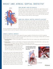
What are Atrial Septal Defects?The heart and its wallsThe adult heart is an organ with four chambers, two upper and two lower.The two upper chambers are known as the atria which are a holding areafor blood and are minor pumps. The two lower chambers are known asventricles, which are the major pumps devoted to pumping blood out of theheart. There is a wall that separates the two atria and a wall that separatesthe two ventricles. These walls are called septums.HOW DO ATRIAL SEPTAL DEFECTS OCCUR?During fetal development, the heart begins as one pumping chamber withno inner walls. As the heart grows inner walls form from the outer walls intoward the center creating four chambers. Since the fetus does not use itslungs, there is an opening between the upper chambers which allows bloodto pass directly from the right side to the left, bypassing the lungs. Normallyafter a baby is born, this opening closes with a flap of tissue that acts like aone-way swinging door, sealing the opening shut. In the lower portion of the heart, as the fetus grows, the septumeventually forms to create two separate ventricles which are the major pumping chambers of the heart.Atrial Septal defectIn some people, the atrial septum which separates the upper chambers of the heart does not fill in completelyleaving a hole. This condition is called atrial septal defect (<strong>ASD</strong>). There are several types of <strong>ASD</strong>s which are labeledbased on where in the septum the hole is located:• Secundum defect is a hole in the middle of the septum. This is the most common type, representing 80 percentof all <strong>ASD</strong>s.• Primum defect is a hole low in the septum. It frequently includes defects in the mitral valve, a one-way valvelocated between the upper and lower chamber on the left side of the heart.• Sinus venosus defect is located high in the septum and frequently involves misplacement of one or more of theveins from the lungs. As a result, these veins,which usually bring oxygenated blood fromthe lungs back to the left side of the heart, maysend it to the right side of the heart instead.In some cases, the hole in the septum allows bloodto flow directly from one atria to the other. Thisis referred to as a shunt. If too much of the bloodmoves from the left to the right side of the heart,it can cause pressure in the lungs to rise. If a shuntis small enough, it can cause very few problemsand may not be noticed until much later in life,sometimes never at all. However, in severe casesof <strong>ASD</strong>, where the shunt is large, it can affect thelungs and lead to significant shortness of breath.
How are <strong>ASD</strong>s Diagnosed?<strong>ASD</strong> SymptomsAtrial septal defects may be present for years causing no symptoms at all. In some cases,blood from the high pressure left side of the heart flows across the hole into the lowpressure right side of the heart resulting in an increase in right heart pressures. Continualblood flow from one side of the heart is called a shunt. <strong>ASD</strong>s with a shunt can causerising pressure in the lungs. Over time, the rising pressure can produce symtoms thatmay include:• difficulty breathing• fatigue• shortness of breath that limits activity• the heart skipping, jumping or palpitating in the chest• a build up of fluid in the feet, ankles and legsDiagnosisThe earliest signs that an <strong>ASD</strong> may be present are appearance of a heart murmur on physical exam or of symptoms.When either are detected, a more complete evaluation is needed. Your cardiologist will perform various tests to helpdiagnose the type and size of the defect.The evaluation for a septal defect may include:• History and Physical: A detailed history of symptoms, when they began and howthey have affected activities, as well as family health history are essential. Duringthe physical exam, your heart and lungs will be carefully assessed.• Chest X-ray: Shows the size of the heart, and outer shape of the chambers.• Electrocardiogram: Helps identify if the heart is enlarged or if the hole hasaffected the electrical current that travels through the heart.• Echocardiogram (Echo) and Doppler Ultrasound: Use sound waves to view theheart. The Echo shows how thick the walls are, if there are openings, how the bloodflows and in what direction. The Echo also shows the direction and flow of bloodacross the hole.• Cardiac Catheterization: A minimally invasive procedure that uses a long thintube travelling through a blood vessel into the heart to measure pressures andoxygen levelsin the heartchambers. This provides information on thedirection of the blood flow. Sometimes, theopening can be closed during a catheterizationprocedure using a non-surgical closure device.• Magnetic Resonance Imaging (MRI): Anoninvasive technique used to gain a detailedimage of your heart’s structure and show howmuch blood is backing up into the lungs.
How are <strong>ASD</strong>s treated?<strong>ASD</strong> TreatmentThe type of treatment depends on the location of the defect and the associated symptoms. If the <strong>ASD</strong> is small, thereare no symptoms or risks, you and your cardiologist may choose to follow the opening for changes or developmentof symptoms. Larger defects – those that are showing changes in the heart or lungs or those that produce symptoms– may need to be repaired either surgically or through a non-surgical catheterization procedure. The type and sizeof <strong>ASD</strong> will determine which approach will be most successful.Medication management<strong>ASD</strong>s can only be corrected with a procedure on the heart either with surgery or througha catheter intervention. Medications may be used to help the heart work better, to reducethe pressures in the heart, to control irregular heart rhythms, or to prevent complicationsthat may occur with the following:• Beta blockers or another heart rate medication which may be used to help keep the heart rhythm regular.• Water pills (diuretics) which may be used to help the kidneys remove excess water to reduce pressures inthe heart.• Anticoagulants like warfarin (Coumadin) or antiplatelets like aspirin or clopidogrel (Plavix) which may be usedto prevent blood clots and reduce the chance of stroke.Non-surgical or Percutaneous ClosureOstium secundum <strong>ASD</strong>s can usually be closed using a non-surgical or percutaneous procedure (meaning through theskin) performed in the cardiac catheterization laboratory. This procedure uses a catheter that has a special closuredevice called a septal occluder mounted on the tip.During the procedure:• Thin flexible tubes or sheaths are inserted into the large blood vessels in each leg. One is for a catheter that hasthe closure device on the end. The second is for a small internal ultrasound probe that helps guide the procedurewith views of the heart, the hole in the septum and the closure device.• A catheter with a dual-umbrella shaped closure device is inserted into one of the sheaths, and threaded up amajor vessel into the right side of the heart and across the hole in the septum.• When the device has crossed through the septum, the device is positioned and opened so that it closes the hole.• The device is checked to make sure it is secure. The small internal ultrasound catheter is used to verifypositioning of the device.• After themovement ofthe heart, thevalves and thedevice positionare checked thecatheters areremoved from thebody.There are two types of septal closure devices:Amplatzer Septal Occluder (left) or Gore Helex Device (right).Continued on next page
Continued from previous pageSurgical RepairThere are some types of <strong>ASD</strong> that are best repaired with open-heart surgery. These are usually primum and sinusvenosus defects, secundum defects that are very large or abnormally shaped, or when there are other cardiacabnormalities that also need to be repaired.• Before the procedure begins, you are put completely to sleep by anesthesia.• During the procedure, your heart is stopped and a heart-lung bypass machine is used to do all the work of yourheart and lungs.• An incision is made in the right upper chamber and the hole in the septum is repaired. If the hole is small, itmay require only stitches to repair, larger holes are closed with a patch. The patch may be made of man-madematerial or from a piece of your own heart muscle tissue taken from the outside of the heart.• Once the <strong>ASD</strong> is repaired, the surgeon makes sure there is no bleeding or leftover air in the heart.• The heart lung machine is stopped and the heart and lungs are allowed to start working again.• After the procedure, there is a recovery period in the intensive care unit that lasts two to three days, followed bya few days in a step-down unit before returning home.Open-heart surgery usually requires staying in the hospital for five to seven days, a period of recovery at home andexercise rehabilitation therapy.For more information, visit henryford.com/heart or call (313) 916-1534.
henryford.com/heart80148