Natural infection of periwinkle (Catharanthus roseus ... - CIMAP Staff
Natural infection of periwinkle (Catharanthus roseus ... - CIMAP Staff
Natural infection of periwinkle (Catharanthus roseus ... - CIMAP Staff

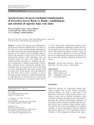
Create successful ePaper yourself
Turn your PDF publications into a flip-book with our unique Google optimized e-Paper software.
Cucumber mosaic virus on <strong>periwinkle</strong> Australasian Plant Disease Notes 31Fig. 2. <strong>Natural</strong>ly infected C. <strong>roseus</strong> twig showing mosaic, leaf and flowerdeformation.natural occurrence <strong>of</strong> CMV on C. <strong>roseus</strong> has not been previouslyrecorded.Infected plant tissues were homogenised in 0.1 M phosphatebuffer (pH 7.2) containing 0.02% thioglycollic acid and sodiumsulfite, squeezed through muslin cloth and mechanically appliedonto leaves using carborundum (600 mesh) as an abrasive.Seedlings treated with buffer only served as negative controlsfor inoculation. Both inoculated and control plants were keptunder observation in an insect-pro<strong>of</strong> glasshouse (Noordam 1973).The virus isolate was efficiently sap transmitted from naturallyinfected <strong>periwinkle</strong> to healthy <strong>periwinkle</strong> and a variety <strong>of</strong> plantspecies mainly from Chenopodiaceae, Cucurbitaceae andSolanaceae. Our studies used a single lesion from <strong>periwinkle</strong>obtained by passage through C. amaranticolor and furtherpropagated in Nicotiana benthamiana. Periwinkle plantsinoculated mechanically reacted with symptoms similar tothose observed on naturally infected plants. The mechanicallyinoculated plant species that showed local and/or systemicsymptoms included; Gomphrena globosa, N. benthamiana,N. glutinosa, N. occidentalis, N. rustica, N. tabacum cv.DR-1(Harrison Special), N. tabacum Samsun NN, N. tabacum WhiteBurley, N. xanthi, Cucumis melo, C. sativus, Pisum sativum,Capsicum annuum, Solanum lycopersicum, Lycopersicumesculentum, Petunia hybrida, Chenopodium album,C. amaranticolor, C. quinoa, Cucurbita pepo, Daturastramonium, D. inoxia, Glycine max, Solanum melongena,Vigna mungo and Solanum nigrum. However, the followingplants failed to develop either local or systemic symptoms;Arachis hypogaea, Brassica campestris, Dahlia hybrida,Chrysanthemum cinerariifolium, Dianthus barbatus,Helianthus annuus, Luffa cylindrical, Mirabilis jalapa,Tagetes erecta, Zinnia elegans, Mentha arvensis, M. spicata,M. citrata, Withania somnifera and Physalis floridana. Selectedhosts <strong>of</strong> the <strong>periwinkle</strong> CMV isolate are compared with the hostrange studies <strong>of</strong> others CMV isolates (Table 1) from vanilla(Madhubala et al. 2005), Egyptian and black henbane (Zaim andKhan1988; Samad et al. 2000), R. serpentina (Zaim and Khan1988), banana (Aglave et al. 2007) and lily (Terami et al. 2004).Groups <strong>of</strong> five aphids (Myzus persicae) were transferred toglass tubes and starved for 2 h. They were allowed to feed for15 min on infected <strong>periwinkle</strong> or N. benthamiana leaves andtransferred to a healthy <strong>periwinkle</strong> or N. benthamiana plant for a24 h inoculation/feeding period. Myzus persicae transmitted thevirus in a non-persistent manner to healthy <strong>periwinkle</strong>(5/10 plants) and N. benthamiana (7/10 plants). Symptomsdeveloped after 15--20 days <strong>of</strong> feeding. For soil transmission,<strong>periwinkle</strong> plants bearing viral symptoms along with soilcollected from infested fields were planted individually inpots. Two weeks later, three healthy <strong>periwinkle</strong> seedlings atthe five to six leaf stage were planted next to the symptombearing diseased plants. Control arrangements were set upusing apparently healthy <strong>periwinkle</strong> in autoclaved soil. Plantswere grown in a glasshouse and observed daily for symptomdevelopment over 6 weeks. Neither disease symptoms nor virusparticles were detected in the seedlings transplanted adjacent toinfected plants.Serial 10-fold dilutions <strong>of</strong> crude sap in 0.1 M phosphate buffer(pH 7.0) were prepared from infected N. benthamiana fordetermination <strong>of</strong> bio-physical studies, dilution end point(DEP), thermal inactivation point (TIP) and longevity in vitro(LIV) <strong>of</strong> the virus (Noordam 1973). Infectivity was assayed onC. amaranticolor. Virus retained infectivity in crude sap up to aTable 1. Comparison <strong>of</strong> selected hosts with different isolates <strong>of</strong> CMVB, blister; CLL, chlorotic local lesion; LD, leaf distortion; LI, latent <strong>infection</strong>; LS, leaf shedding; M, mosaic; NLL, necrotic local lesion; NS, no symptoms;SN, systemic necrosis; SS, systemic symptoms; StS, streaks on stem; VC, vein clearing; --, not testedHost testedIsolates <strong>of</strong> CMVPeriwinkle Vanilla Henbane Rauvolfia Banana LilyC amaranticolor CLL CLL, NLL NLL NLL CLL CLL, NLLC. annuum M M, LC NS -- M --C. sativus M VC M SS CLL, M, VC MD. stramonium M, B -- CLL, M LS, LD CLL -- NSG. globosa NLL, LD -- -- NLL -- SNG. max M, VC NS -- -- CLL, M LIL. esculentum M, LD StS, LD M, LD SS M, LD MN. glutinosa M M, B NLL, M -- M, LD MZ. elegans NS -- -- SS -- M
32 Australasian Plant Disease Notes A. Samad et al.bp2122651484973426835302027190415841375947831Amplified product~657 bp564Fig. 3. Electron micrograph <strong>of</strong> a purified virus preparation stained in 2%uranyl acetate showing CMV particles (bar = 55 nm).Fig. 4. RT-PCR amplification <strong>of</strong> CMV coat protein gene (~657 bp). Lane 1,l DNA/HindIII/Eco R1 marker; lanes 2--5, amplified DNA fragment frominfected samples; lanes 6--7, healthy samples.Table 2. Comparison <strong>of</strong> coat protein gene sequence identityThe sequence <strong>of</strong> the India C18 isolate (EU310928) is compared at the nucleotide (nt) and amino acid (aa) level using the BLAST and DiAlign tools with isolates<strong>of</strong> Cucumber mosaic virus (CMV) reported from India and elsewhereAccession Strain <strong>Natural</strong> host Location Subgroup %IdentityntaaEU310928 CMV-C18 <strong>Catharanthus</strong> <strong>roseus</strong> Lucknow, India Ib 100 100X89652 CMV-PM Physalis minima Lucknow, India Ib 98 98EF593024 CMV-DI Datura innoxia Lucknow, India Ib 98 96AF281864 CMV-DI Datura innoxia Lucknow, India Ib 98 96EF593023 CMV-AT Amaranthus tricolour Lucknow, India Ib 98 96AF198622 CMV-AS Amaranthus sp. Lucknow, India Ib 98 96EF153733 CMV-CM Chrysanthemum mor. Lucknow, India Ib 97 95EF593026 CMV-JC Jatropha curcus Lucknow, India Ib 98 98EF153739 CMV-JC Jatropha curcus Lucknow, India Ib 98 98EF593025 CMV-RS Rauvolfia serpentina Lucknow, India Ib 98 99DQ914877 CMV-RS Rauvolfia serpentina Lucknow, India Ib 98 99AF350450 CMV-HM Hyoscyamus muticus Lucknow, India Ib 98 98AJ810260 CMV-Ch Capsicum annum India Ib 96 97AY754359 CMV-VP Vanilla planifolia Kerala, India Ib 95 98AY545924 CMV-PN Piper nigrum Kerala, India Ib 95 99AY861397 CMV-WP Vanilla tahitensis New Zealand Ib 94 97EF153737 CMV-LG Lemon grass Lucknow, India Ib 95 96AY125575 CMV-ban Musa paradisiacal Kerala, India Ib 95 96AY690620 CMV-PB Piper betle Kerala, India Ib 95 97AY690621 CMV-PL Piper longum Kerala, India Ib 95 97D43800 CMV-Pepo -- Japan Ia 90 94L36251 CMV-Kor -- Korea Ia 89 91AJ006988 CMV-Pl -- China Ia 91 95U43888 CMV-MA Musa acuminata Israel Ia 92 95D10538 CMV-Fny -- NY, USA Ia 92 95AJ585086 CMV-Li Lilium longifolium HP, India II 75 79D00463 CMV-Wl -- USA II 76 81L15336 CMV-Trk7 -- Hungary II 76 80NC_002040 PSV Vigna unguiculata USA Out-group 46 46
34 Australasian Plant Disease Notes A. Samad et al.AMVRTase (10U) at 42 C for 90 min, in a total volume <strong>of</strong> 20 mLcontaining RNase inhibitor (25U), 1 mM each dNTP and adownstream primer (5 0 GCAT GGTACCTCAAACTGGGAGCAC-3 0 ). The PCR (25 mL) was performed using 2 mL <strong>of</strong> cDNA andprimers specific to CMV coat protein (CP, Srivastava et al. 2004)(F: 5 0 -GCATTCTAGA TGGACAAATCTGAATC-3 0 and R:5 0 GCATGGTACCTCA AACTGGGAGCAC-3 0 ) in anautomated thermal cycler (Perkin Elmer-Gene Amp PCRsystem 2400). The PCR cycles were, one initial cycle <strong>of</strong>denaturation at 94 C for 5 min followed by denaturation at94 C (1 min), annealing at 52 C (1 min) and extension at 72 Cfor 1.30 min. The final extension at 72 C was maintained for5 min. The PCR amplification product was separated byelectrophoresis in 1.2% agarose gel in 1X TAE and stainedwith ethidium bromide revealing an amplified DNA fragment<strong>of</strong> the expected size (~650 bp) from virus infected samplesonly but not from healthy samples (Fig. 4). DNA markerl/EcoRI+HindIII double digests were used as size markers.The amplified PCR product was cloned into pGEM-T Easyvector kit (Promega, USA), recombinant clones were identifiedby restriction digestion using the restriction endonucleaseenzyme EcoR1 and selected clones were sequenced using XL3130 Genetic Analyser (ABS, USA). The sequenced region wassubmitted to GenBank (Accession number EU310928) andcontained a single open reading frame, which comprised 657bases <strong>of</strong> nucleotides potentially coding for 218 amino acids. Thenew sequence was compared using bioinformatics tools DiAlign(Genomatrix) MEGA 4.0 with CP gene sequences <strong>of</strong> all availableCMV isolates from India as well as representative isolates fromother parts <strong>of</strong> the world belonging to both the subgroups (I and II)(Table 2). Nucleotide and deduced amino acid sequence <strong>of</strong> the CPgene <strong>of</strong> CMV infecting <strong>periwinkle</strong> showed closest identity (98%)with the subgroup IB CMV isolates from Rauvolfia serpentina(EF 593025) and with GenBank accession numbers. DQ640743and EF593026 (Fig. 5). On the basis <strong>of</strong> these biological andmolecular studies it is concluded that the virus naturally infecting<strong>periwinkle</strong> is a subgroup IB CMV. The high sequence identitiesobserved between Indian isolates <strong>of</strong> CMV compared with isolatesfrom other localities indicate that <strong>periwinkle</strong> isolate <strong>of</strong> CMVoriginated locally.AcknowledgementsThe authors are thankful to Dr S.P.S. Khanuja (Director, <strong>CIMAP</strong>) forencouragement and providing necessary facilities. Thanks also toDr AK Gupta, National Gene Bank <strong>of</strong> Medicinal and Aromatic Plants,<strong>CIMAP</strong>, Lucknow for providing the germplasm <strong>of</strong> <strong>periwinkle</strong> (C. <strong>roseus</strong>)for this study.ReferencesAglave BA, Krishnareddy M, Patil FS, Andhale MS (2007) MolecularIdentification <strong>of</strong> a Virus Causing Banana Chlorosis Disease fromMarathwada Region. International Journal <strong>of</strong> Biotechnology andBiochemistry 3, 13--23.Chatzivassiliou EK, Weekes R, Morris J, Wood KR, Barker I, Katis NI (2000)Tomato spotted wilt virus (TSWV) in Greece: its incidence following theexpansion <strong>of</strong> Frankliniella occidentalis, and characterisation <strong>of</strong> isolatescollected from various hosts. The Annals <strong>of</strong> Applied Biology 137,127--144. doi: 10.1111/j.1744-7348.2000.tb00044.xGibbs AJ, Harrison BD (1976) Physical and chemical methods <strong>of</strong> assay andanalysis. In ‘Plant virology -- The principles’. (Edward Arnold: London)Huang CH, Chang YC (2005) Identification and molecular characterization <strong>of</strong>Zantedeschia mild mosaic virus (ZaMMV), a new calla lily-infectingpotyvirus. Archives <strong>of</strong> Virology 150, 1221--1230. doi: 10.1007/s00705-004-0488-3Madhubala R, Bhadramurthy V, Bhat AI, Hareesh PS, Pretheesh ST,Bhai RS (2005) Occurrence <strong>of</strong> Cucumber mosaic virus on vanilla(Vanilla planifolia Andrews) in India. Journal <strong>of</strong> Biosciences 30,339--350. doi: 10.1007/BF02703671Noordam D (1973) ‘Identification <strong>of</strong> plant viruses: Methods andexperiments.’ (Centre for Agricultural Publishing and Documentation:Wageningen, The Netherlands)Palukaitis P, Roossinck MJ, Dietzgen RG, Francki RIB (1992) Cucumbermosaic virus. Advances in Virus Research 41, 281--348.Prince WC (1934) Isolation and study <strong>of</strong> some yellow strains <strong>of</strong> cucumbermosaic virus. Phytopathology 24, 743--761.Raj SK, Srivastava A, Chandra G, Singh BP (2002) Characterisation <strong>of</strong>Cucumber mosaic virus isolate infecting Gladiolus cultivars andcomparative analysis <strong>of</strong> serological and molecular methods forsensitive diagnosis. Current Science 83, 1132--1136.Raj SK, Kumar S, Pratap D, Vishnoi R, Snehi SK (2007) <strong>Natural</strong> Occurrence<strong>of</strong> Cucumber mosaic virus on Rauvolfia serpentina, a new record. PlantDisease 91, 322. doi: 10.1094/PDIS-91-3-0322CRaj SK, Kumar S, Snehi SK (2008) First Report <strong>of</strong> Cucumber mosaic viruson Jatropha curcas in India. Plant Disease 92, 171. doi: 10.1094/PDIS-92-1-0171CRoossinck MJ (1999) Cucumoviruses (Bromoviridae) -- general features.In ‘Encyclopedia <strong>of</strong> Virology’. 2nd edn (Eds L Grano<strong>of</strong>, RG Webster)pp. 315--320. (Academic Press: San Diego)Salazar LF, Muller G, Querci M, Zapata JL, Owens RA (2000) Potato yellowvein virus: its host range, distribution in South America and identificationas a crinivirus transmitted by Trialeurodes vaporariorum. The Annals <strong>of</strong>Applied Biology 137, 7--19. doi: 10.1111/j.1744-7348.2000.tb00052.xSamad A, Raj SK, Srivastava A, Chandra G, Ajayakumar PV, Zaim M,Singh BP (2000) Characterization <strong>of</strong> a cucumber mosaic virusisolates infecting Egyptian henbane (Hyoscyamus muticus L.) inIndia. Acta Virologica. English Ed. 44, 131--136.Sambrook J, Russell DW (2001) ‘Molecular cloning: A laboratory manual.’(Cold Spring Harbor Laboratory Press: New York)Singh HP, Hallan V, Raikhy G, Kulshrestha S, Sharma ML, Ram R, Garg ID,Zaidi AA (2005) Characterization <strong>of</strong> an Indian isolate <strong>of</strong> carnation mottlevirus infecting carnations. Current Science 88, 594--601.Srivastava A, Chandra G, Raj SK (2004) Molecular characterization <strong>of</strong> a strain<strong>of</strong> cucumber mosaic virus based on coat protein and movement proteingenes. Acta Virologica. English Ed. 48, 229--239.Stearn WT (1975) A synopsis <strong>of</strong> the genus <strong>Catharanthus</strong> (Apocynaceae).In ‘<strong>Catharanthus</strong> alkaloids’. (Eds WL Taylor, NR Fransworth) pp. 9--44.(Marcel Dekker: New York)Sudhakar N, Nagendra-Prasad D, Mohan N, Murugesan K (2006) First Report<strong>of</strong> Cucumber mosaic virus Subgroup II Infecting Lycopersiconesculentum in India. Plant Disease 90, 1457. doi: 10.1094/PD-90-1457BSvoboda GH, Blake DA (1975) The phytochemistry and pharmacology <strong>of</strong><strong>Catharanthus</strong> <strong>roseus</strong>. In ‘<strong>Catharanthus</strong> alkaloids’. (Eds WL Taylor,NR Fransworth) pp. 45--124. (Marcel Dekker: New York)Terami F, Fukumoto F, Handa K (2004) Cucumber mosaic virus isolated fromAmazon lily (Eucharis grandiflora). Journal <strong>of</strong> General Plant Pathology70, 192--193. doi: 10.1007/s10327-004-0104-0Verma N, Mahinghara BK, Ram R, Zaidi AA (2006) Coat proteinsequence shows that Cucumber mosaic virus isolate from geraniums(Pelargonium spp.) belongs to subgroup II. Journal <strong>of</strong> Biosciences31, 47--56. doi: 10.1007/BF02705234Zaim M, Khan MMAA (1988) Green mosaic <strong>of</strong> Hyoscyamus niger L. andRauvolfia serpentina Benth. Indian Journal <strong>of</strong> Plant Pathology 6,152--157.Manuscript received 30 January 2008, accepted 19 March 2008http://www.publish.csiro.au/journals/apdn