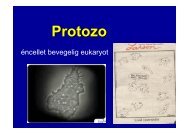
Journal of Translational Medic<strong>in</strong>e 2008, 6:13http://www.translational-medic<strong>in</strong>e.com/content/6/1/13Site specific methylation with<strong>in</strong> the MAL promoterFigure 2Site specific methylation with<strong>in</strong> the MAL promoter. Bisulphite sequenc<strong>in</strong>g of the MAL promoter verifies methylationstatus assessed by methylation-specific polymerase cha<strong>in</strong> reaction. The upper part of the figure is a schematic presentation ofthe CpG sites successfully amplified by the two analyzed bisulphite sequenc<strong>in</strong>g fragments, A (-68 to +168; to the right) <strong>and</strong> B (-427 to -85; to the left). The transcription start site is represented by +1 <strong>and</strong> the vertical bars <strong>in</strong>dicate the location of <strong>in</strong>dividualCpG sites. The two arrows <strong>in</strong>dicate the location of the MSP primers <strong>in</strong> the present study <strong>and</strong> a previously published study analyz<strong>in</strong>gpromoter methylation of MAL [31]. For the lower part of the figure, filled circles represent methylated CpGs; open circlesrepresent unmethylated CpGs; <strong>and</strong> open circles with a slash represent partially methylated sites (the presence ofapproximately 20–80% cytos<strong>in</strong>e, <strong>in</strong> addition to thym<strong>in</strong>e). The column of U, M <strong>and</strong> U/M at the right side of this lower part liststhe methylation status of the respective cell l<strong>in</strong>es as assessed by us us<strong>in</strong>g MSP analyses. Abbreviations: MSP, methylation-specificPCR; s, sense; as, antisense; U, unmethylated; M, methylated; U/M, presence of both unmethylated <strong>and</strong> methylated b<strong>and</strong>.Real-time quantitative gene expressionThe level of MAL mRNA expression <strong>in</strong> cell l<strong>in</strong>es (n = 46),primary colorectal carc<strong>in</strong>omas (n = 16), <strong>and</strong> <strong>no</strong>rmalmucosa (n = 3) was assessed by quantitative real timePCR. There was a strong association between MAL promoterhypermethylation <strong>and</strong> reduced or lost gene expressionamong cell l<strong>in</strong>es (P = 0.041; Figure 4). Furthermore,the gene expression of MAL was up-regulated <strong>in</strong> coloncancer cell l<strong>in</strong>es after promoter demethylation <strong>in</strong>duced bythe comb<strong>in</strong>ed treatment 5-aza-2'-deoxycytid<strong>in</strong>e <strong>and</strong> trichostat<strong>in</strong>A (Figure 5). Treatment with the deacetylase<strong>in</strong>hibitor trichostat<strong>in</strong> A alone did <strong>no</strong>t <strong>in</strong>crease MALexpression, whereas treatment with the DNA demethylat<strong>in</strong>g5-aza-2'-deoxycytid<strong>in</strong>e led to high expression <strong>in</strong> HT29cells, but more moderate levels <strong>in</strong> HCT15 cells (Figure 5).Among primary colorectal carc<strong>in</strong>omas, those harbour<strong>in</strong>gpromoter hypermethylation of MAL (n = 13) expressedsomewhat lower levels of MAL mRNA compared with theunmethylated tumours (n = 3), although <strong>no</strong>t statisticallysignificant (Figure 4).MAL prote<strong>in</strong> expression is lost <strong>in</strong> colorectal carc<strong>in</strong>omasTo evaluate the immu<strong>no</strong>histochemistry analyses of MAL,kidney <strong>and</strong> heart muscle tissues were <strong>in</strong>cluded as positive<strong>and</strong> negative controls, respectively (Figure 6A–B) [30].From the 231 scorable colorectal tissue cores, i.e. thoseconta<strong>in</strong><strong>in</strong>g malignant colorectal epithelial tissue, 198were negative for MAL sta<strong>in</strong><strong>in</strong>g (Figure 6C–D). Twentyn<strong>in</strong>eof these had positive sta<strong>in</strong><strong>in</strong>g <strong>in</strong> <strong>no</strong>n-epithelial tissuecomponents with<strong>in</strong> the same tissue cores, ma<strong>in</strong>ly <strong>in</strong> neurons<strong>and</strong> blood vessels (<strong>no</strong>t shown). In comparison, allthe sections of <strong>no</strong>rmal colon tissue conta<strong>in</strong>ed positivesta<strong>in</strong><strong>in</strong>g for MAL <strong>in</strong> the epithelial cells (Figure 6E–F).DiscussionIn the present study, we have demonstrated that asequence with<strong>in</strong> the MAL promoter close to the transcriptionstart is hypermethylated <strong>in</strong> the vast majority of malignant,as well as <strong>in</strong> benign colorectal tumours, <strong>in</strong> contrastto <strong>no</strong>rmal colon mucosa samples which are unmethylated,<strong>and</strong> we contend that MAL rema<strong>in</strong>s a promis<strong>in</strong>g diag<strong>no</strong>sticbiomarker for early colorectal tumorigenesis [12].The ade<strong>no</strong>mas <strong>and</strong> carc<strong>in</strong>omas analyzed <strong>in</strong> the presentstudy are from unselected cl<strong>in</strong>ical series <strong>and</strong> are thereforerepresentative for the average risk population. However,the equal distribution between MSI <strong>and</strong> MSS carc<strong>in</strong>omas<strong>in</strong> the present study is <strong>no</strong>t representative for a consecutiveseries.Hypermethylation of MAL has, by quantitative methylation-specificpolymerase cha<strong>in</strong> reaction (MSP), previouslybeen shown by others to be present only <strong>in</strong> a small fraction(6%, 2/34) of colon carc<strong>in</strong>omas [31], even thoughthe expression of MAL was reported to be reduced/lost <strong>in</strong>the majority of colorectal tumours [11,17,31]. In contrast,we report here a significantly higher methylation fre-Page 6 of 11(page number <strong>no</strong>t for citation purposes)
Journal of Translational Medic<strong>in</strong>e 2008, 6:13http://www.translational-medic<strong>in</strong>e.com/content/6/1/13The "bisulphite sequence" of the MAL promoterFigure 3The "bisulphite sequence" of the MAL promoter. Representative bisulphite sequenc<strong>in</strong>g electropherograms of the MALpromoter <strong>in</strong> colon cancer cell l<strong>in</strong>es. A subsection of the bisulphite sequence electropherogram, cover<strong>in</strong>g CpG sites +11 to +15relative to transcription start. Cytos<strong>in</strong>es <strong>in</strong> CpG sites are <strong>in</strong>dicated by a black arrow, whereas cytos<strong>in</strong>es that have been convertedto thym<strong>in</strong>es are underl<strong>in</strong>ed <strong>in</strong> red. The MAL promoter sequenc<strong>in</strong>g electropherograms illustrated here, are from theunmethylated V9P cell l<strong>in</strong>e <strong>and</strong> the hypermethylated ALA <strong>and</strong> HCT116.quency of MAL <strong>in</strong> both benign <strong>and</strong> malignant colorectaltumours (71% <strong>in</strong> ade<strong>no</strong>mas <strong>and</strong> 80% <strong>in</strong> carc<strong>in</strong>omas). Thediscrepancy <strong>in</strong> methylation frequencies between thepresent report <strong>and</strong> the previous study by Mori <strong>and</strong> coworkers[31] is probably a consequence of study design.From direct bisulphite sequenc<strong>in</strong>g of colon cancer celll<strong>in</strong>es, we have <strong>no</strong>w shown that the DNA methylation ofMAL is unequally distributed with<strong>in</strong> the CpG isl<strong>and</strong> of itspromoter (Figure 2). CpG isl<strong>and</strong>s often span more tha<strong>no</strong>ne kilobase of the gene promoter, <strong>and</strong> the methylationstatus with<strong>in</strong> this region is sometimes mistakenlyassumed to be equally distributed. This is exemplified bythe MLH1 gene <strong>in</strong> which hypermethylation of a limitednumber of CpG sites approximately 200 base pairsupstream of the transcription start po<strong>in</strong>t <strong>in</strong>variably correlateswith the lack of gene expression, while other sites do<strong>no</strong>t [32,33]. S<strong>in</strong>ce the results of an MSP analysis rely onthe match or mismatch of the unmethylated <strong>and</strong> methylatedprimer sequences to bisulphite treated DNA, oneshould ensure that the primers anneal to relevant CpGsites <strong>in</strong> the gene promoter. In the present study, wedesigned the MSP primers close to the transcription startpo<strong>in</strong>t of the gene (-72 to +70) <strong>and</strong> found, by bisulphitesequenc<strong>in</strong>g, concordance between the overall methylationstatus of MAL as assessed by MSP <strong>and</strong> the methylationstatus of the <strong>in</strong>dividual CpG sites covered by our MSPprimer set (Figure 2). This part of the CpG isl<strong>and</strong> washypermethylated <strong>in</strong> the majority of colon cancer cell l<strong>in</strong>es(95%). We also found that these cell l<strong>in</strong>es, as well as thoseof other tissues, showed loss of MAL RNA expression fromquantitative real time analyses, <strong>and</strong> that removal of DNAhypermethylation by the comb<strong>in</strong>ed treatment of 5-aza-2'-deoxycytid<strong>in</strong>e <strong>and</strong> Trichostat<strong>in</strong> A re-<strong>in</strong>duced the expressio<strong>no</strong>f MAL <strong>in</strong> colon cancer cell l<strong>in</strong>es (Figure 5). Furthermore,by analyz<strong>in</strong>g a large series of cl<strong>in</strong>icallyrepresentative samples by prote<strong>in</strong> immu<strong>no</strong>histochemistrywe confirmed that the expression of MAL was lost <strong>in</strong>malignant colorectal epithelial cells as compared to <strong>no</strong>rmalmucosa.We have further analyzed the same region of the MAL promoteras Mori et al., which is located -206 to -126 basepairs upstream of the transcription start po<strong>in</strong>t [31]. Bydirect bisulphite sequenc<strong>in</strong>g, we showed that only am<strong>in</strong>ority of the CpG sites covered by the Mori antisenseprimer were methylated <strong>in</strong> the 19 colon cancer cell l<strong>in</strong>esPage 7 of 11(page number <strong>no</strong>t for citation purposes)
- Page 1 and 2:
Novel genetic and epigenetic altera
- Page 3 and 4:
TABLE OF CONTENTSACKNOWLEDGEMENTS .
- Page 5 and 6:
ACKNOWLEDGEMENTSThe present work ha
- Page 7 and 8:
Prefacetechnology[3]. This new tech
- Page 10 and 11:
SummaryThe subgroup of carcinomas w
- Page 12 and 13:
Introduction“Epigenetic inheritan
- Page 14 and 15:
Introductionamino acid change it is
- Page 16 and 17:
Introductionmethylation during embr
- Page 18 and 19:
IntroductionDNA is most of the time
- Page 20 and 21:
IntroductionFigure 5. DNA methylati
- Page 22 and 23:
IntroductionFigure 6. Incidence rat
- Page 24 and 25:
IntroductionFigure 8. Tumor staging
- Page 26 and 27:
Introductioninasmuch as 80% of colo
- Page 28 and 29:
IntroductionInstabilities involved
- Page 30 and 31:
Introductionthere seems to be a fid
- Page 32 and 33:
Introductionsevere alterations are
- Page 34 and 35:
Introductionpopulation-wide screeni
- Page 36 and 37:
IntroductionFigure 12. Present and
- Page 38 and 39:
RESULTS IN BRIEFPaper Ia. “DNA hy
- Page 40 and 41:
Results in Briefinstability, and se
- Page 42 and 43:
Results in BriefUnivariate survival
- Page 44 and 45:
Discussionseveral factors, and full
- Page 46 and 47:
Discussionlow threshold, we increas
- Page 48 and 49: DiscussionIt may seem like unnecess
- Page 50 and 51: Discussionthan 96% DHPLC do not sta
- Page 52 and 53: DiscussionFigure 13. Mutation detec
- Page 54 and 55: DiscussionClinical impact of molecu
- Page 56 and 57: Discussionmarkers with a very high
- Page 58 and 59: Discussionchromosomes in metaphase[
- Page 60 and 61: DiscussionThese examples underline
- Page 62 and 63: Discussiongenes. One is based on mu
- Page 64 and 65: CONCLUSIONSWe have identified novel
- Page 66 and 67: Future PerspectivesMolecular risk a
- Page 68 and 69: REFERENCES1. Breasted J (1930) The
- Page 70 and 71: References29. Deng G, Chen A, Pong
- Page 72 and 73: References57. Al-Sukhni W, Aronson
- Page 74 and 75: References84. Kunkel TA (1993) Nucl
- Page 76 and 77: ReferencesLeggett B, Levine J, Kim
- Page 78 and 79: References133. Lind GE, Thorstensen
- Page 80 and 81: References156. Meling GI, Lothe RA,
- Page 82 and 83: ReferencesT, Song X, Day RH, Sledzi
- Page 84 and 85: References196. Honda S, Haruta M, S
- Page 86 and 87: ORIGINAL ARTICLESAPPENDIXAppendix I
- Page 89 and 90: GASTROENTEROLOGY 2007;132:1631-1639
- Page 91: Paper IbGuro E Lind, Terje Ahlquist
- Page 94 and 95: Journal of Translational Medicine 2
- Page 96 and 97: Journal of Translational Medicine 2
- Page 100 and 101: Journal of Translational Medicine 2
- Page 102 and 103: Journal of Translational Medicine 2
- Page 105: Paper IITerje Ahlquist, Guro E Lind
- Page 108 and 109: BackgroundMost cases of colorectal
- Page 110 and 111: ADAMTS1 CDKN2A CRABP1 HOXA9 MAL MGM
- Page 112 and 113: pseudogene, leading to a high rate
- Page 114 and 115: strands. Proc Natl Acad Sci U S A 1
- Page 116 and 117: concomitant absence of transcript a
- Page 119 and 120: Volume 10 Number 7 July 2008 pp. 68
- Page 121 and 122: 682 RAS Signaling in Colorectal Car
- Page 123 and 124: 684 RAS Signaling in Colorectal Car
- Page 125 and 126: 686 RAS Signaling in Colorectal Car
- Page 127: Table W2. Detailed Somatic Events o
- Page 131 and 132: Identification of RCC2 as a prognos
- Page 133 and 134: INTRODUCTIONMicrosatellite instabil
- Page 135 and 136: unselected series of primary tumors
- Page 137 and 138: specificity, i.e. that they only am
- Page 139 and 140: On the assumption that DNA repair a
- Page 141 and 142: In order to ensure that gene mutati
- Page 143 and 144: Figure 2. Mutation frequency differ
- Page 145 and 146: and TAF1B (0.50), ACVR2A and ASTE1
- Page 147 and 148: Multivariate analysesA multivariate
- Page 149 and 150:
When comparing our findings of muta
- Page 151 and 152:
The test series included a low numb
- Page 153 and 154:
entering M-phase remains to be seen
- Page 155 and 156:
12. Duval A, Reperant M, Hamelin R
- Page 157 and 158:
34. Martineau-Thuillier S, Andreass
- Page 159:
AppendicesAppendix I:List of abbrev
- Page 163 and 164:
Critical Reviews TM in Oncogenesis,
- Page 165 and 166:
TARGET GENES OF MSI COLORECTAL CANC
- Page 167 and 168:
TARGET GENES OF MSI COLORECTAL CANC
- Page 169 and 170:
TARGET GENES OF MSI COLORECTAL CANC
- Page 171 and 172:
TARGET GENES OF MSI COLORECTAL CANC
- Page 173 and 174:
TARGET GENES OF MSI COLORECTAL CANC
- Page 175 and 176:
TARGET GENES OF MSI COLORECTAL CANC
- Page 177 and 178:
TARGET GENES OF MSI COLORECTAL CANC
- Page 179 and 180:
TARGET GENES OF MSI COLORECTAL CANC
- Page 181 and 182:
TARGET GENES OF MSI COLORECTAL CANC
- Page 183 and 184:
TARGET GENES OF MSI COLORECTAL CANC
- Page 185 and 186:
TARGET GENES OF MSI COLORECTAL CANC
- Page 187 and 188:
TARGET GENES OF MSI COLORECTAL CANC
- Page 189 and 190:
TARGET GENES OF MSI COLORECTAL CANC
- Page 191:
TARGET GENES OF MSI COLORECTAL CANC