ab128503 PI3 Kinase Assay Kit - Abcam
ab128503 PI3 Kinase Assay Kit - Abcam
ab128503 PI3 Kinase Assay Kit - Abcam

You also want an ePaper? Increase the reach of your titles
YUMPU automatically turns print PDFs into web optimized ePapers that Google loves.
<strong>ab128503</strong><strong>PI3</strong> <strong>Kinase</strong> <strong>Assay</strong> <strong>Kit</strong>Instructions for UseFor the rapid, sensitive and accuratequantification of <strong>PI3</strong> <strong>Kinase</strong> activity in varioussamplesThis product is for research use only and is notintended for diagnostic use.
Table of Contents1. Introduction 32. <strong>Assay</strong> Summary 43. Components and Storage 54. Preparation of Reagents 65. <strong>Assay</strong> Procedure 72
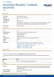
1. IntroductionPhosphoinositide 3-kinases (<strong>PI3</strong>Ks) are a family of enzymes thatplay a pivotal role in important cellular regulatory mechanisms.<strong>PI3</strong>Ks are capable of phosphorylating the 3’-OH position ofphosphoinositide lipids (PIs) generating lipid second messengers.Their function has been linked to the regulation of numerousbiological processes including cell growth, differentiation, survival,proliferation, migration. On the basis of structural similarities andsubstrate specificity, the <strong>PI3</strong>K family is divided into three classestermed I, II, and III.All human class I members are heterodimers consisting of a catalyticsubunit (MW approx. 110 kDa) and a non-catalytic subunit (MW 50,55, 85, 87 or 101 kDa). They are known to phosphorylatephosphatidylinositol (PI), phosphatidylinositol-4-mono-phosphate(PIP) and phosphatidylinositol-4,5-bisphosphate (PIP2) in vitro buthave a strong preference for PIP2 in vivo.<strong>Abcam</strong>’s <strong>PI3</strong> <strong>Kinase</strong> <strong>Assay</strong> <strong>Kit</strong> provides a highly efficient andeasy-to-handle tool for the sensitive detection and quantification ofPI 3-<strong>Kinase</strong> activity or the screening for PI 3-<strong>Kinase</strong> inhibitors.The activity of <strong>PI3</strong>-<strong>Kinase</strong>s is measured by the amount ofradioactively labeled [γ 32 P]-ATP incorporated into lipid substrate. Thesubstrate is thereafter separated using thin layer chromatography(TLC) and its radioactivity detected in a scintillation counter.3
2. <strong>Assay</strong> SummaryPrepare [γ 32 P] ATP MixPrepare Lipid Substrate VesiclesPrepare <strong>Kinase</strong> <strong>Assay</strong> MixAdd [γ 32 P] ATP MixIncubate for 15 min at 37°CStop ReactionExtract LipidsQuantify Using a Scintillation Counter4
3. Components and StorageA. <strong>Kit</strong> ContentsItemQuantityReaction Buffer2 x 1.5 ml1 mM ATP 400 µlLipid Substrate8 tubesControl <strong>PI3</strong> <strong>Kinase</strong> 5 µgStore at -20°C for up to 12 months. Avoid freeze/thaw cycles.B. Additional Materials Required• (γ 32 P)-ATP• 1M HCl• Chloroform / Methanol 1:1• 2M Acetic acid / Isopropanol 1:2• Silica gel TLC plates• Scintillation counter5
4. Preparation of ReagentsA. Preparation of [γ 32 P] ATP MixPrepare a mix of ATP and [γ 32 P]-ATP to obtain an activity of 1 µCi ata concentration of 240 µM.10 µl [γ 32 P]-ATP Mix are required perassay point.B. Preparation of Lipid Substrate VesiclesEach Lipid Substrate vial contains the amount of PI required for thepreparation of 12 assay points. Fill up the Lipid Substrate vial with125 µl water per vial and vortex thoroughly. Sonicate the emulsionfor 1 hour in a water bath sonicator.6
5. <strong>Assay</strong> ProcedureA. <strong>Kinase</strong> <strong>Assay</strong>1. To prepare the <strong>Kinase</strong> <strong>Assay</strong>, mix the following componentsfor each assay point in separate vials:Reaction Buffer 25 µlLipid Substrate Vesicle 10 µlPI 3-<strong>Kinase</strong> Sample5 µl(diluted as appropriate)2. Incubate 5 min at room temperature3. Add 10 µl [γ 32 P]-ATP Mix to start the reaction4. Incubate 15 min at 37°C5. Stop the reaction by addition of 100 µl HCl (1 M) and vortexB. Lipid Extraction1. Add 300 µl of chloroform / methanol (1:1)2. Vortex the mix and centrifuge for 1 min to separate thephases3. Remove the upper aqueous phase4. Wash the organic layer with 400 µl HCl (1 M) and removethe water phase7
C. Quantification1. Transfer 100 µl of the organic phase onto a silica gel TLCplate and run it in 2 M acetic acid / isopropanol (1:2)2. Expose the plate for visualization and quantification of<strong>PI3</strong> <strong>Kinase</strong> spots in a scintillation counter3. Alternatively, a 100 µl aliquot of the washed chloroformphase may be counted by liquid scintillationFor further technical questions please do not hesitate tocontact us by email (technical@abcam.com) or phone(select “contact us” on www.abcam.com for the phonenumber for your region).8
UK, EU and ROWEmail: technical@abcam.comTel: +44 (0)1223 696000www.abcam.comUS, Canada and Latin AmericaEmail: us.technical@abcam.comTel: 888-77-ABCAM (22226)www.abcam.comChina and Asia PacificEmail: hk.technical@abcam.comTel: 108008523689 ( 中 國 聯 通 )www.abcam.cnJapanEmail: technical@abcam.co.jpTel: +81-(0)3-6231-0940www.abcam.co.jpCopyright © 2012 <strong>Abcam</strong>, All Rights Reserved. The <strong>Abcam</strong> logo is a registered trademark. 11All information / detail is correct at time of going to print.