The Identification of MicroRNAs in a Genomically Unstable Region ...
The Identification of MicroRNAs in a Genomically Unstable Region ...
The Identification of MicroRNAs in a Genomically Unstable Region ...

Create successful ePaper yourself
Turn your PDF publications into a flip-book with our unique Google optimized e-Paper software.
<strong>The</strong> <strong>Identification</strong> <strong>of</strong> <strong>MicroRNAs</strong> <strong>in</strong> a <strong>Genomically</strong> <strong>Unstable</strong><strong>Region</strong> <strong>of</strong> Human Chromosome 8q24Konrad Huppi, 1 Natalia Volfovsky, 3 Timothy Runfola, 1 Tamara L. Jones, 1 Mark Mackiewicz, 1Scott E. Mart<strong>in</strong>, 1 J. Frederic Mush<strong>in</strong>ski, 2 Robert Stephens, 3 and Natasha J. Caplen 11 Gene Silenc<strong>in</strong>g Section, Genetics Branch and 2 Laboratory <strong>of</strong> Cancer Biology and Genetics, Center for CancerResearch, National Cancer Institute, NIH, Bethesda, Maryland; and 3 Advanced Biomedical Comput<strong>in</strong>g Center,National Cancer Institute-Frederick/Science Applications International Corporation-Frederick, Inc.,NIH, Frederick, MarylandAbstract<strong>The</strong> PVT1 locus is identified as a cluster <strong>of</strong> T(2;8) andT(8;22) ‘‘variant’’ MYC-activat<strong>in</strong>g chromosomaltranslocation breakpo<strong>in</strong>ts extend<strong>in</strong>g 400 kb downstream<strong>of</strong> MYC <strong>in</strong> a subset (20%) <strong>of</strong> Burkitt’s lymphoma (vBL).Recent reports that microRNAs (miRNA) may be associatedwith fragile sites and cancer-associated genomicregions prompted us to <strong>in</strong>vestigate whether the PVT1region on chromosome 8q24may conta<strong>in</strong> miRNAs.Computational analysis <strong>of</strong> the genomic sequencecover<strong>in</strong>g the PVT1 locus and experimental verificationidentified seven miRNAs. One miRNA, hsa-miR-1204,resides with<strong>in</strong> a previously described PVT1 exon (1b)that is <strong>of</strong>ten fused to the immunoglobul<strong>in</strong> light cha<strong>in</strong>constant region <strong>in</strong> vBLs and is present <strong>in</strong> high copynumber <strong>in</strong> MYC/PVT1 –amplified tumors. Like its humancounterpart, mouse mmu-miR-1204 represents theclosest miRNA to Myc (f50 kb) and is found only1 to 2 kb downstream <strong>of</strong> a cluster <strong>of</strong> retroviral <strong>in</strong>tegrationsites. Another miRNA, mmu-miR-1206, is close to acluster <strong>of</strong> variant translocation breakpo<strong>in</strong>ts associatedwith mouse plasmacytoma and exon 1 <strong>of</strong> mouse Pvt1.Virtually all the miRNA precursor transcripts areexpressed at higher levels <strong>in</strong> late-stage B cells (<strong>in</strong>clud<strong>in</strong>gplasmacytoma and vBL cell l<strong>in</strong>es) compared withimmature B cells, suggest<strong>in</strong>g possible roles <strong>in</strong>lymphoid development and/or lymphoma. In addition,lentiviral vector-mediated overexpression <strong>of</strong> the miR-1204 precursor (human and mouse) <strong>in</strong> a mousepre–B-cell l<strong>in</strong>e <strong>in</strong>creased expression <strong>of</strong> Myc. High levels<strong>of</strong> expression <strong>of</strong> the hsa-miR-1204 precursor is alsoReceived 3/1/07; revised 10/4/07; accepted 10/22/07.Grant support: Intramural Research Program [Center for Cancer Research,National Cancer Institute (NCI)] <strong>of</strong> the NIH. Contract N01-CO-012400 supportedwork conducted by N. Volfovsky and R. Stephens, Advanced BiomedicalComput<strong>in</strong>g Center NCI-Frederick/Science Applications International Corporation-Frederick,Inc., NCI, NIH.<strong>The</strong> costs <strong>of</strong> publication <strong>of</strong> this article were defrayed <strong>in</strong> part by the payment <strong>of</strong>page charges. This article must therefore be hereby marked advertisement <strong>in</strong>accordance with 18 U.S.C. Section 1734 solely to <strong>in</strong>dicate this fact.Note: Supplementary data for this article are available at Molecular CancerResearch Onl<strong>in</strong>e (http://mcr.aacrjournals.org/).Requests for repr<strong>in</strong>ts: Konrad Huppi, Gene Silenc<strong>in</strong>g Section, Genetics Branch,Center for Cancer Research, National Cancer Institute, NIH, Build<strong>in</strong>g 37, Room6128, Bethesda, MD 20892. Phone: 301-402-7506; Fax: 301-402-3241. E-mail:huppi@helix.nih.govCopyright D 2008 American Association for Cancer Research.doi:10.1158/1541-7786.MCR-07-0105seen <strong>in</strong> several epithelial cancer cell l<strong>in</strong>es withMYC/PVT1 coamplification, suggest<strong>in</strong>g a potentiallybroad role for these miRNAs <strong>in</strong> tumorigenesis.(Mol Cancer Res 2008;6(2):212–21)IntroductionFor years, noncod<strong>in</strong>g RNAs have represented an exceptionto the ‘‘Central Dogma’’ theory that RNA predom<strong>in</strong>atelyencodes prote<strong>in</strong>. Recently, a new series <strong>of</strong> noncod<strong>in</strong>g RNAshave been discovered <strong>in</strong> a broad range <strong>of</strong> species, termedprimary microRNAs (miRNA) that are processed <strong>in</strong>to maturemiRNAs <strong>of</strong> f21 to 22 nucleotides (nt) via an <strong>in</strong>termediateprecursor miRNA (1). Analysis <strong>of</strong> the distribution <strong>of</strong> miRNAgenes <strong>in</strong> the human genome has suggested that many are foundwith<strong>in</strong> genomically unstable regions. This f<strong>in</strong>d<strong>in</strong>g has strengthenedthe argument that deregulated expression <strong>of</strong> miRNAsand/or their targets could be critical components <strong>in</strong> manydisease processes, <strong>in</strong>clud<strong>in</strong>g cancer (2). For example, a cluster<strong>of</strong> miRNAs resides on human chromosome 13q14, a frequentlydeleted chromosomal region <strong>in</strong> cancer (3). <strong>The</strong> absence <strong>of</strong><strong>in</strong>volvement <strong>of</strong> any prote<strong>in</strong>-encod<strong>in</strong>g gene <strong>in</strong> this regioncoupled with the location <strong>of</strong> hsa-miR-15 and hsa-miR-16with<strong>in</strong> a 30-kb – deleted segment <strong>in</strong> chronic lymphocyticleukemia has suggested that, at least <strong>in</strong> this context, thesemiRNAs can function as tumor suppressors (4). Other miRNAsthat have been implicated <strong>in</strong> cancer <strong>in</strong>clude hsa-let-7, hsamir-155,and a cluster <strong>of</strong> miRNAs found with<strong>in</strong> chromosome13q31 that are frequently amplified <strong>in</strong> tumors from patientswith various forms <strong>of</strong> lymphoma (5-9).An early example <strong>of</strong> a connection between cancer andchromosomal breakpo<strong>in</strong>ts are translocations associated withhuman chromosome 8q24 conta<strong>in</strong><strong>in</strong>g the gene encod<strong>in</strong>g theproto-oncogene transcription factor MYC (SupplementaryFig. S1A; ref. 10). In 80% <strong>of</strong> Burkitt’s lymphoma (BL), aT(8;14) chromosomal translocation juxtaposes MYC to the IGHlocus. Variant translocations [T(2;8) or T(8;22)] are found <strong>in</strong>the rema<strong>in</strong><strong>in</strong>g 20% <strong>of</strong> BLs, with breakpo<strong>in</strong>ts extend<strong>in</strong>g 400 kbdownstream <strong>of</strong> MYC <strong>in</strong> a region referred to as PVT1. Becausesimilar clusters <strong>of</strong> PVT1-associated chromosomal breakpo<strong>in</strong>tsor retroviral <strong>in</strong>tegration sites are found <strong>in</strong> mouse plasmacytomas(PCT) and rat immunocytomas, it has been suggested thataltered expression <strong>of</strong> MYC is the target <strong>of</strong> this genomic<strong>in</strong>stability (11). For example, PVT1-<strong>in</strong>volved translocations are<strong>of</strong>ten accompanied by a change <strong>in</strong> the <strong>in</strong>itiation <strong>of</strong> MYCtranscription from the normal P2 promoter to the stronger P1212Mol Cancer Res 2008;6(2). February 2008
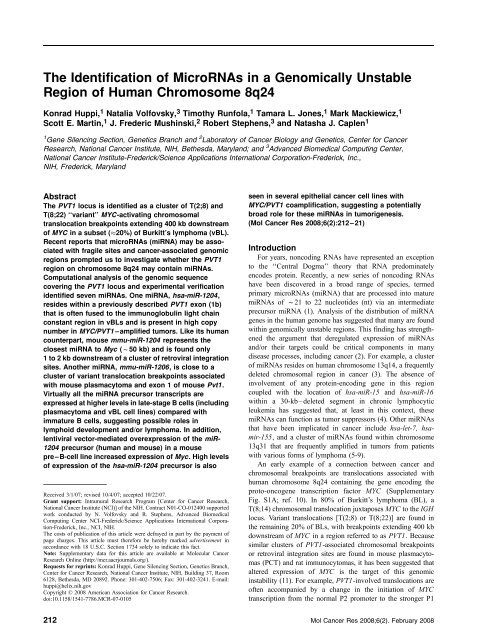
216Huppi et al.However, the seed sequence for hsa-miR-1205 is identicalto the seed sequence for hsa-miR-17-3p, which is expressed<strong>in</strong> HeLa cells (34). A sequence with<strong>in</strong> hsa-miR-2PVT1 is thesame as the seed sequence <strong>of</strong> the miRNA mmu-miR-698,a recently cloned miRNA from mouse embryos (35). <strong>The</strong>hsa-miR-1207-3p sequence is related to a group <strong>of</strong> mammalianmiRNAs with overlapp<strong>in</strong>g seeds [rno-miR-337 (28), mmu-miR-763 (36), and hsa-miR-565 (37)]. <strong>The</strong> lengths <strong>of</strong> the predictedprecursor sequences are between 58 and 72 nt for most <strong>of</strong> themiRNAs. However, two miRNAs hsa-miR-1207-5p and hsamiR-1207-3p,which we orig<strong>in</strong>ally considered to be <strong>in</strong>dependentoverlapp<strong>in</strong>g precursors, may reside on complementarystrands <strong>of</strong> a s<strong>in</strong>gle, longer (87 nt) hairp<strong>in</strong> structure (Table 1).As mentioned above, both these miRNAs are expressedsimilarly across the cell l<strong>in</strong>es when exam<strong>in</strong>ed by Northernblot analysis (Fig. 1A), suggest<strong>in</strong>g a common promoter andtranscriptional regulatory unit. Although such a precursorstructure is relatively rare, previous reports have identifiedother examples <strong>of</strong> complementary overlapp<strong>in</strong>g miRNAs (e.g.,let-7d and let-7d*; ref. 38). How these miRNA species areprocessed and used is probably complex and will require further<strong>in</strong>vestigation.FIGURE 3. Quantitative RT-PCR analysis <strong>of</strong> miR-1204 and miR-1206 precursor expression <strong>in</strong> human and mouse B-cell l<strong>in</strong>es and lymphoid tumors (A).<strong>The</strong> expression <strong>of</strong> mmu-miR-1204, mmu-miR-1206, and MYC <strong>in</strong> mouse B-cell l<strong>in</strong>es; pro-B (HAFTL1), pre-B (ABLS5, ABLS103, ABLS105), small B (SJL4,BAL17), Ly1+ B (NFS3), mature PCTs (M104E M315, T1033), and mature vPCTs (ABPC4, ABPC105; ref. 46). B. <strong>The</strong> expression <strong>of</strong> hsa-miR-1204, hsa-miR-1206, and MYC <strong>in</strong> the human BL l<strong>in</strong>es; Namalwa, CA46, and the vBL l<strong>in</strong>e PA682 compared with normal peripheral blood lymphocytes. Expression levelswere normalized to act<strong>in</strong> and the log 2 expression levels were determ<strong>in</strong>ed from the ratio <strong>of</strong> the sample value to the normal tissue value (cDNA synthesis andPCR amplification were repeated three to four times).Mol Cancer Res 2008;6(2). February 2008
220Huppi et al.mature miRNA sequences derived from this region. With theexception <strong>of</strong> hsa-miR-2PVT1 (which is expressed at muchlower levels than the other miRNAs identified <strong>in</strong> this study), all<strong>of</strong> the miRNAs identified are transcribed <strong>in</strong> the same directionas both MYC and PVT1.A number <strong>of</strong> studies have shown that the normal expression<strong>of</strong> miRNAs can be affected by chromosomal translocation,amplification, or deletion and that this deregulated expressionhas the potential to have a direct consequence on the function <strong>of</strong>these miRNAs (3, 4). Recently, the oncogenic potential <strong>of</strong>deregulated miRNA expression has also been revealed throughthe use <strong>of</strong> a retroviral mutagenesis approach, which shows thatretroviral <strong>in</strong>tegration <strong>of</strong> a mur<strong>in</strong>e leukemia virus <strong>in</strong>to themir-17-92 cluster on mouse chromosome 14 <strong>in</strong>creasedexpression <strong>of</strong> the primary miRNA transcript and <strong>in</strong>duceddevelopment <strong>of</strong> T-cell lymphomas (46). <strong>The</strong> miRNAs identified<strong>in</strong> this study may also be susceptible to some <strong>of</strong> thetranscriptional alterations previously described for MYC andPVT1 as a result <strong>of</strong> the chromosomal translocations, amplifications,or retroviral <strong>in</strong>tegrations commonly associated with thechromosome 8q24 region (and the syntenic region <strong>in</strong> mouse).To address the possible effects <strong>of</strong> previously def<strong>in</strong>ed genomicand transcriptional alterations with<strong>in</strong> this region on the miRNAsidentified <strong>in</strong> this study, we focused <strong>in</strong>itially on the expression <strong>of</strong>miR-1204 as this miRNA maps closest to MYC <strong>in</strong> human andmouse, partially overlaps with a previously def<strong>in</strong>ed exon <strong>of</strong>human PVT1 and resides close to a cluster <strong>of</strong> retroviral<strong>in</strong>tegration site <strong>in</strong> mouse. We observed high levels <strong>of</strong> the mmumiR-1204precursor <strong>in</strong> all vBL and vPCT samples. Interest<strong>in</strong>gly,we have observed that the highest levels <strong>of</strong> miR-1204transcription are found <strong>in</strong> many samples that abundantlyexpress IG E as their light cha<strong>in</strong> (NFS3, M315, M104E, andCA46). If this correlation can be confirmed <strong>in</strong> additionalsamples, this may suggest a role miR-1204 <strong>in</strong> controll<strong>in</strong>gexpression <strong>of</strong> IG E or IG light cha<strong>in</strong> genes <strong>in</strong> general. We alsoasked whether genomic and transcriptional changes associatedwith this region seen <strong>in</strong> solid tumors could be extended to thePVT1-based miRNAs. An <strong>in</strong>crease <strong>in</strong> copy number andexpression for hsa-miR-1204, consistent with that seen forMYC, was also observed <strong>in</strong> several breast and colon cell l<strong>in</strong>es.To further substantiate a connection between the PVT1 basedmiRNAs and MYC, we constitutively overexpressed miR-1204(mouse and human) <strong>in</strong> mouse pro–B-cell or pre–B-cell l<strong>in</strong>es.Increased Myc transcription was observed <strong>in</strong> the pre–B-celll<strong>in</strong>e only, suggest<strong>in</strong>g that a stage-specific down-regulation <strong>of</strong> atarget(s) must have some <strong>in</strong>direct effect on Myc expression.Although further studies will be required to determ<strong>in</strong>e thepossible function <strong>of</strong> the miRNAs identified <strong>in</strong> this study andtheir role <strong>in</strong> tumorigenesis, the current study illustrates thevalue <strong>of</strong> look<strong>in</strong>g for the presence <strong>of</strong> regulatory RNAs <strong>in</strong>chromosomal regions associated with cancer where no prote<strong>in</strong>encod<strong>in</strong>ggene candidate has formally been identified.Materials and MethodsCell L<strong>in</strong>es<strong>The</strong> BL cell l<strong>in</strong>es Raji, Namalwa, PA682, and CA46 werema<strong>in</strong>ta<strong>in</strong>ed <strong>in</strong> RPMI 1640 plus 10% fetal bov<strong>in</strong>e serum and arethe generous gifts <strong>of</strong> Drs. G. Tosato, W. Shi, M. Zajac-Kaye,and K. Bhatia [National Cancer Institute (NCI), Bethesda, MD].<strong>The</strong> breast cancer cell l<strong>in</strong>es MDA-MB-231, MCF7, SK-BR3,and the colon cancer cell l<strong>in</strong>e HCT-116, 11 were ma<strong>in</strong>ta<strong>in</strong>ed <strong>in</strong>DMEM plus 5% fetal bov<strong>in</strong>e serum. Mouse PCTs and cell l<strong>in</strong>esrepresent<strong>in</strong>g various stages <strong>of</strong> B-cell development have beendescribed previously (42). Mouse pro-B (BAF3) and pre-B(v-abl-PreB1) cell l<strong>in</strong>es were ma<strong>in</strong>ta<strong>in</strong>ed <strong>in</strong> RPMI 1640supplemented with <strong>in</strong>terleuk<strong>in</strong>-3 (10%) for the BAF-3 cellsand 2-hME (5mmol/L) for v-abl-PreB1.Molecular Analysis <strong>of</strong> miRNAsNorthern analysis was conducted us<strong>in</strong>g small RNA (lessthan 30 nt) extracted from total RNA accord<strong>in</strong>g to themanufacturer’s recommendation (Ambion, Inc.) and standardgel electrophoresis, Northern blot transfer, and hybridizationto 32 P-labeled riboprobes. For additional details, see SupplementaryMethods. <strong>The</strong> methods used for clon<strong>in</strong>g PVT1 exons,primer extension, and 5 RACE studies <strong>of</strong> precursor miRNAsequences and quantitative PCR analysis <strong>of</strong> miRNA copynumber and expression are detailed <strong>in</strong> SupplementaryMethods. <strong>The</strong> generation <strong>of</strong> lentiviral-based plasmids express<strong>in</strong>ghsa-miR-1204 and mmu-miR-1204, viral production, andthe selection <strong>of</strong> stably transduced cell populations is described<strong>in</strong> Supplementary Methods. <strong>The</strong> sequences <strong>of</strong> the oligonucleotideprimers used <strong>in</strong> this study are shown <strong>in</strong> SupplementaryTable S1.AcknowledgmentsWe thank Dr. Paul Meltzer (Genetics Branch, Center for Cancer Research, NCI,NIH) for shar<strong>in</strong>g unpublished data; Brady Wahlberg for expert assistance <strong>in</strong>clon<strong>in</strong>g the human and mouse miRNA constructs; James Owens and Leigh-AnnCruz <strong>of</strong> the Laboratory <strong>of</strong> Cancer Biology and Genetics, CCR, NCI, NIH; and B.Conde and B. Crise (Advanced Technology Program, NCI-Frederick/ScienceApplications International Corporation-Frederick, Inc.) for expert assistance.Statement <strong>of</strong> authorship: K. Huppi, R. Stephens, N. Volfovsky, and N.J. Caplenconceived and designed research. K. Huppi, N. Volfovsky, T. Runfola, T.L. Jones,M. Mackiewicz, and S.E. Mart<strong>in</strong> acquired data. J.F. Mush<strong>in</strong>ski contributedanalytic tools. K. Huppi, N. Volfovsky, R. Stephens, and N.J. Caplen analyzeddata. K. Huppi, N. Volfovsky, J.F. Mush<strong>in</strong>ski, and N.J. Caplen drafted themanuscript.11 http://dtp.nci.nih.gov/References1. Du T, Zamore PD. microPrimer: the biogenesis and function <strong>of</strong> microRNA.Development 2005;132:4645 – 52.2. Huppi K, Volfovsky N, Mackiewicz M, et al. <strong>MicroRNAs</strong> and genomic<strong>in</strong>stability. Sem<strong>in</strong> Cancer Biol 2007;17:65 – 73.3. Cal<strong>in</strong> GA, Dumitru CD, Shimizu M, et al. Frequent deletions and downregulation<strong>of</strong> micro-RNA genes miR15 and miR16 at 13q14 <strong>in</strong> chroniclymphocytic leukemia. Proc Natl Acad Sci U S A 2002;99:15524 – 9.4. Cimm<strong>in</strong>o A, Cal<strong>in</strong> GA, Fabbri M, et al. miR-15 and miR-16 <strong>in</strong>duce apoptosisby target<strong>in</strong>g BCL2. Proc Natl Acad Sci U S A 2005;102:13944 – 9.5. Johnson SM, Grosshans H, Sh<strong>in</strong>gara J, et al. RAS is regulated by the let-7microRNA family. Cell 2005;120:635 – 47.6. Takamizawa J, Konishi H, Yanagisawa K, et al. Reduced expression <strong>of</strong> the let-7 microRNAs <strong>in</strong> human lung cancers <strong>in</strong> association with shortened postoperativesurvival. Cancer Res 2004;64:3753 – 6.7. Yanaihara N, Caplen N, Bowman E, et al. Unique microRNA molecularpr<strong>of</strong>iles <strong>in</strong> lung cancer diagnosis and prognosis. Cancer Cell 2006;9:189 – 98.8. Vol<strong>in</strong>ia S, Cal<strong>in</strong> GA, Liu CG, et al A microRNA expression signature <strong>of</strong>Mol Cancer Res 2008;6(2). February 2008
miRNAs <strong>in</strong> a <strong>Genomically</strong> <strong>Unstable</strong> <strong>Region</strong> <strong>of</strong>8q24221human solid tumors def<strong>in</strong>es cancer gene targets. Proc Natl Acad Sci U S A 2006;103:2257 – 61.9. He L, Thomson JM, Hemann MT, et al. A microRNA polycistron as apotential human oncogene. Nature 2005;435:828 – 33.10. Marcu KB, Bossone SA, Patel AJ. myc function and regulation. Annu RevBiochem 1992;61:809 – 60.11. Lazo PA, Lee JS, Tsichlis PN. Long-distance activation <strong>of</strong> the Mycprotooncogene by provirus <strong>in</strong>sertion <strong>in</strong> Mlvi-1 or Mlvi-4 <strong>in</strong> rat T-cell lymphomas.Proc Natl Acad Sci U S A 1990;87:170 – 3.12. Hummel M, Bent<strong>in</strong>k S, Berger H, et al. A biologic def<strong>in</strong>ition <strong>of</strong> Burkitt’slymphoma from transcriptional and genomic pr<strong>of</strong>il<strong>in</strong>g. N Engl J Med 2006;354:2419 – 30.13. Shtivelman E, Hengle<strong>in</strong> B, Groitl P, Lipp M, Bishop JM. <strong>Identification</strong> <strong>of</strong> ahuman transcription unit affected by the variant chromosomal translocations 2;8and 8;22 <strong>of</strong> Burkitt lymphoma. Proc Natl Acad Sci U S A 1989;86:3257 – 60.14. Huppi K, Siwarski D, Skurla R, Kl<strong>in</strong>man D, Mush<strong>in</strong>ski JF. Pvt-1 transcriptsare found <strong>in</strong> normal tissues and are altered by reciprocal(6;15) translocations <strong>in</strong>mouse plasmacytomas. Proc Natl Acad Sci U S A 1990;87:6964 – 8.15. Shtivelman E, Bishop JM. Effects <strong>of</strong> translocations on transcription fromPVT. Mol Cell Biol 1990;10:1835 – 9.16. Huppi K, Siwarski D. Chimeric transcripts with an open read<strong>in</strong>g frame aregenerated as a result <strong>of</strong> translocation to the Pvt-1 region <strong>in</strong> mouse B-cell tumors.Int J Cancer 1994;59:848 – 51.17. Palumbo AP, Boccadoro M, Battaglio S, et al. Human homologue <strong>of</strong>Moloney leukemia virus <strong>in</strong>tegration-4 locus (MLVI-4), located 20 kilobases 3<strong>of</strong> the myc gene, is rearranged <strong>in</strong> multiple myelomas. Cancer Res 1990;50:6478 – 82.18. Borg A, Baldetorp B, Ferno M, Olsson H, Sigurdsson H. c-myc amplificationis an <strong>in</strong>dependent prognostic factor <strong>in</strong> postmenopausal breast cancer. Int J Cancer1992;51:687 – 91.19. Shtivelman E, Bishop JM. <strong>The</strong> PVT gene frequently amplifies with MYC <strong>in</strong>tumor cells. Mol Cell Biol 1989;9:1148 – 54.20. Feo S, Di Liegro C, Jones T, Read M, Fried M. <strong>The</strong> DNA region around thec-myc gene and its amplification <strong>in</strong> human tumour cell l<strong>in</strong>es. Oncogene 1994;9:955 – 61.21. Lancaster JM, Dressman HK, Whitaker RS, et al. Gene expression patternsthat characterize advanced stage serous ovarian cancers. J Soc Gynecol Investig2004;11:51 – 9.22. Bhattacharya N, Sabbir MG, Roy A, Dam A, Roychoudhury S, Panda CK.Approximately 580 Kb surround<strong>in</strong>g the MYC gene is amplified <strong>in</strong> head andneck squamous cell carc<strong>in</strong>oma <strong>of</strong> Indian patients. Pathol Res Pract 2005;201:691 – 7.23. Bakkus MH, Brakel-van Peer KM, Michiels JJ, van ’t Veer MB, Benner R.Amplification <strong>of</strong> the c-myc and the pvt-like region <strong>in</strong> human multiple myeloma.Oncogene 1990;5:1359 – 64.24. Huppi K, Siwarski D, Shaughnessy JD, Jr., Mush<strong>in</strong>ski JF. Co-amplification<strong>of</strong> c-myc/pvt-1 <strong>in</strong> immortalized mouse B-lymphocytic cell l<strong>in</strong>es results <strong>in</strong> a novelpvt-1/AJ-1 transcript. Int J Cancer 1993;53:493 – 8.25. Asker C, Mareni C, Coviello D, et al. Amplification <strong>of</strong> c-myc and pvt-1homologous sequences <strong>in</strong> acute nonlymphatic leukemia. Leuk Res 1988;12:523 – 7.26. Storlazzi CT, Fioretos T, Paulsson K, et al. <strong>Identification</strong> <strong>of</strong> a commonlyamplified 4.3 Mb region with overexpression <strong>of</strong> C8FW, but not MYC <strong>in</strong> MYCconta<strong>in</strong><strong>in</strong>gdouble m<strong>in</strong>utes <strong>in</strong> myeloid malignancies. Hum Mol Genet 2004;13:1479 – 85.27. Valencia-Sanchez MA, Liu J, Hannon GJ, Parker R. Control <strong>of</strong> translationand mRNA degradation by miRNAs and siRNAs. Genes Dev 2006;20:515 – 24.28. Cal<strong>in</strong> GA, Sevignani C, Dumitru CD, et al. Human microRNA genes arefrequently located at fragile sites and genomic regions <strong>in</strong>volved <strong>in</strong> cancers.Proc Natl Acad Sci U S A 2004;101:2999 – 3004.29. Lim LP, Glasner ME, Yekta S, Burge CB, Bartel DP. Vertebrate microRNAgenes. Science 2003;299:1540.30. Lim LP, Lau NC, We<strong>in</strong>ste<strong>in</strong> EG, et al. <strong>The</strong> microRNAs <strong>of</strong> Caenorhabditiselegans. Genes Dev 2003;17:991 – 1008.31. Altuvia Y, Landgraf P, Lithwick G, et al. Cluster<strong>in</strong>g and conservation patterns<strong>of</strong> human microRNAs. Nucleic Acids Res 2005;33:2697 – 706.32. Xie X, Lu J, Kulbokas EJ, et al. Systematic discovery <strong>of</strong> regulatory motifs <strong>in</strong>human promoters and 3 UTRs by comparison <strong>of</strong> several mammals. Nature 2005;434:338 – 45.33. H<strong>of</strong>acker IL. Vienna RNA secondary structure server. Nucleic Acids Res2003;31:3429 – 31.34. Lagos-Qu<strong>in</strong>tana M, Rauhut R, Meyer J, Borkhardt A, Tuschl T. NewmicroRNAs from mouse and human. RNA 2003;9:175 – 9.35. M<strong>in</strong>eno J, Okamoto S, Ando T, et al. <strong>The</strong> expression pr<strong>of</strong>ile <strong>of</strong> microRNAs<strong>in</strong> mouse embryos. Nucleic Acids Res 2006;34:1765 – 71.36. Berezikov E, van Teter<strong>in</strong>g G, Verheul M, et al. Many novel mammalianmicroRNA candidates identified by extensive clon<strong>in</strong>g and RAKE analysis.Genome Res 2006;16:1289 – 98.37. Cumm<strong>in</strong>s JM, He Y, Leary RJ, et al. <strong>The</strong> colorectal microRNAome. ProcNatl Acad Sci U S A 2006;103:3687 – 92.38. Houbaviy HB, Murray MF, Sharp PA. Embryonic stem cell-specificmicroRNAs. Dev Cell 2003;5:351 – 8.39. Axelson H, Panda CK, Silva S, et al. A new variant 15;16 translocation <strong>in</strong>mouse plasmacytoma leads to the juxtaposition <strong>of</strong> c-myc and immunoglobul<strong>in</strong> E.Oncogene 1991;6:2263 – 70.40. Smale ST, Baltimore D. <strong>The</strong> ‘‘<strong>in</strong>itiator’’ as a transcription control element.Cell 1989;57:103 – 13.41. Cal<strong>in</strong> GA, Ferrac<strong>in</strong> M, Cimm<strong>in</strong>o A, et al. A MicroRNA signature associatedwith prognosis and progression <strong>in</strong> chronic lymphocytic leukemia. N Engl J Med2005;353:1793 – 801.42. Mush<strong>in</strong>ski JF, Davidson WF, Morse HC III. Activation <strong>of</strong> cellularoncogenes <strong>in</strong> human and mouse leukemia-lymphomas: spontaneous and <strong>in</strong>ducedoncogene expression <strong>in</strong> mur<strong>in</strong>e B lymphocytic neoplasms. Cancer Invest 1987;5:345 – 68.43. Adhikary S, Eilers M. Transcriptional regulation and transformation by Mycprote<strong>in</strong>s. Nat Rev Mol Cell Biol 2005;6:635 – 45.44. Cost<strong>in</strong>ean S, Zanesi N, Pekarsky Y, et al. Pre-B cell proliferation andlymphoblastic leukemia/high-grade lymphoma <strong>in</strong> E(A)-miR155 transgenic mice.Proc Natl Acad Sci U S A 2006;103:7024 – 9.45. Zhelev Z, Bakalova R, Ohba H, et al. Suppression <strong>of</strong> bcr-abl synthesis bysiRNAs or tyros<strong>in</strong>e k<strong>in</strong>ase activity by Glivec alters different oncogenes, apoptotic/antiapoptotic genes and cell proliferation factors (microarray study). FEBS Lett2004;570:195 – 204.46. Wang CL, Wang BB, Bartha G, et al. Activation <strong>of</strong> an oncogenicmicroRNA cistron by provirus <strong>in</strong>tegration. Proc Natl Acad Sci U S A 2006;103:18680 – 4.Mol Cancer Res 2008;6(2). February 2008