You also want an ePaper? Increase the reach of your titles
YUMPU automatically turns print PDFs into web optimized ePapers that Google loves.
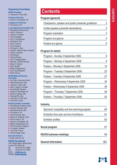
Organising CommitteesISUOG President● Lawrence D. Platt, USACongress Chairman● Kypros H. Nicolaides, UKCongress Co-Chairman● Tom Bourne, UKLocal Organising Committee, UK● Sabaratnam Arulkumaran● Alan D. Cameron● Julene S. Carvalho● Trish Chudleigh● Roy G. Farquharson● Nicholas M. Fisk● Davor Jurkovic● Lorin Lakasing● Christoph C. Lees● Fergal D. Malone, Rep. of Ireland● Darryl J. Maxwell● Karl Murphy● Pranav Pandya● Aris T. Papageorghiou● Nicholas J. Raine-Fenning● Steve C. Robson● Peter Soothill● Basky Thilaganathan● Guy Thorpe-Beeston● Peter TwiningISUOG Meeting PlanningCommittee● Ann Tabor, Denmark (Chair)● Alfred Abuhamad, USA● Joshua A. Copel, USA● Alain L Gagnon, Canada● Karel Mars˘ál, Sweden● Kypros H. Nicolaides, UK● David A. Nyberg, USA● Gianluigi Pilu, Italy● Lawrence D. Platt, USA● Yves Ville, FranceISUOG Scientific Committee● Karel Mars˘ál, Sweden (Chair)● Ahmet A. Baschat, USA● Beryl Benacerraf, USA● Tom Bourne, UK● Enrico Ferrazzi, Italy● Steven R. Goldstein, USA● Davor Jurkovic, UK● Kypros H. Nicolaides, UK● Basky Thilaganathan, UK● Dirk Timmerman, Belgium● Lil Valentin, Sweden● Yves Ville, France● Simcha Yagel, IsraelExecutive Director● Sarah Johnson, UKISUOG SecretariatUnit 4 Blythe Mews, Blythe Road,London W14 0HW, UKTel: +44 (0) 20 7471 9955Fax: +44 (0) 20 7471 9959Email: info@<strong>isuog</strong>.orgWebsite: www.<strong>isuog</strong>.orgContents<strong>Program</strong> (general)Chairperson, speaker and poster presenter guidelines 2Invited speaker presenter declarations 3<strong>Program</strong> orientation 3<strong>Program</strong> at a glance 4Posters at a glance 6<strong>Program</strong> (in detail)<strong>Program</strong> – Sunday 3 September 2006 8<strong>Program</strong> – Monday 4 September 2006 9Posters – Monday 4 September 2006 14<strong>Program</strong> – Tuesday 5 September 2006 22Posters – Tuesday 5 September 2006 27<strong>Program</strong> – Wednesday 6 September 2006 34Posters – Wednesday 6 September 2006 39<strong>Program</strong> – Thursday 7 September 2006 48Posters – Thursday 7 September 2006 53IndustrySponsors’ hospitality and live scanning program 60Exhibition floor plan and list of exhibitors 61Exhibitor profiles 62Social program 67ISUOG business meetings 68General informationIBCCONTENTS1
Invited speaker presenter declarationsThe following declarations and affiliations have been notified:J.A. Copel Equipment loans from and Consultant for Aloka, GE and Philips.T. D’Hooghe Current/past advisor or Consultant for Ferring, Genentech, Pfizer, Organon and Serono. Serono Chair for Reproductive Medicine atLeuven University, Belgium.H.P. Dietz Has spoken at events held by GE Australia, the total income from such engagements in 2005 totally Aus$750.N.M. FiskDirector of RevealCyte Ltd, Consultant for Ferring International and Trustee of Multiple Births Foundation.S.R. Goldstein Serves on the Gynecology Advisory Boards of Eli Lilly, GlaxoSmithKline, Merck, Pfizer and Proctor & Gamble, and is one of the Boardof Directors for SonoSite, Inc.W. Lee Serves on the Advisory Board and Speakers’ Bureau and is a Consultant for GE Healthcare, with limited research support. Consultantfor Philips Medical and Siemens Medical Solutions, with limited research support from both companies.K. Murphy Serves on the Council of the Independent Doctors’ Forum UK and MDU London. Founder Member/Director of the Irish Medical Society.J. Trinder Received a £20,000 grant from Exelgyn for the continuation of the MIST Trial.B. Tutschek Has a consulting contract with Philips Medical Systems in Germany for a project unrelated to and not presented at this Congress.List of invited facultyA. Abuhamad, USAJ.L. Alcázar, SpainL.D. Allan, UKA.A. Baschat, USAM. Bazot, FranceB.R. Benacerraf, USAT. Bourne, UKP. Calda, Czech RepublicA. Cameron, UKS. Campbell, UKP. Carter, UKJ.S. Carvalho, UKR. Chaoui, GermanyG. Condous, AustraliaG. Conway, UKJ.A. Copel, USAT. D’Hooghe, BelgiumJ. Deprest, BelgiumG.R. DeVore, USAH.P. Dietz, AustraliaS.H. Eik-Nes, NorwayJ. Elson, UKE.E. Epstein, SwedenC. Exacoustos, ItalyE. Ferrazzi, ItalyN.M. Fisk, UKR. Frydman, FranceA.L. Gagnon, CanadaH.M. Gardiner, UKS.R. Goldstein, USAE. Gratacós, SpainJ.G. Grudzinskas, UKJ. Gupta, UKK. Hecher, GermanyW. Holzgreve, SwitzerlandI. Jacobs, UKE. Jauniaux, UKD. Jurkovic, UKK. Kagan, UKK.D. Kalache, UKN. Kametas, UKV. Khullar, UK<strong>Program</strong> orientationE. Kirk, UKT. Kiserud, NorwayL. Lakasing, UKW. Lee, USAC.C. Lees, UKF.P.G. Leone, ItalyD. Levine, USAA.M. Lower, UKF.D. Malone, Rep of IrelandI. Manyonda, UKH. Marret, FranceK. Mars˘ál, SwedenD. Maulik, USAD.J. Maxwell, UKM. Meyer-Wittkopf, SwitzerlandB. Mol, NetherlandsR. Moshy, UKK. Murphy, UKG. Nargund, UKT.R. Nelson, USAP. Neven, BelgiumA.C.C. Ngu, AustraliaK.H. Nicolaides, UKD.A. Nyberg, USAE. Okaro, UKD. Paladini, ItalyP. Pandya, UKA.T. Papageorghiou, UKJ. Parsons, UKG. Pilu, ItalyL.D. Platt, USAS. Quenby, UKR.A. Quintero, USAN.J. Raine-Fenning, UKL. Regan, UKR.H. Reznek, UKS.C. Robson, UKR. Romero, USAJ. Rymer, UKM.V. Senat, Italy (TBC)W. Sepulveda, ChileP.W. Soothill, UKA. Sultan, UKA. Tabor, DenmarkA.C. Testa, ItalyB. Thilaganathan, UKG. Thorpe-Beeston, UKD. Timmerman, BelgiumI.E. Timor-Tritsch, USAJ. Trinder, UKB. Tutschek, USAP. Twining, UKL. Valentin, SwedenT. Van Den Bosch, BelgiumT. Van Gorp, BelgiumC. Van Holsbeke, BelgiumY. Ville, FranceM. Weston, UKM. Whittaker, UK (TBC)S. Yagel, IsraelI. Zalud, USAINVITED SPEAKER PRESENTER DECLARATIONS / PROGRAM ORIENTATION● The program at a glance gives you an outline to each day’sevents including session titles and times, and all refreshmentbreaks.● The posters at a glance gives you an outline to each day'sposter sessions.● The program pages give you the detailed chronological listingof events – please note that most sessions run in parallel so youwill need to read through all sessions of similar time to decidewhich best suits your needs.● In all poster and oral presentation listings the presentingauthors names are underlined.● All lectures which are supported by free communicationabstracts have an ‘OC’ number listed next to them, you can usethis to refer to the Abstract book in your Congress bag and thedetailed abstract.● Some of the sessions include ‘live scan’ or ‘demonstration’ time.These are live or video updates on the latest techniques andtechnologies in the field which support the scientific data in thesame subject area.● The poster titles are listed in the pages immediately followingthe day of oral sessions. Oral posters are referenced to as ‘OP’numbers and view-only posters are referenced to as ‘P’numbers – you can use these both to locate the originalabstracts in the Abstract book and to find the relevant posters inthe dedicated poster areas. For the sake of easy reference,posters are listed in chronological, numerical order by day. Youcan also find all individual PPT posters by their poster numberon the poster pods.● There are allocated chaired poster discussion times for some ofthe themed poster sessions (‘OPs’). Poster authors are askedto be present during the relevant discussion session (these arelisted at the appropriate times in the program pages) and to beavailable during session breaks on the day of their presentationto answer questions.● The exhibition hall opens at 09:30 each day in time for themorning coffee break. The only exception to this is on Sunday 3September when the hall will open at 12:15 in time for the lunchbreak. All refreshment breaks are located within this room.● All oral poster (chaired discussion) sessions will be judgedwithin their subject categories. The best oral poster presentersin each session will receive certificates and will be formallyacknowledged in the Journal Ultrasound in Obstetrics &Gynecology. Presenting authors are advised that results ofjudging will be announced at the closing ceremony on Thursday7 September and are therefore encouraged to attend.3
<strong>Program</strong> at a glanceTuesday 5 September 2006Mezzanine RoomsKing’s Suite Palace Suite Exhibition Windsor & Westminster Waterloo & Chelsea & St James’Kensington & Buckingham Hall Palace Suites Suite Tower RichmondPoster viewing all day07:30–08:15 Poster discussion08:30–10:00 Magnetic resonance Early pregnancy II – 09:00–17:00: Sponsor private hospitality suitesimagingmiscarriage10:00–10:30 CoffeeMEDISON SIEMENS PHILIPS10:30–12:00 Central nervous systemEarly pregnancy III –Live demonstrationectopic pregnancysponsored by Philips12:00–14:00 12:00–12:45: Poster discussion12:45–13:45: GE Lunchsatellite symposium14:00–15:30 Risk assessment inmultiple pregnanciesLive demonstrationChronic pelvic painsponsored by Medison15:30–16:00 Coffee16:00–17:15 Twin–twin transfusionsyndromeGynecology case examples19:00–24:00 Congress Party (London Eye chamagne sunset flight and River Thames boat cruise)PROGRAM AT A GLANCEWednesday 6 September 2006King’s Suite Palace Suite Exhibition Windsor & Westminster Waterloo & Chelsea & St James’Kensington & Buckingham Hall Palace Suites Suite Tower RichmondPoster viewing all day07:30–08:15 Poster discussion08:30–10:00 Maternal circulation and 09:00–17:00: Sponsor private hospitality suitesprediction of pregnancycomplication Reproductive medicine –MEDISON SIEMENS GEovarian functionLive demonstrationsponsored by Toshiba10:00–10:30 Coffee10:30–12:00 Highest scored obstetricabstracts II Reproductive medicine –Live demonstrationmanagementsponsored by Toshiba12:00–14:00 12:00–12:45: Poster discussion12:45–13:45: Philips Lunchsatellite symposium14:00–15:30 Fetal haemodynamicsand IUGRAbnormal uterine bleedingLive demonstrationand menorrhagiasponsored by Siemens15:30–16:00 Coffee16:00–17:30 Neural tube defectsLive demonstrationThe menopausalsponsored by SiemensendometriumSession ends at 17:00Thursday 7 September 2006King’s Suite Palace Suite Exhibition Windsor & WestminsterKensington & Buckingham Hall Palace Suites SuitePoster viewing all day07:30–08:15 Poster discussion08:30–10:00 Fetal heart I – methodologyScanning asymptomaticLive demonstrationwomensponsored by Philips10:00–10:30 Coffee10:30–12:00 Fetal heart IILessons from theLive demonstrationIOTA trialsponsored by Aloka12:00–14:00 12:00–12:45: Poster discussion12:45–13:45: Medison Lunchsatellite symposium14:00–15:30 Fetal heart IIIManaging ovarianLive demonstrationpathologysponsored by Toshiba15:30–16:00 Coffee16:00–17:15 Prediction and prevention Imaging and the oncologyof preterm deliverypatientClosingMezzanine Rooms5
POSTERS AT A GLANCEPosters at a glanceMezzanine Rooms(1st floor, East Wing)Windsor Suite(Basement B2, East Wing)All posters are on display in continuous rolling loops throughout the day of presentation from 09:00 to 17:00.Day Poster session Poster numbers Poster Room ScreensessiondiscussiontimesOP OP02: Fetal anomaly screening I OP02.01 – OP02.12 12:00–12:45 CadoganP P02: Fetal anomaly screening I P02.01 – P02.25 Viewing only BerkeleyOP OP13: Reference values and miscellaneous OP13.01 – OP13.15 12:00–12:45 BelgraveP P13: Reference values and miscellaneous P13.01 – P13.30 Viewing only ClarenceOP OP15: Early pregnancy OP15.01 – OP15.08 07:30–08:15 Lancaster & York 2Monday 4 P P15: Early pregnancy P15.01 – P15.15 Viewing only Lancaster 2September OP OP16: Urogynecology OP16.01 – OP16.07 12:00–12:45 Lancaster 1P P16: Urogynecology P16.01 – P16.03 Viewing only Lancaster 1OP OP03: Fetal central nervous system I OP03.01 – OP03.11 12:00–12:45 York 1P P03: Fetal central nervous system I P03.01 – P03.17 Viewing only York 2OP OP01: Screening for chromosomal abnormalities Ia OP01.01 – OP01.11 07:30–08:15 Blenheim 1OP OP01: Screening for chromosomal abnormalities Ib OP01.12 – OP01.25 12:00–12:45 Blenheim 2P P01: Screening for chromosomal abnormalities I P01.01 – P01.23 Viewing only Blenheim 3OP OP05: Twins OP05.01 – OP05.14 12:00–12:45 CadoganP P05: Twins P05.01 – P05.26 Viewing only BerkeleyOP OP01: Screening for chromosomal abnormalities II OP01.26 – OP01.31 12:00–12:20 BelgraveP P01: Screening for chromosomal abnormalities II P01.25 – P01.46 Viewing only ClarenceOP OP12: Controversies OP12.01 – OP12.05 12:20–12:45 BelgraveTuesday 5 OP OP17: Chronic pain OP17.01 – OP17.08 07:30–08:15 Lancaster & York 2September OP OP18: Abnormal bleeding OP18.01 – OP18.07 12:00–12:45 Lancaster 1P P18: Abnormal bleeding P18.01 – P18.17 Viewing only Lancaster 1OP OP06: Fetal lungs OP06.01 – OP06.09 12:00–12:45 York 1P P02: Fetal anomaly screening II P02.26 – P02.49 Viewing only York 2OP OP03: Fetal central nervous system II OP03.12 – OP03.20 07:30–08:15 Blenheim 1OP OP04: Fetal heart I OP04.01 – OP04.14 12:00–12:45 Blenheim 2P P04: Fetal heart I P04.01 – P04.14 Viewing only Blenheim 36
Posters at a glancePalace Suite(Basement B3, East Wing)POSTERS AT A GLANCEDay Poster session Poster numbers Poster Room ScreensessiondiscussiontimesOP OP09: 3D/4D ultrasound I OP09.01 – OP09.14 12:00–12:45 BerkeleyP P09: 3D/4D ultrasound I P09.01 – P09.14 Viewing only CadoganOP OP02: Fetal anomaly screening III OP02.13 – OP02.23 12:00–12:45 BelgraveP P02: Fetal anomaly screening III P02.50 – P02.74 Viewing only ClarenceOP OP06: Fetal therapy / Anaemia OP06.10 – OP06.25 12:00–12:45 Regent’sP P06: Fetal therapy / Anaemia P06.01 – P06.11 Viewing only Regent’sWednesday 6 OP OP19: Endometrium OP19.01 – OP19.09 07:30–08:15 Lancaster & York 2September P P19: Endometrium P19.01 – P19.04 Viewing only Lancaster 2OP OP20: Fertility OP20.01 – OP20.08 12:00–12:45 Lancaster 1P P20: Fertility P20.01 – P20.15 Viewing only Lancaster 1OP OP04: Fetal heart II OP04.15 – OP04.28 12:00–12:45 York 1P P04: Fetal heart II P04.15 – P04.27 Viewing only York 2OP OP07: IUGR I – prediction of pre-eclampsia OP07.01 – OP07.13 07:30–08:15 Blenheim 1P P07: IUGR I – prediction of pre-eclampsia P07.01 – P07.27 Viewing only Blenheim 3OP OP07: IUGR II – Doppler ultrasound in IUGR OP07.14 – OP07.28 12:00–12:45 Blenheim 2OP OP09: 3D/4D ultrasound II OP09.15 – OP09.27 12:00–12:45 BerkeleyP P09: 3D/4D ultrasound II P09.15 – P09.28 Viewing only CadoganOP OP11: Labour OP11.01 – OP11.14 12:00–12:45 BelgraveP P11: Labour P11.01 – P11.17 Viewing only ClarenceOP OP08: Fetal macrosomia OP08.01 – OP08.12 12:00–12:45 Regent’sOP OP21: Imaging in oncology OP21.01 – OP21.10 07:30–08:15 Lancaster & York 1Thursday 7 P P21: Imaging in oncology P21.01 – P21.03 Viewing only Lancaster 1September OP OP22: Ovarian pathology OP22.01 – OP22.08 12:00–12:45 Lancaster 2P P22: Ovarian pathology P22.01 – P22.04 Viewing only Lancaster 2OP OP10: Placenta OP10.01 – OP10.15 12:00–12:45 York 1P P14: Fetal magnetic resonance imaging P14.01 – P14.10 Viewing only York 2OP OP02: Fetal anomaly screening IV OP02.24 – OP02.40 07:30–08:15 Blenheim 1P P02: Fetal anomaly screening IV P02.75 – P02.99 Viewing only Blenheim 3OP OP04: Fetal heart III OP04.29 – OP04.37 12:00–12:45 Blenheim 27
SUNDAY PROGRAM<strong>Program</strong> – Sunday 3 September 200610:30–12:00 Opening plenaries Room: King’s SuiteChairs: L.D. Platt (USA); K.H. Nicolaides (UK)10:30 Opening addressL.D. Platt (USA)10:35 Welcome to LondonK.H. Nicolaides (UK)10:45 Introduction of the Ian Donald Gold Medal winner and presentation of medalK. Mars˘ál (Sweden)10:50 Ian Donald Gold Medal lecture: Doppler – more or lessB. Trudinger (Australia)11:10 Genetic conditions detectable from fetal material in the maternal circulationW. Holzgreve (Switzerland)11:25 Characterising ovarian tumoursD. Timmerman (Belgium)11:40 Too many twinsY. Ville (France)12:00–12:30 ISUOG Annual General Meeting Room: King’s Suite12:30–13:30 Lunch Exhibition area13:30–14:50 Plenary lectures Room: King’s SuiteChairs: Y. Ville (France); S.H. Eik-Nes (Norway)13:30 Live scan demonstration: New developments in fetal heart examination (*Sponsored by GE)G.R. DeVore (USA)13:50 The assessment and classification of obstetric pelvic floor trauma (including video demo) OC01H.P. Dietz; O. Lekskulchai (Australia)14:10 Can 3D volume sets alone be used to detect fetal malformations? (including video demo) OC02B.R. Benacerraf; B. Bromley; T.S. Shipp (USA)14:30 Stuart Campbell lecture: Screening in pregnancyK.H. Nicolaides (UK)15:00–15:30 Coffee Exhibition area15:30–17:30 Plenary lectures Room: King’s SuiteChairs: S. Campbell (UK); K. Hecher (Germany)15:30 Ovarian ageingR. Frydman (France)15:45 Doppler in the assessment of severe fetal compromiseE. Gratacós (Spain)16:00 Discussant for above lectureA.A. Baschat (USA)16:10 MR volumetry in fetuses with ventriculomegaly OC03D. Levine; J. Kazan; V. Dialani; G. Chiang; H. Feldman (USA)16:25 Stump the ProfessorPanel of Professors: B.R. Benacerraf 1 ; S. Campbell 2 ; S.H. Eik-Nes 3 ; S.R. Goldstein 1 ; K. Hecher 4 ;K.H. Nicolaides 2 ; G. Pilu 5 ; L.D. Platt 1 ; I.E. Timor-Tritsch 1 ; Y. Ville 61(USA); 2 (UK); 3 (Norway); 4 (Germany); 5 (Italy); 6 (France)18:00–21:30 Welcome reception Room: King’s & Monarch Suites8*Live scan demonstration sessions are generously supported by our sponsoring partners. Sponsors provide systems and support for all technicalservices to bring live scan sessions to you. Scientific content is defined by ISUOG.
<strong>Program</strong> – Monday 4 September 200607:30–08:15 Oral poster discussions07:30–08:15 Chairs: G. Condous (Australia); I.E. Timor-Tritsch (USA) Room: Lancaster & YorkOP15: Early pregnancyOP15.01–OP15.0807:30–08:15 Chairs: D.A. Nyberg (USA); P. Pandya (UK) Room: BlenheimOP01: Screening for chromosomal abnormalities IaOP01.01–OP01.11Poster viewing is available all day, all poster rooms, all sessions (see pages 14–21 for details)08:30–09:20 Parallel: Fetal face / Fetal behaviour (*Sponsored by GE) Room: King’s SuiteChairs: S. Campbell (UK); L.D. Platt (USA)08:30 Assessment of fetal to neonatal behavioral continuity by 4D ultrasonography OC04M.S. Stanojevic; A.K. Kurjak; W.A. Andonotopo (Croatia)08:35 3D and 4D ultrasound for evaluation of fetal face anomalies OC05E. Merz; Ch. Welter; A. Oberstein (Germany)08:40 Improving cleft palate/cleft lip antenatal diagnosis by 3D ultrasound: the ‘flipped face view’ OC06L.D. Platt; G.R. DeVore; D.H. Pretorius (USA)08:45 Live scan demonstration: A novel way of imaging the fetal soft palateB.R. Benacerraf (USA)09:05 DiscussionMONDAY PROGRAM09:20–10:00 Parallel: Highest scored obstetric abstracts I Room: King’s SuiteChairs: S. Campbell (UK); L.D. Platt (USA)09:20 Transabdominal B-mode and V-mode vs B-mode/ and V-mode/sono-MRI in achievement and examination of fetal OC07physiologic mid-sagittal scan: the experience in 1998–2006 periodE. Varvarigos 1 ; M. Iaccarino 1 ; S. Iaccarino 1 ; R.N. Laurini 21(Italy); 2 (Sweden)09:25 Discussion09:30 Relative increase in lung volume in fetuses with congenital diaphragmatic hernia treated by fetoscopic endoluminal OC08tracheal occlusion (FETO)M. Cannie; J. Jani; D. Van Schoubroeck; F. De Keyzer; S. Dymarkowski; J. Deprest (Belgium)09:35 Discussion09:40 Selectivity of laser coagulation of chorionic plate anastomoses in twin–twin transfusion syndrome – a correlation with OC09fetal and perinatal outcomeJ.S. Stirnemann; B. Nasr; E.Q. Quarello; M. Nassar; Y. Ville (France)09:45 Discussion09:50 Pregnancy outcome after enlarged nuchal translucency and normal mid-trimester scan: practical guidelines in OC10counselling parentsC.M. Bilardo; M.A. Muller; E. Pajkrt (Netherlands)09:55 Discussion08:30–10:00 Parallel: Controversies in gynecology – a series of discussions Room: Palace SuiteChairs: D. Timmerman (Belgium); T. Bourne (UK)08:30 Debate: Early pregnancy units: have we created a monster?For the motion: D. Jurkovic (UK)Against the motion: R. Moshy (UK)09:00 Debate: Postmenopausal women should undergo an annual scan to assess the endometrium and ovariesFor the motion: E. Ferrazzi (Italy)Against the motion: L. Valentin (Sweden)09:30 Debate: Laparoscopy is unnecessary for the majority of women with pelvic painFor the motion: E. Okaro (UK)Against the motion: M. Whittaker (UK)10:00–10:30 Coffee and PowerPoint poster viewing Exhibition area10:30–12:00 Parallel: Screening for chromosomal abnormalities Room: King’s Suite(*Sponsored by Medison)Chairs: K.H. Nicolaides (UK); F.D. Malone (Republic of Ireland)10:30 OverviewK.H. Nicolaides (UK)10:35 Live scan demonstration: Nuchal translucency screeningK.H. Nicolaides; K. Kagan (UK)*Live scan demonstration sessions are generously supported by our sponsoring partners. Sponsors provide systems and support for all technicalservices to bring live scan sessions to you. Scientific content is defined by ISUOG.9
<strong>Program</strong> – Monday 4 September 200610:55 Impact of a national first-trimester Down syndrome screening policy OC11A. Tabor (Denmark)11:00 First-trimester nuchal translucency and nasal bone assessment for Down syndrome screening at a single centre OC12G. Monni; M.A. Zoppi; R.M. Ibba; M. Floris; F. Manca; C. Axiana (Italy)11:05 Fetal nasal bone and ductus venosus blood flow assessed transvaginally at the 11–14 week scan – new data including OC13unselected and high risk pregnanciesA. Borrell; V. Borobio; A. Gonce; I. Mercade; A. Sanchez; V. Penalva; B. Puerto; E. Gratacós (Spain)11:10 Integrated screening for Down syndrome in routine clinical practice OC14P. Pandya; P. Jones; W. Huttly; C. Rodeck; N. Wald (UK)11:15 The use of fetal echocardiography when performing the genetic sonogram: 98% detection rate for trisomy 21 in OC15high-risk womenG.R. DeVore (USA)11:20 Fetal loss rate after first-trimester screening for chromosomal anomalies and after diagnostic procedures for OC16akaryotyping in women aged 36 years or olderP. Robles de Medina; A.L. van der Nooij; C.M. Bilardo (Netherlands)11:25 Facial angle in fetuses with trisomy 21 at 11–13+6 weeks OC16bJ. Sonek 1 ; M. Borenstein 2 ; T. Dagklis 2 ; N. Persico 2 ; K.H. Nicolaides 21(USA); 2 (UK)11:30 DiscussionMONDAY PROGRAM10:30–12:00 Parallel: Early pregnancy I – pregnancies of unknown location (PUL) Room: Palace SuiteChairs: S.R. Goldstein (USA); D. Jurkovic (UK)10:40 The development and role of discriminatory zonesB. Mol (Netherlands)10:55 The role of progesterone for the assessment of PULJ. Elson (UK)11:10 Does curettage have a role for the evaluation of PUL?G. Condous (Australia)11:25 The hCG ratio and mathematical models for the evaluation of PUL OC17–OC20E. Kirk 1 ; P. Alnaes-Katjavivi 2 ; G. Condous 1 ; C. Bottomley 1 ; B. Van Calster 3 ; S. Van Huffel 3 ; O. Istre 2 ;D. Timmerman 3 ; T. Bourne 11(UK); 2 (Norway); 3 (Belgium)11:40 Round table discussionG. Condous 1 ; J. Elson 2 ; E. Kirk 2 ; B. Mol 31(Australia); 2 (UK); 3 (Netherlands)12:00–14:00 Lunch Exhibition area12:00–12:45 Oral poster discussions12:00–12:45 Chairs: A.L. Gagnon (Canada); P. Twining (UK) Room: CadoganOP02: Fetal anomaly screening IOP02.01–OP02.1212:00–12:45 Chairs: B. Tutschek (USA); W. Lee (USA) Room: BelgraveOP13: Reference values and miscellaneousOP13.01–OP13.1512:00–12:45 Chairs: A. Sultan (UK); V. Khullar (UK) Room: LancasterOP16: UrogynecologyOP16.01–OP16.0712:00–12:45 Chairs: G. Pilu (Italy); W. Sepulveda (Chile) Room: YorkOP03: Fetal central nervous system IOP03.01–OP03.1112:00–12:45 Chairs: K. Murphy (UK); A.T. Papageorghiou (UK) Room: BlenheimOP01: Screening for chromosomal abnormalities IbOP01.12–OP01.25Poster viewing is available all day, all poster rooms, all sessions (see pages 14–21 for details)12:45–13:45 **Siemens satellite symposium: The fetal brain by MRI and Room: King’s Suiteultrasound – diagnostic conundra case studiesChair: D.J. Maxwell (UK)Speakers: L. Guibaud (France); G. Hackett (UK); C.C. Lees (UK); E. Whitby (UK)14:00–15:30 Parallel: 11–14 week anomaly scan (*Sponsored by Philips) Room: King’s SuiteChairs: A. Tabor (Denmark); P.W. Soothill (UK)14:00 Live scan demonstration: Early anomaly scanB. Thilaganathan (UK)*Live scan demonstration sessions are generously supported by our sponsoring partners. Sponsors provide systems and support for all technicalservices to bring live scan sessions to you. Scientific content is defined by ISUOG**Lunchtime satellite symposia are brought to you by our sponsors. ISUOG takes no responsibility for scientific or clinical content.11
MONDAY PROGRAM<strong>Program</strong> – Monday 4 September 200614:20 Comparison of nuchal scan and detailed morphology scan in the first-trimester screening for fetal structural OC21abnormalitiesM. Chen; Y.H. Lam; C.P. Lee; R. Tang; B. Chan; S.F. Wong; H.Y. Tse; M.H.Y. Tang; K.Y. Leung (China)14:25 Detection of fetal anomalies in the first and second trimesters OC22A. Kang; H. Struben; W. Holzgreve; O. Lapaire; S. Doht; S. Tercanli (Switzerland)14:30 What information on fetal anatomy can a single transabdominal first-trimester three-dimensional sweep provide? OC23R.J. Benzie; D.E.V. Fauchon; D. Wye; R. Thavaravy (Australia)14:35 Discussion14:45 First-trimester intrauterine growth restriction in fetuses with chromosomal abnormalities OC24A.T. Papageorghiou; K. Avgidou; K.H. Nicolaides (UK)14:50 Fetal growth and congenital malformations OC25A. Nikkilä; B. Källén; K. Mars˘ál (Sweden)14:55 Discussion15:05 Detection of ARSA at early screening – limitations in routine workup OC26M. Entezami; K.S. Heling; R. Chaoui; A. Hagen; M. Albig; R.D. Wegner; M. Stumm (Germany)15:10 The predictive values of ultrasonographic measurement of the fetal cardiothoracic ratio in pregnancies affected by OC28homozygous O thalassemiaK.Y. Leung 1 ; C. Liao 2 ; C.M. Li 2 ; S.Y. Ma 2 ; M.H.Y. Tang 1 ; C.P. Lee 1 ; Y.H. Lam 1 ; V. Chan 11(Hong Kong); 2 (China)15:15 Discussion14:00–15:30 Parallel: Managing ovarian pathology in practice Room: Palace SuiteChairs: T. Bourne (UK); B.R. Benacerraf (USA)14:00 What the morphology of a mass tells you about its natureC. Exacoustos (Italy)14:15 Does Doppler add anything to the assessment?I. Zalud (USA)14:30 Managing ovarian pathology in pregnancyG. Condous (Australia)14:45 Subjective impression of common massesD. Timmerman (Belgium)15:00 Case examples: Benign or malignant pathology? (Group 1)C. Exacoustos (Italy)Case examples: Pattern recognition (Group 2)A.C. Testa (Italy)Case examples: Acute pathology – torsion, haemorrhagic cysts and cyst rupture (Group 3)L. Valentin (Sweden)15:30–16:00 Coffee and PowerPoint poster viewing Exhibition area16:00–17:40 Parallel: Fetal lungs (*Sponsored by Medison) Room: King’s SuiteChairs: J.A. Copel (USA); A. Cameron (UK)16:00 OverviewA. Cameron (UK)16:10 Live scan demonstration: 3D lung volumesK.D. Kalache (UK)16:30 Video demonstration: Tracheal occlusion – how to remove the balloonJ. Deprest (Belgium)16:40 Outcome of features with diaphragmatic hernia treated with FETO and announcement of OC29–OC30randomised studiesJ. Deprest 1 ; J. Jani 1 ; E. Gratacós 2 ; S. Salcedo 2 ; A. Greenough 3 ; K. Allegaert 1 ; O. Moreno 2 ; S. Patel 3 ;K.H. Nicolaides 31(Belgium); 2 (Spain); 3 (UK)16:45 2D and 3D in the assessment of fetal lung volume in fetuses with diaphragmatic hernia OC31–OC33J. Jani 1 ; C.F.A. Peralta 2 ; D. Van Schoubroeck 1 ; J. Deprest 1 ; T. Cos 1 ; K.H. Nicolaides 2 ; A. Benachi 31(Belgium); 2 (UK); 3 (France)16:55 Lung volume measurements by 3D ultrasound are not superior to biometry by 2D ultrasound to predict pulmonary OC34hypoplasia in fetuses with musculoskeletal disordersL.F. Gonçalves; J.P. Kusanovic; J. Espinoza; W. Lee; N. McNamee; M.L. Schoen; O. Erez; M. Treadwell;R. Romero (USA)17:00 Changes in lung blood perfusion in congenital diaphragmatic hernia treated with FETO and association with clinical OC35outcomeO. Moreno-Alvarez 1 ; J. Jani 2 ; E. Hernández-Andrade 1 ; T. Jansson 3 ; J. Deprest 2 ; E. Gratacós 11(Spain); 2 (Belgium); 3 (Sweden)12*Live scan demonstration sessions are generously supported by our sponsoring partners. Sponsors provide systems and support for all technicalservices to bring live scan sessions to you. Scientific content is defined by ISUOG.
<strong>Program</strong> – Monday 4 September 200617:05 Predicting postnatal pulmonary arterial hypertension using the vascular indices estimated by three-dimensional power OC36Doppler ultrasonography in isolated congenital diaphragmatic herniaR. Ruano 1 ; M.C. Aubry 2 ; B. Barthe 2 ; M. Zugaib 1 ; Y. Dumez 2 ; A. Benachi 21(Brazil); 2 (France)17:10 The hyperoxygenation test for fetal pulmonary artery reactivity before and after in utero unplugging in fetuses OC37with severe congenital diaphragmatic hernia that underwent fetoscopic endoluminal tracheal occlusionE. Done; J. Jani; D. Van Schoubroeck; A. Debeer; J. Deprest (Belgium)17:15 Preventive collagen plugging of fetal membrane defects following fetoscopic endoluminal tracheal occlusion (FETO) OC38R. Devlieger; L. Lewi; J. Jani; D. Van Schoubroeck; M. Cannie; J. Deprest (Belgium)17:20 DiscussionMONDAY PROGRAM16:00–17:30 Parallel: Urogynecology Room: Palace SuiteChairs: S. Yagel (Israel); V. Khullar (UK)16:00 The role of MRI and radiology in urogynecologyJ. Deprest (Belgium)16:15 Ultrasound imaging of the pelvic floorH.P. Dietz (Australia)16:30 Ultrasound imaging in anal incontinenceA. Sultan (UK)16:45 Imaging-based management in urogynecologyV. Khullar (UK)17:00 Postpartum evaluation of the anal sphincter after surgical repair of third-degree tear by 3D transperineal ultrasound OC39in primiparous womenD.V. Valsky; B. Messing; D. Rosenak; D. Hochner-Celnikier; S.M. Cohen; S. Yagel (Israel)17:05 Volume of the internal and external anal sphincters assessed by three-dimensional endoanal ultrasound technique OC40I.P. Olsen; T. Wilsgaard; T. Kiserud (Norway)17:10 Detrusor wall thickness is of limited use as a test for detrusor overactivity OC41O. Lekskulchai 1 ; H.P. Dietz 21(Thailand); 2 (Australia)17:15 Visualization of the vaginal high-pressure zone by three-dimensional ultrasound images in the pelvic floor OC42S.A. Jung; D.H. Pretorius; B.S. Padda; M.M. Weinstein; C.W. Nager; D. Den Boer; R.K. Mittal (USA)17:20 Discussion13
MONDAY POSTERSPowerPoint poster sessionsMezzanine Rooms(1st floor, East Wing)Windsor Suite(Basement B2, East Wing)Palace Suite(Basement B3, East Wing)Note re. abstract numbers below:‘OP’ indicates an oral poster presentation, to be discussed during early morning and lunch time chaired discussion sessions‘P’ indicates a view-only posterMonday 4 September 2006Abstract no.OP01: Screening for chromosomal abnormalities Ia Room: Blenheim, screen 1Likelihood ratio for trisomy 21 in fetuses with abnormal ductus venosus flow at the 11–14 week scanC.G.V. Murta (Brazil)Association of first-trimester nasal bone length with free beta hCG, PAPP-A and nuchal translucencyT.W. Hallahan (USA)First-trimester PAPP-A is a predictor of adverse pregnancy outcomeL. Krofta (Czech Republic)Should isolated choroid plexus cyst(s) or intracardiac echogenic foci detected in the low-risk population be referred for tertiary scans?T.N. Leung (Hong Kong)Screening for chromosomal abnormalities during the second-trimester scan in a general population without termination of pregnancyM.C. Parra-Cordero (Chile)The genetic sonogram for women at increased risk for Down syndrome (DS)M.C. Van den Hof (Canada)Subjective appraisal of fetal nasal bridge (Nbr) and/or bone (NB) for the risk of Down syndrome (DS) after 18 weeks’ gestationM.C. Van den Hof (Canada)Interobserver variability of the subjective evaluation of the nasal bone (NB) and nasal bridge (Nbr) for the risk of Down syndrome (DS)after 18 weeks’ gestationD.C. Young (Canada)Nasal bone evaluation in prenatal screening for trisomy 21: a reviewJ.D. Sonek (USA)Understanding the decision-making process for prenatal testing in women over 34 years' old; a provincial perspectiveA.L. Gagnon (Canada)Prevalence of an aberrant right subclavian artery (ARSA) in fetuses with chromosomal aberrationsR. Chaoui (Germany)OP01.01OP01.02OP01.03OP01.04OP01.05OP01.06OP01.07OP01.08OP01.09OP01.10OP01.11OP01: Screening for chromosomal abnormalities Ib Room: Blenheim, screen 2Maternal age-specific detection rates and false-positive rates for first- and second-trimester screening for Down syndromeG.R. DeVore (USA)Non-invasive prenatal diagnosis of fetal sex using free fetal DNA in the maternal circulation – changing obstetric managementL.S. Chitty (UK)The significance of echogenic bowelA. Saba (UK)Significance of ultrasound markers for the detection of chromosomal abnormalities in a high-risk Saudi populationW. Kurdi (Saudi Arabia)The isolated echogenic intracardiac focus in fetuses with trisomy 21: the role of echocardiographyG.R. DeVore (USA)Cerebral ventriculomegaly: is the prevalence increased for fetuses with trisomy 21?M. Albig (Germany)Combining fetal nuchal fold thickness with second-trimester biochemistry to screen for trisomy 21A. Borrell (Spain)OP01.12OP01.13OP01.14OP01.15OP01.16OP01.17OP01.1814
MONDAY POSTERSPowerPoint poster sessionsAbstract no.OP02: Fetal anomaly screening IRoom: CadoganFetal anomaly scan: is 30 seconds enough?OP02.01M. Entezami (Germany)The 11–14 week anatomy scan: impact of sonographer trainingOP02.02K.W. Fong (Canada)Audit of outcome and accuracy of prenatal diagnosis in a tertiary fetal medicine unitOP02.03P.M. Kyle (New Zealand)Ultrasound examinations for detecting congenital defects in diabetic pregnanciesOP02.04A. Galindo (Spain)Correlation between prenatal diagnosis ultrasound and fetal examination: comparison of 112 cases resulting in termination of pregnancyOP02.05due to fetal abnormalitiesM.V. Senat (France)Correlation of ultrasonographic, pathological and cytogenetic findings in structural abnormalities diagnosed in first trimester during five-year OP02.06periodI. Kucerova (Czech Republic)Interdisciplinary ethical decision-making by ethics forum following prenatal diagnosis of major fetal anomaliesOP02.07M. Meyer-Wittkopf (Switzerland)Effectiveness of ultrasound in the antenatal prediction of symptomatic congenital CMV infectionOP02.08G. Simonazzi (Italy)The dynamics of the silent focal myometral contraction in pregnancyOP02.09Y. Romem (Israel)Sonographically detected accessory placental lobe and pregnancy outcomeOP02.10M.C. Hoffman (USA)The spectrum and outcome of prenatally diagnosed fetal tumorsOP02.11D. Kamil (Germany)Fetal structural abnormalities detected at the 11–14 week scanOP02.12L.C.S. Bussamra (Brazil)P02: Fetal anomaly screening I Room: BerkeleyTo evaluate the value of systematic ultrasound examination in detecting fetal malformation in the second trimester P02.01C. Xinlin (Chile)Review of fetal abnormalities review in an ultrasound and prenatal diagnosis unit in Barcelona P02.02C. Rueda (Spain)Abstract withdrawn P02.03Our experience as a third-level center for prenatal diagnosis P02.04A.S. Simionescu (Romania)Outcome of pregnancies with nuchal translucency more than 3mm P02.05M. Dharmalingam (UK)First-trimester cystic hygroma: diagnosis, management and outcomes in an Irish tertiary referral centre P02.06C.M. Lynch (Republic of Ireland)Abstract withdrawn P02.07Is the accuracy in ultrasonographic prenatal diagnosis of congenital anomaly affected by fetal anatomical systems? P02.08K.W. Kim (Republic of Korea)Isolated placental aneuploidy associated with spontaneous resolution of hydrops fetalis P02.09C.G.V. Murta (Brazil)The obstetrical significance of single umbilical artery P02.10T.H. Kwon (Republic of Korea)The outcome of fetal isolated pleural effusion P02.11S.J. Lee (Republic of Korea)Isolated short femur – what does this mean? P02.12A. Kennedy (USA)Prenatal diagnosis of diaphragmatic hernia: an early sonographic finding for second-trimester diagnosis P02.13S. Gabrielli (Italy)Unilateral short fetal femur: case report and review of the literature P02.14M. Ben-Ami (Israel)Abstract withdrawn P02.1516
PowerPoint poster sessionsAbstract no.Pseudocyst of the umbilical cord: prenatal ultrasound characteristics and clinical significance P02.16S.A. Abdullah (Saudi Arabia)Late sonographic appearance of fetal anophthalmia/microphthalmia P02.17M.L. Pisaturo (Italy)Early second-trimester amniotic shelf – a common, transient, benign finding on early second trimester sonography P02.18E. Mazaki (Israel)Prenatal diagnosis of cleft lip and palate in an ICSI singleton pregnancy P02.19I. Korkontzelos (Greece)Anencephaly in Singapore P02.20B.L. Tan (Singapore)Acrania – the same phenotypic expression caused by different etiological factors P02.21R. Mota (Portugal)Ultrasonographic diagnosis of the Dandy-Walker malformation. Additional ultrasound findings and perinatal outcome P02.22E. Cordioli (Brazil)Holoprosencephaly at 9 weeks 6 days in a triploid fetus: 2D and 3D ultrasound findings P02.23W. Sepulveda (Chile)Congenital anaplastic ependymoma: an uncommon cause of hydrocephalus diagnosed prenatally – a case report P02.24J. Lange (Germany)Fetal intracerebral hemorrhage – a case report P02.25S. Boito (Italy)MONDAY POSTERSOP03: Fetal central nervous system I Room: York, screen 1Intrauterine sonographic measurement of embryonic brain mantleH. Tanaka (Japan)Abnormal opercular formation on prenatal cerebral imaging significance and correlation with fetopathological and postnatal dataL. Guibaud (France)Fetal medicine physicians and the communication of ‘bad news’: parent evaluations of being informed of their child’s major abnormalityM. Meyer-Wittkopf (Switzerland)Echography of the fetal brainstem: a biometric and anatomical studyC. Courtiol (France)Visualisation of the fetal optic chiasmJ.P.B. Bault (France)Size of fetal cerebral ventricles: a non parametric approachL.J. Salomon (France)3D virtual longitudinal plane to identify the fetal corpus callosumS. Yagel (Israel)Measurement of the volume of the fetal corpus callosum using 3D sonographyR. Achiron (Israel)Cavum septum pellucidium mimics in agenesis of the corpus callosum and septo-optic dyplasia. Look at the ventricles not the cavumP. Twining (UK)Interest of systematic cerebellar transverse and great cistern measurement during routine ultrasonography examinationO. Morel (France)3D virtual longitudinal plane to identify the fetal vermisS. Yagel (Israel)OP03.01OP03.02OP03.03OP03.04OP03.05OP03.06OP03.07OP03.08OP03.09OP03.10OP03.11P03: Fetal central nervous system I Room: York, screen 2Prenatal diagnosis of closed spinal dysraphism P03.01D. Pugash (Canada)The importance of genetic counselling in holoprosencephaly P03.02A.L. David (UK)Fetal holoprosencephaly – associated malformations and chromosomal disorders P03.03A. Geipel (Germany)The influence of mode of conception, fetal gender and twin pregnancy in the development of cerebral structures: a fetal brain P03.043D ultrasound studyF.F. Correa (Spain)10-year (1995–2004) outcome of fetal neural tube defects P03.05S. Pathak (UK)Lissencephaly revisited: sonographic challenge of diagnosing abnormal cortical development – a case report P03.06U. Braig (Germany)Early prenatal diagnosis of a vein of Galen aneurysm P03.07M.W. Bebbington (USA)Ventriculomegaly and megacisterna magna maybe associated with fetal toxoplasmosis P03.08A.V. Carmo (Brazil)17
MONDAY POSTERSPowerPoint poster sessionsAbstract no.Reversed end-diastolic wave of middle cerebral artery in prenatal diagnosis of holoprosencephaly P03.09C.Y. Chen (Taiwan)Third ventricle teratoma – case report and literature review P03.10P. Anastassopoulos (UK)Fetal hydrocephalus: prenatal diagnosis and postnatal management P03.11J.L. Hernandez (Spain)Large fetal encephalocele – prenatal diagnosis and neonatal management P03.12A. Reitter (Germany)Intrauterine diagnosis of diastematomyelia: 2 case reports P03.13Y. Uyar (Turkey)Fetal hydrocephalus in a pregnancy complicated by idiopathic thrombocytopenic purpura P03.14H.M. Choi (Republic of Korea)Prenatal diagnosis of hydranencephaly using 2D and 3D ultrasound P03.15S.J. Kim (Republic of Korea)Antenatal diagnosis from six cases of ventriculomegaly P03.16S. Marta (Portugal)Prenatal diagnosis of schizencephalia – a case report P03.17A.S. Cerdeira (Portugal)OP13: Reference values and miscellaneousFetal thymus size as a predictor of intra-amniotic infection in women with preterm premature rupture of membranesR. Achiron (Israel)The pregnancy outcome of women with congenital uterine anomaly exceeding 20 weeks of gestationK.W. Kim (Republic of Korea)Assessment of palpable nodules of the abdominal wall with postpartum onsetF. Prefumo (Italy)Congenital CMV infection: ultrasound abnormalities in a non-selected populationE. Sleurs (Belgium)Pregnancy on intensified hemodialysis: fetal surveillance and perinatal outcomeC. Bamberg (Germany)Can ultrasound estimation of the central venous pressure replace invasive measurement to predict a low central venous pressure?H.A. Lombaard (South Africa)Nomograms of axial fetal cerebellar hemisphere circumference and area throughout gestationM. Dalloul (USA)Longitudinal reference ranges for flow velocities and waveform indices of the ductus venosusJ. Kessler (Norway)New reference ranges for serial measurements of middle cerebral artery Doppler velocities and indices based on longitudinal dataC. Ebbing (Norway)Variability in the fetal middle cerebral artery peak systolic blood flow velocity at three different segmentsT.M. Manrique (Spain)Uterine artery blood flow volume: ranges in uncomplicated human pregnanciesS. Rigano (Italy)Predicting adverse neonatal outcome in fetuses with severe placental insufficiency: a pivotal role for birth weight Z-scoresR.A. Moreira de Sà (Brazil)Customized centiles and perinatal morbidityF. Figueras (Spain)Modelling french birth biometricsL.J. Salomon (France)Evaluation of clinical performance of obstetric residents: ultrasonographic estimation of fetal weightY.D. Kang (Republic of Korea)Room: BelgraveOP13.01OP13.02OP13.03OP13.04OP13.05OP13.06OP13.07OP13.08OP13.09OP13.10OP13.11OP13.12OP13.13OP13.14OP13.15P13: Reference values and miscellaneous Room: ClarenceDoes the introduction of a policy to date pregnancies from ultrasound alone decrease the rate of IOL for prolonged pregnancy? P13.01B. Singhania (UK)Is early fetal growth affected by method of conception? P13.02A.C. Gjerris (Denmark)First-trimester growth in fetuses from assisted reproduction and spontaneous pregnancies P13.03P.W. Hui (Hong Kong)Effect of fetal gender, maternal smoking and maternal height on crown-rump length P13.04L. Rode (Denmark)Accuracy of second-trimester fetal head circumference and biparietal diameter for predicting time of spontaneous birth P13.05S.L. Johnsen (Norway)18
PowerPoint poster sessionsAbstract no.The effect of second-trimester fetal morphometry on duration of pregnancy P13.06S.L. Johnsen (Norway)Comparison of last menstrual period and crown-rump length in gestational dating – the effect of fetal gender, maternal smoking and maternal P13.07heightL. Rode (Denmark)Comparison of reference charts for estimated fetal weight and for actual birthweight P13.08L.J. Salomon (France)Customized fetal weight estimation at term P13.09S. Fiore (Italy)Impact of the modelling method on reference charts and equations for biometry P13.10L.J. Salomon (France)Quality control of fetal biometry: an integrated and automated approach P13.11L.J. Salomon (France)Is there a place for a single biometry check at the start of the third trimester of pregnancy for the early detection of IUGR or macrosomia? P13.12P.A.O.M. De Reu (Netherlands)Accuracy of qualitative measurement of amniotic fluid volume at term P13.13L. Smarkusky (USA)The dynamics of cervical length measurements P13.14M. Meijer-Hoogeveen (Netherlands)Umbilical vein flow and its relation to gestational age and estimated weight P13.15M. Yamamoto (Chile)Reproducibility and audit: important parameters for participation in venous Doppler sonography studies P13.16C. Hofstaetter (Germany)Intra- and interobserver reliability of umbilical vein blood flow P13.17S. Fernandez (Spain)The fetal portal vein – normal blood flow development during the second half of pregnancy P13.18J. Kessler (Norway)Reference ranges for Doppler-assessed aortic isthmus blood flow velocitiy and pulsatility indices in normal human fetuses P13.19B. Puerto (Spain)Real time velocity profile of fetal umbilical arteries (FUA) by global acquisition and signal processing (GASP) software for multigate spectral P13.20Doppler analysis (MSDA)G. Urban (USA)Velocity profile characteristics of fetal descending aorta (FDAo) by global acquisition and signal processing (GASP) software for multigate P13.21spectral Doppler analysis (MSDA)G. Urban (USA)Real time velocity profile of uterine artery (UtA) and vein (UtV) by global acquisition and signal processing (GASP) software for multigate P13.22spectral Doppler analysis (MSDA)G. Urban (USA)Relationship between umbilical artery Doppler resistance with placenta and neonatal birth weight in normal pregnancies P13.23J. Hartung (Germany)Longitudinal study of uterine artery Doppler after normal vaginal delivery P13.24A. Mulic-Lutvica (Sweden)Vascular resistance analysis during L-arginine therapy P13.25M. Ropacka (Poland)Detectation of human parvovirus B19 in cases of hydrops fetalis in Sao Paulo, Brazil P13.26J.Q. Andrade (Brazil)Gastroschisis: factors influencing neonatal outcome P13.27D. Subramanian (UK)Fetal intracranial hemorrhage and maternal anticoagulation with warfarin in patient with mechanical prosthetic heart valve – a case report P13.28G. Simonazzi (Italy)Location of the conus medullaris in nomal fetuses P13.29C. Xinlin (Chile)Quantitative ultrasound measurement of bone mineral density changes in pregnancy and the association with back pain symptoms during P13.30and 24 months after pregnancyM.W.N. Wong (Hong Kong)MONDAY POSTERSOP15: Early pregnancy Room: Lancaster, screen 2What are the causes of deficient uterine scars following Cesarean section?OP15.01D. Ofili-Yebovi (UK)Can ultrasound parameters predict the outcome of medical abortion?OP15.02S. Yagel (Israel)The prediction of outcome of expectant management of ectopic pregnanciesOP15.03E. Sawyer (UK)19
MONDAY POSTERSPowerPoint poster sessionsAdnexal findings, serum b-hcg and progesterone levels in ectopic pregnancyC.N. Nzewi (UK)Surgical intervention for diagnosis of ectopic pregnancy: are we doing the right thing?N. Dixit (UK)The value of ultrasound for the diagnosis of gestational trophoblastic diseaseC. Bottomley (UK)A potential new approach to the monitoring and management of Cesarean scar ectopic pregnanciesC. Bottomley (UK)Sonomorphology of ectopic pregnancy: an experience in a university hospitalA.P. Manjunath (India)Abstract no.OP15.04OP15.05OP15.06OP15.07OP15.08P15: Early pregnancy Room: Lancaster, screen 2Transvaginal ultrasound and human chorionic gonadotropin in pregnancies of unknown location P15.01C.M. Mulcahy (Republic of Ireland)A clinical study of cornual pregnancy; comparative study according to the timing of diagnosis P15.02G.S.R. Lee (Republic of Korea)Cesarean scar pregnancy with expectant management to full term: a case report P15.03A. El-Matary (UK)Role of maternal age in sonographic prediction of miscarriage in pregnant women with 1st-trimester bleeding P15.04T. Ghi (Italy)Bilateral ectopic pregnancy P15.05M. Rasidaki (Greece)A case report of cervical pregnancy P15.06T.A. Hantoushzadeh (Islamic Republic of Iran)Color Doppler study of middle cerebral artery blood flow in early normal pregnancy P15.07T. Urbaniak (Poland)Ultrasound and histological correlation of retained product of pregnancy P15.08A. Jamal (Islamic Republic of Iran)First-trimester Doppler assessment of spiral arteries in normal and abnormal pregnancies P15.09S. Nagy (Hungary)Yolk sac size and embryonic heart rate in the first trimester: findings in an Asian population P15.10L.K. Ng (Singapore)Subsequent pregnancy outcome after embolisation of uterine arteriovenous malformation P15.11C.F. Phoon (Singapore)Laminaria (L) dilatation and curettage in Cesarean scar pregnancy (CSP) with a literature review P15.12T. Hoshino (Japan)Can ultrasound miss the diagnosis of ectopic pregnancy in rudimentary horn? P15.13S. Sen (UK)Heterotopic pregnancy – a case report P15.14A. Jetti (UK)A case of placenta increta presenting as delayed postabortal hemorrhage P15.15C.Y. Kim (Republic of Korea)OP16: Urogynecology Room: Lancaster, screen 1Trauma after instrumental delivery – the use of 3D/4D ultrasound in the evaluation of levator ani muscleL. Krofta (Czech Republic)Maternal age at first vaginal delivery is associated with the prevalence of major levator traumaH.P. Dietz (Australia)Use of 3D ultrasound to assess puborectalis muscle measurementsD.H. Pretorius (USA)Translabial 3D/4D ultrasound of transobturator mesh implantsH.P. Dietz (Australia)Posterior pelvic floor assessment: a prospective comparison of functional vaginal endosonography and colpocistodefecographyS. Piciucchi (Italy)The efficacy and mechanism of action of the transobturator tapeJ.W. Ross (USA)Assessement of selected transvaginal sonography parameters in incontinent womenW. Sawicki (Poland)OP16.01OP16.02OP16.03OP16.04OP16.05OP16.06OP16.0720
PowerPoint poster sessionsAbstract no.P16: Urogynecology Room: Lancaster, screen 1The relationship between prolapse severity and symptoms P16.01H.P. Dietz (Australia)Ultrasonographic study on uterine hemodynamic changes following transcervical endometrial resection P16.02D. Zhang (Chile)To study the complication following hysteroscopic endometrial resection by ultrasonography P16.03D. Zhang (Chile)MONDAY POSTERS21
TUESDAY PROGRAM<strong>Program</strong> – Tuesday 5 September 200607:30–08:15 Oral poster discussions07:30–08:15 Chairs: E. Okaro (UK); T. D’Hooghe (Belgium) Room: Lancaster & YorkOP17: Chronic painOP17.01–OP17.0807:30–08:15 Chairs: A. Abuhamad (USA); D. Paladini (Italy) Room: BlenheimOP03: Fetal central nervous system IIOP03.12–OP03.20Poster viewing is available all day, all poster rooms, all sessions (see pages 27–33 for details)08:30–10:00 Parallel: Magnetic resonance imaging (MRI) Room: King’s SuiteChairs: D.A. Nyberg (USA); S. Yagel (Israel)08:30 OverviewD.A. Nyberg (USA)08:40 Preliminary feasibility of fetal cardiac MRI (including video demonstration) OC43G. Gorincour; J. Bouvenot; B. Bonello; A. Fraisse; A. Potier; B. Kreitmann; B. Bourliere-Najean (France)08:50 Expert neurosonography and magnetic resonance to assess the neurologic risk of mild fetal cerebral OC44ventriculomegalyA. Carletti; M. Segata; T. Ghi; G. Tani; G. Pilu; N. Rizzo (Italy)08:55 Third-trimester fetal MRI in isolated 10–12 mm ventriculomegaly: is it worth it? OC45L.J. Salomon; J. Ouahba; A.L. Delezoide; E. Vuillard; J.F. Oury; G. Sebag; C. Garel (France)09:00 Discussion09:15 Prenatal diagnosis of placenta accreta by ultrasound and magnetic resonance imaging OC46B. Dwyer; A. Rao; L. Tran; V. Belogolovkin; I. Carroll; R. Barth; U. Chitkara (USA)09:20 Placental functional MRI: a comprehensive mouse model OC47L.J. Salomon; N. Siauve; F. Taillieu; D. Balvay; C. Vayssettes; G. Frija; C.A. Cuenod; O. Clement; Y. Ville (France)09:25 Comparison of 3D ultrasound and MRI in assessing lung volumes in fetuses with diaphragmatic hernia OC48J. Jani 1 ; M. Cannie 1 ; C.F.A. Peralta 2 ; S. Dymarkowski 1 ; P. Lewi 1 ; K.H. Nicolaides 2 ; J. Deprest 11(Belgium); 2 (UK)09:30 Prenatal diagnosis and treatment of fetal hypothyroidism and goiter: the role of MRI OC49A. Gulraze; A. Mahrous; Z. Patay; W. Kurdi (Saudi Arabia)09:35 Use of a confidence scale in reporting normal and abnormal fetal anatomy on postmortem MRI OC50A.C.G. Breeze; J.J. Cross; P.A.K. Set; A.L. Whitehead; C.C. Lees; G.A. Hackett; I. Joubert; D.J. Lomas (UK)09:40 Discussion08:30–10:00 Parallel: Early pregnancy II – miscarriage Room: Palace SuiteChairs: D. Jurkovic; I. Manyonda (UK)08:30 Recurrent miscarriage – the scienceL. Regan (UK)08:45 Conservative and surgical management of miscarriage – the MIST studyJ. Trinder (UK)09:00 Clinical management of recurrent miscarriageS. Quenby (UK)09:15 Early diagnosis and management of molar miscarriagesE. Jauniaux (UK)09:30 Transvaginal ultrasound-guided evacuation of retained products of conception OC51C.C.T. Lee; J. Ben-Nagi; D. Ofili-Yebovi; J. Ross; D. Jurkovic (UK)09:35 Routine ultrasound scanning in a termination of pregnancy clinic setting promotes choice and improves OC52patient managementV.A. Rodie; A.J. Thomson (UK)09:40 The effect of low dose aspirin and omega-3 fatty acids on uterine artery Doppler flow velocity in women OC53with impaired uterine perfusion having a history of recurrent abortionN. Lazzarin; E. Vaquero; C. Exacoustos; G. Di Pierro; C. Amoroso; D. Arduini (Italy)09:45 Do women with hyperemesis gravidarum need an ultrasound scan? OC54A.T. Papageorghiou; E. Kirk; G. Condous; C. Bottomley; T. Bourne (UK)09:50 Discussion10:00–10:30 Coffee and PowerPoint poster viewing Exhibition area10:30–12:00 Parallel: Central nervous system (*Sponsored by Philips) Room: King’s SuiteChairs: G. Pilu (Italy); P. Calda (Czech Republic)10:30 OverviewG. Pilu (Italy)22*Live scan demonstration sessions are generously supported by our sponsoring partners. Sponsors provide systems and support for all technicalservices to bring live scan sessions to you. Scientific content is defined by ISUOG.
<strong>Program</strong> – Tuesday 5 September 200610:40 Live scan demonstration: Ultrasound of the fetal brainG. Pilu (Italy)11:00 Developmental anatomy and morphology of the cerebellar vermis – essential knowledge for the OC56imaging assessment of posterior fossa anomaliesA.J. Robinson; S.I. Blaser; A. Toi; S. Pantazi; D. Chitayat; G. Ryan (Canada)11:05 The cisterna magna septa – a vestigial remnant of the roof of the rhombencephalic vesicle OC57A.J. Robinson 1 ; R.B. Goldstein 21(Canada); 2 (USA)11:10 Clinical significance of fetal posterior fossa malformations OC58A. Carletti; G. Tani; T. Ghi; G. Gandolfi Colleoni; G. Contratti; M. Segata; D. Santini; P. Bonasoni; N. Rizzo; G. Pilu (Italy)11:15 Discussion11:30 Comparison among B-Mode, V-Mode and V-Mode sono MRI in differential diagnosis between cerebellar vermis OC59rotation and DW complexE. Varvarigos 1 ; M. Iaccarino 1 ; S. Iaccarino 1 ; G. De Chiara 1 ; R.N. Laurini 21(Italy); 2 (Sweden)11:35 Diagnosis of midline anomalies of the fetal brain with the three-dimensional median view OC60G. Pilu; M. Segata; T. Ghi; A. Carletti; A. Perolo; D. Santini; P. Bonasoni; G. Tani; N. Rizzo (Italy)11:40 Transfrontal 3D ‘shot’: the best 3D approach to the visualization of the fetal midline cerebral structures OC61F. Vinals; R. Naveas; A. Giuliano (Chile)11:45 DiscussionTUESDAY PROGRAM10:30–12:00 Parallel: Early pregnancy III – ectopic pregnancy Room: Palace SuiteChairs: E.E. Epstein (Sweden); S.R. Goldstein (USA)10:30 The ultrasound features of ectopic pregnancyG. Condous (Australia)10:40 Decision-tree analysis and selecting women for conservative management of ectopic pregnancyJ. Elson (UK)10:50 Selecting women for conservative management of tubal and interstitial ectopic pregnancies and protocols formedical treatmentE. Kirk (UK)11:05 Conservative or surgical management of early cervical and Caesarean scar actopics?D. Jurkovic (UK)11:15 The efficacy of Shirodkar cervical suture in securing hemostasis following evacuation of Cesarean scar ectopic OC62pregnancyJ. Ben-Nagi; J. Yazbek; D. Ofili-Yebovi; E. Sawyer; S. Helmy; D. Jurkovic (UK)11:20 A comparison of fertility outcomes following surgical and expectant management of tubal ectopic pregnancy OC63S. Helmy; E. Sawyer; D. Ofili-Yebovi; J. Yazbek; J. Ben-Nagi; D. Jurkovic (UK)11:25 Triage of patients with early pregnancy complications before ultrasound OC64H. Zingenberg; E. Dreisler (Denmark)11:30 Does a pregnancy of unknown location protocol work in a district general hospital? OC65C.L. Facey; M. Chetty; J. Edmondson; J. Elson (UK)11:35 Discussion12:00–14:00 Lunch Exhibition area12:00–12:45 Oral poster discussions12:00–12:45 Chairs: K. Hecher (Germany); Y. Ville (France) Room: CadoganOP05: TwinsOP05.01–OP05.14Chairs: S.C. Robson (UK); F.D. Malone (Republic of Ireland)Room: Belgrave12:00–12:20 OP01: Screening for chromosomal abnormalities II OP01.26–OP01.3112:20–12:45 OP12: Controversies OP12.01–OP12.0512:00–12:45 Chairs: A.M. Lower (UK); F.P.G. Leone (Italy) Room: LancasterOP18: Abnormal bleedingOP18.01–OP18.0712:00–12:45 Chairs: J. Deprest (Belgium); L. Lakasing (UK) Room: YorkOP06: Fetal lungsOP06.01–OP06.0912:00–12:45 Chairs: J.A. Copel (USA); L.D. Allan (UK) Room: BlenheimOP04: Fetal heart IOP04.01–OP04.14Poster viewing is available all day, all poster rooms, all sessions (see pages 27–33 for details)23
<strong>Program</strong> – Tuesday 5 September 200612:45–13:45 **GE satellite symposium Room: King’s SuiteSpeakers: A. Abuhamad – VCAD (Volume computer-aided diagnosis)B. Benoit – Early pregnancyR. Chaoui – Early pregnancy / heartN.J. Raine-Fenning – Gynecology14:00–15:30 Parallel: Risk assessment in multiple pregnancies Room: King’s Suite(*Sponsored by Medison)Chairs: N.M. Fisk (UK); K. Murphy (UK)14:00 Detection of twin–twin transfusion syndrome OC66L. Sperling; L.U. Larsen; I. Qvist; C. Jorgensen; A. Tabor (Denmark)14:05 Classification of selective intrauterine growth restriction in monochorionic twins according to umbilical artery Doppler OC67of the smaller fetusE. Gratacós 1 ; L. Lewi 2 ; B. Munoz 1 ; E.R. Acosta-Rojas 1 ; J.M. Martinez-Crespo 1 ; E. Carreras 1 ; J. Deprest 21(Spain); 2 (Belgium)14:10 Perinatal outcome in monochorionic twin pregnancies presenting with incomplete diagnostic criteria for twin–twin OC68transfusion syndromeM. Nassar; B. Nasr; J.S. Stirnemann; Y. Ville (France)14:15 Percent absent end-diastolic velocity in the umbilical artery as a predictor of fetal demise of the donor twin after laser OC69therapy in twin–twin transfusion syndromeE.V. Kontopoulos; R.A. Quintero; R. Chmait; P.W. Bornick; M. Allen (USA)14:20 Discussion14:35 Live scan demonstration: Risk assessment in multiple pregnanciesW. Sepulveda (Chile)14:55 Cardiac function in staging twin–twin transfusion syndrome in relation to Quintero OC70F. Proulx; J.S. Stirnemann; Y. Ville (France)15:00 Fetal cardiac function evaluated with the modified myocardial performance index in twin–twin transfusion syndrome OC71and impact of laser therapyE. Gratacós; H. Figueroa; B. Munoz; O. Moreno; L. Cabero; E. Hernandez-Andrade (Spain)15:05 Cardiac output and blood flow volume in central vessels after fetoscopic coagulation of cord vessels in fetal sheep OC72M. Tchirikov; M. Strohner; H.J. Schröder; K. Hecher (Germany)15:10 Umbilical venous volume flow in untreated and treated twin–twin transfusion syndrome (TTTS) OC73A.A. Baschat 1 ; M. Tchirikov 2 ; A. Huber 2 ; P. Glosemeier 2 ; K. Hecher 21(USA); 2 (Germany)15:15 DiscussionTUESDAY PROGRAM14:00–15:30 Parallel: Chronic pelvic pain Room: Palace SuiteChairs: I.E. Timor-Tritsch (USA); A.C.C. Ngu (Australia)14:00 OverviewI.E. Timor-Tritsch (USA)14:10 Ultrasound based “soft markers” for the prediction of pelvic pathology in women with pelvic painE. Okaro (UK)14:25 Ultrasound and the diagnosis of deep endometriosisM. Bazot (France)14:40 The pathophysiology and current strategies for the diagnosis and management of endometriosis-associated painT. D’Hooghe (Belgium)14:55 Ultrasound-guided aspiration for treatment of tubo-ovarian abscess OC74K. Gjelland 1 ; E. Ekerhovd 2 ; T. Kiserud 1 ; S. Granberg 21(Norway); 2 (Sweden)15:00 Differential diagnosis of uterine myomas and adenomyosis by color Doppler (CD) and contrast-enhanced OC75ultrasound (CEU)G. Serafini; F. Prefumo; N.G. Gandolfo; L. Crocetti; N.M. Gandolfo (Italy)15:05 Angiogenesis in ovarian endometrioma and pelvic pain OC76J.L. Alcázar; M. García-Manero; C. Laparte (Spain)15:10 Sonographic evaluation of posterior deep pelvic endometriosis: role of three-dimensional ultrasound to assess the OC77extension of the diseaseC. Exacoustos; E. Zupi; B. Szabolcs; C. Amoroso; A. Amadio; M.E. Romanini; D. Arduini (Italy)15:15 Adenomyosis: diagnosis by transabdominal-ultrasound-guided transcervical myometrial needle biopsy for suspicious OC78lesion on transvaginal ultrasonographyJ.H. Nam (Republic of Korea)15:20 Discussion*Live scan demonstration sessions are generously supported by our sponsoring partners. Sponsors provide systems and support for all technicalservices to bring live scan sessions to you. Scientific content is defined by ISUOG.**Lunchtime satellite symposia are brought to you by our sponsors. ISUOG takes no responsibility for scientific or clinical content.25
TUESDAY PROGRAM<strong>Program</strong> – Tuesday 5 September 200615:30–16:00 Coffee and PowerPoint poster viewing Exhibition area16:00–17:10 Parallel: Twin–twin transfusion syndrome (TTTS) Room: King’s SuiteChairs: R.A. Quintero (USA); P. Pandya (UK)16:00 Twin–twin transfusion syndrome before 18 weeks of gestation OC79B. Nasr; J.S. Stirnemann; M. Nassar; L. Ortqvist; Y. Ville (France)16:05 Severe twin–twin transfusion syndrome (TTTS) – is there a role for laser beyond the conventional gestational age OC80guidelines?G. Ryan; R. Windrim; F. Alkazaleh; C. Pennell; O. Beresovska; E.N. Kelly; P.G.R. Seaward (Canada)16:10 Selective intrauterine growth restriction in monochorionics with intermittent absent/reverse diastolic flow: laser treatment OC81vs elective delivery at 32 weeksE. Gratacós 1 ; L. Lewi 2 ; B. Munoz 1 ; E.R. Acosta-Rojas 1 ; J.M. Martinez-Crespo 1 ; E. Carreras 1 ; J. Deprest 21(Spain); 2 (Belgium)16:15 Is laser treatment of twin–twin transfusion syndrome as effective in triplets as in twins? OC82R.A. Quintero; R. Chmait; P.W. Bornick; M. Allen (USA)16:20 Discussion16:35 Incidence, mechanisms and patterns of fetal cerebral lesions in twin–twin transfusion syndrome OC83E.Q. Quarello; M.M. Molho; Y. Ville (France)16:40 How early do imaging changes occur in MC/DA surviving twins following co-twin demise? OC84E.A. Dunn; R. Windrim; F. Alkazaleh; C. Pennell; P.G.R. Seaward; E.N. Kelly; S. Blaser; G. Ryan (Canada)16:45 Accurate neurosonographic prediction of brain injury in the surviving fetus after the death of a OC85monochorionic co-twinG. Simonazzi; G. Pilu; M. Segata; F. Sandri; G. Ancora; G. Tani; T. Ghi; N. Rizzo (Italy)16:50 Is there a role for intrauterine rescue transfusions in anemic monochorionic survivors? OC86E.Q. Quarello; J.S. Stirnemann; J.P. Bernard; F.L. Leleu; Y. Ville (France)16:55 Discussion16:00–17:15 Parallel: Gynecology case examples Room: Palace SuiteChairs: A.C.C. Ngu (Australia); N.J. Raine-Fenning (UK)16:00 Acute appendicitis and bowel pathologyM. Weston (UK)16:20 The endometrial cavityE.E. Epstein (Sweden)16:35 Early pregnancy complicationsG. Condous (Australia)16:55 Doppler in assisted conception managementG. Nargund (UK)26
PowerPoint poster sessionsMezzanine Rooms(1st floor, East Wing)Windsor Suite(Basement B2, East Wing)Palace Suite(Basement B3, East Wing)TUESDAY POSTERSNote re. abstract numbers below:‘OP’ indicates an oral poster presentation, to be discussed during early morning and lunch time chaired discussion sessions‘P’ indicates a view-only posterTuesday 5 September 2006OP01: Screening for chromosomal abnormalities IIAbstract no.Room: BelgraveThe value of nuchal translucency measurements in inherited metabolic disordersOP01.26M.M. Altuwaijri (Saudi Arabia)First-trimester nuchal translucency and ductus venosus measurement: are they independent markers?OP01.27J. Airoldi (US)Quality control of an image-scoring method for nuchal translucency ultrasonographyOP01.28N. Fries (France)The 11–14 week scan course: a comparison of the live vs Internet courseOP01.29J.M. Johnson (Canada)First report on the use of nuchal translucency and first-trimester serum screen in SingaporeOP01.30A. Tan (Singapore)Incorporation of nasal bone assessment into first-trimester Down syndrome screening with free beta hCG, PAPP-A and nuchal translucency OP01.31F. Orlandi (Italy)P01: Screening for chromosomal abnormalities II Room: ClarenceAbstract withdrawn P01.24Prenatal diagnosis of tetraploidy in a 13-week fetus with omphalocele and normal nuchal translucency P01.25L.C.S. Bussamra (Brazil)Outcomes of chromosomally normal fetuses with elevated nuchal translucency measurements P01.26R.L. Arvon (USA)The role of ductus venosus blood flow assessment in screening for chromosomal abnormalities at 10–14 weeks of gestation P01.27C.G.V. Murta (Brazil)Analysis of nuchal translucency measurements in pregnancies achieved by assisted reproduction techniques P01.28C. Lara (Spain)Left atrioventricular valve spectral Doppler in first-trimester fetuses with enlarged nuchal translucency P01.29G. Monni (Italy)Ultrasonographic search of aneuploidy markers in early pregnancy P01.30A. Corda (Italy)Comparison of gray-scale and B-color ultrasound images in nasal bone measurement P01.31C.G.V. Murta (Brazil)Ethnic variation of fetal nasal bone length between 11–14 weeks’ gestation in Brazilian population P01.32C.G.V. Murta (Brazil)Volume of sampled amniotic fluid and prenatal cytogenetic diagnosis: results of retrospective study P01.33M.A. Guven (Turkey)Evaluation of complications after the genetic amniocentesis P01.34K. Jalinik (Poland)Ultrasound screening for chromosomal abnormalities by fetal nuchal translucency measurement between 11–14 weeks of gestation P01.35C.G.V. Murta (Brazil)Indications, results and complications of chorionic villus sampling and amniocentesis in our new perinatology unit P01.3627
TUESDAY POSTERSPowerPoint poster sessionsAbstract no.A. Jamal (Islamic Republic of Iran)Reference range of fetal nasal bone length at 11–14 weeks of gestation P01.37L.C.S. Bussamra (Brazil)Chromosomal discrepancy in amnion and fetal blood P01.38K. Maeda (Japan)Experience on 1,282 cases of genetic amniocentesis: fetal loss and complications P01.39M. Angiolucci (Italy)Prenatal diagnosis of trisomy 21 in a fetus with normal nuchal translucency thickness and reversed end-diastolic ductus venosus flow P01.40C.G.V. Murta (Brazil)Ultrasonographic findings in complete trisomy 9: report of two cases P01.41E. Antolín (Spain)Likelihood ratio for trisomy 21 in fetuses with abnormal nuchal translucency measurement at the 11–14-week scan P01.42C.G.V. Murta (Brazil)Prenatal diagnosis of a case with EMANUEL syndrome (supernumary der(22) syndrome) P01.43M.A. Guven (Turkey)First-trimester fetal heart rate and Down syndrome screening P01.44I. Kalelioglu (Turkey)Fetal gender screening by ultrasound at 11–13+6 weeks P01.45C.H. Hsiao (Taiwan)The incidence and characteristics of first-trimester fetal lateral neck cysts P01.46C. Lara (Spain)P02: Fetal anomaly screening II Room: York, screen 2Prenatal diagnosis of fetal spina bifida in a German tertiary centre of prenatal medicine P02.26U. Germer (Germany)A black spine at thirteen weeks – an early and easily recognisable sign of cleido-cranial dysplasia P02.27H.D. Hove (Denmark)Prenatal diagnosis of craniosynostosis: case report and review of the literature P02.28J. Jadaon (Israel)Achondrogenesis type II-hypochondrogenesis: a case report P02.29A.V. Carmo (Brazil)Myoclonic jerks during first trimester as an early sonographic symptom of arthrogryposis multiplex P02.30R. Hershkovitz (Israel)Osteogenesis imperfecta type II a P02.31A. Calvo (Spain)Spinal muscular atrophy and increased nuchal translucency: case report P02.32L.C.S. Bussamra (Brazil)An unusual case of prenatally diagnosed femur-fibula-ulna complex P02.33S.M. Whitten (UK)Prenatal diagnosis of distal trisomy 10q (10q24.3_qter) in a fetus with sacrococcygeal teratoma P02.34M. Basbug (Turkey)Prenatal sonographic findings in a fetus with splenogonadal fusion limb defect syndrome P02.35M. Basbug (Turkey)Prenatal diagnosis of isolated macrodactyly: a case report P02.36A. Yuksel (Turkey)Ultrasound diagnosis of fetal cystic hygroma (analysis of 33 cases) P02.37C. Xinlin (Chile)The prenatal ultrasound diagnostics of isolated absence of the sternum and cartilaginous parts of the ribs: a case report P02.38L. Teregulova (Russian Federation)Nuchal translucency in screening for congenital heart defects in chromosomally normal fetuses P02.39C.G.V. Murta (Brazil)The influence of maternal age on the association of an isolated fetal intracardiac echogenic focus and fetal aneuploidy P02.40C.E. Interthal (Germany)Isolated right pulmonary agenesis in a twin gestation – prenatal diagnosis and postnatal follow-up P02.41P. Dar (USA)Abstract withdrawn P02.42Congenital diaphragmatic hernia diagnosed in the first trimester P02.43G. Daskalakis (Greece)Congenital diaphragmatic hernia in a Saudi population: a 5-year review P02.44S. Sultan (Saudi Arabia)28
PowerPoint poster sessionsAbstract no.Our experience of ten years with nonimmune fetal ascites P02.45H.B. Ferreira (Portugal)Spontaneous resolution of fetal ascites: report of two cases P02.46C. Lara (Spain)Sonographic detection of solitary hepatic cyst in utero P02.47M. Okumura (Brazil)Limb body wall complex (LBW) at 32 weeks of gestation: case report P02.48A. Veduta (Romania)Prenatal diagnosis and clinical management of the body stalk anomaly during gestation – the importance of the precocious diagnostic P02.49G. Lobo (Brazil)TUESDAY POSTERSOP03: Fetal central nervous system II Room: Blenheim, screen 1The six-year experience of a single practice with central nervous system anomaliesI.E. Timor-Tritsch (USA)Sonographic morphology of the fetal fourth ventricle, cerebellar vallecula and Blake’s pouch – potential prognosticators in abnormalitiesof vermian developmentA.J. Robinson (Canada)Different degrees of ventriculomegaly: frequency of chromosomal anomalies, associated malformations and congenital infectionsM. Albig (Germany)Isolated mild cerebral ventriculomegaly: cytogenetic findings and pregnancy outcomeM. Albig (Germany)Prenatal detection of thromboses of the dural sinuses: report of five cases, diagnosis and outcomeH. Laurichesse Delmas (France)The cisterna magna septa – a potential new marker for maldevelopment of the roof of the rhombencephalonA.J. Robinson (Canada)Brain echogenicities in fetuses at risk for preterm birthJ.I.P. De Vries (Netherlands)Fetal ultrasound and magnetic resonance imaging in rhombencephalosynapsisF.M. McAuliffe (Republic of Ireland)Reference values of frontal lobar measurements in early pregnancy by transvaginal scanL. Guariglia (Italy)OP03.12OP03.13OP03.14OP03.15OP03.16OP03.17OP03.18OP03.19OP03.20OP04: Fetal heart I Room: Blenheim, screen 2Pulmonary venous blood flow velocities in the fetal hypoplastic left heart syndrome (HLHS)K. Janiak (Poland)Partial atrioventricular septal defect (pAVSD) in the fetus. Diagnostic features and associations in a multicentre series of 17 casesD. Paladini (Italy)Timing of presentation and outcome of fetuses suspected of having coarctation of the aortaR. Axt-Fliedner (Germany)Prenatal diagnosis and outcome of fetuses with congenital absence of the pulmonary valveA. Galindo (Spain)Is the crux of the heart normal in the conotruncal pathologies? About a series of 40,740 pregnanciesC. Fredouille (France)Prevalence and clinical significance of fetal tricuspid valve regurgitation during the second half of pregnancyF.A. Guerra (Chile)Increased nuchal translucency associated with absent pulmonary valve-like syndrome identified in the first trimesterS. Sairam (UK)Prenatal diagnosis of pulmonary atresia with concomitant cardiac and extracardiac malformations by means of fetal echocardiographyE.S. Sinkovskaya (Germany)Pulmonary blood supply in pulmonary atresia with ventricular septal defect: prenatal identification of systemic to pulmonary collateral arteriesA. Seale (UK)Tetralogy of Fallot in the fetus in the current eraL.C.Y. Poon (UK)Ebstein’s malformation detected in fetal life – associated findings, intrauterine course and outcomeC. Berg (Germany)Prenatal diagnosis of persistent left superior vena cava: associated defects and outcomeA. Galindo (Spain)Fetal echocardiography in the first trimester: intrauterine development of congenital heart defectsK.S. Heling (Germany)Outcome of antenatally diagnosed cardiac rhabdomyoma: case series and review of literatureA.S. Chao (Taiwan)OP04.01OP04.02OP04.03OP04.04OP04.05OP04.06OP04.07OP04.08OP04.09OP04.10OP04.11OP04.12OP04.13OP04.1429
TUESDAY POSTERSPowerPoint poster sessionsAbstract no.P04: Fetal heart I Room: Blenheim, screen 3Evaluation of anatomy and function of cardiovascular system in fetuses at 11,0 to 13,6 weeks of gestation P04.01K. Jalinik (Poland)Early diagnosis of congenital heart disease in fetuses with increased nuchal translucency and normal karyotype P04.02J.M. Martinez (Spain)The role of ductus venosus Doppler waveform in detection of fetus cardiac malformation in the first trimester of pregnancy P04.03C.G.V. Murta (Brazil)Prenatal diagnosis of congenital heart disease at 11–14 week ultrasound examination P04.04H. Munoz (Chile)Atypical inferior vena cava and ductus venous blood flow velocity pattern in the fetus with Ebstein’s anomaly: a case report P04.05T. Kaji (Japan)Prenatal diagnosis of congenital heart disease in a non-selected population P04.06M. Juan (Spain)Congenital heart disease – a series of 95 cases P04.07A. Jetti (UK)Right cardiac isomerism – a more common occurrence in Asian fetuses? P04.08Y.L. Yan (Singapore)Discussion of the relationship between congenital heart defects and other organ abnormalities P04.09C. Xinlin (Chile)Antenatal sonographic small head circumference correlates with a small head circumference at birth for neonates with congenital heart disease P04.10P. Robertson (USA)Trisomy 18 with ambiguous genitalia and congenital heart defect P04.11D. Albu (Romania)A rare variant of the fetal transposition of the great arteries: diagnostic difficulties P04.12J.A. Duyos (Spain)Late diagnosis and delivery of isolated ectopia cordis – a case report P04.13Ch. Karidas (Greece)Nonimmune hydrops fetalis – case report P04.14J. Kucera (Czech Republic)OP05: TwinsRoom: CadoganThe prediction of twin–twin transfusion syndrome and severe discordant growth in monochorionic twin pregnancies by a difference inOP05.01abdominal circumference of >16 mm at 16 weeks GAL. Lewi (Belgium)First-trimester discordance in crown-rump length predicts timing of development of twin–twin transfusion syndromeOP05.02A.Y. El Kateb (France)Impact of intertwin abdominal circumference difference (AC ratio) to predict of birth weight discordance in twin gestationsOP05.03S.Y. Kim (Republic of Korea)Diagnosis of late onset TTTS with umbilical vein flow imbalanceOP05.04M. Yamamoto (Chile)High rate of chorionic blood flow in arteriovenous anastomoses (AVA) in monochorionic placentae (MC) is still less than in normal cotyledons OP05.05L.Y. Wee (UK)Quasi-randomized multicenter international clinical trial of amniocentesis versus laser therapy for stage III-IV twin–twin transfusion syndrome OP05.06R.A. Quintero (USA)Does successful laser coagulation of the vascular anastomoses ‘bichorionize’ the monochorionic placenta: an injection study with radioactively OP05.07labelled red blood cellsL. Lewi (Belgium)Quantitative umbilical venous flow in twin–twin transfusion syndrome (TTTS): intertwin differences and modifications after therapeuticOP05.08proceduresM. Bellotti (Italy)Preliminary experience in Poland with fetoscopic laser surgery for severe TTTSOP05.09D. Borowski (Poland)Endoscopic laser coagulation of placental anastomoses in severe twin–twin transfusion syndrome: perinatal outcome during the learning curve OP05.10W. Sepulveda (Chile)Does the way of conception influence pregnancy outcome after fetal reduction?OP05.11L. De Catte (Belgium)Diffusion weighted MRI can detect acute ischemic fetal cerebral lesions in twin–twin transfusion syndromeOP05.12E.Q. Quarello (France)Fetal loss rate after midtrimester amniocentesis in twin pregnanciesOP05.13M.Y. Kim (Republic of Korea)Interstitial intrafetal laser for selective and non-selective reduction in complicated monochorionic multiple pregnanciesOP05.14L. Pasquini (UK)30
PowerPoint poster sessionsAbstract no.P05: Twins Room: CadoganRole of ultrasound cervical length and vaginal infections in predicting spontaneous preterm delivery in twin pregnancies P05.01A. Fichera (Italy)Is there a relationship between cervical length and fetal weight throughout pregnancy? P05.02I.R. Rocha e Silva (Brazil)Uterine artery Doppler velocimetry in twins: is there a different role between monochorionic and dichorionic pregnancies? P05.03A. Fichera (Italy)Vascular resistance changes during amnioreduction in TTTS P05.04M. Ropacka (Poland)Fetal selective termination P05.05M. Arigita (Spain)Maternal & fetal complications in twin pregnancy P05.06S. Khazardoost (Islamic Republic of Iran)Two cases of ‘Mirror syndrome’ in TTTS with and without fetoscopic treatment P05.07A. Olivella (Spain)Prenatal diagnosis of triplet pregnancy discordant for Edwards syndrome P05.08D.F. Albu (Romania)Case report: anceps acardius, an uncommon form of twin reversed arterial perfusion (TRAP) sequence in triplets P05.09D. Bugallo (Argentina)Twin pregnancy with hydatidiform mole as placenta previa and normal fetus P05.10P. Kretowicz (Poland)A case of complete hydatidiform mole in a quadruplet pregnancy coexisting with one living fetus P05.11M. Basbug (Turkey)Hydrops fetalis of the donor fetus in twin–twin transfusion syndrome after therapeutic amnioreduction P05.12S.J. Choi (Republic of Korea)Selective reduction of conjoined twins at 13 weeks of a triplet pregnancy P05.13L.C.S. Bussamra (Brazil)A novel case of monochorionic diamniotic conjoined twins with genitourinary and gastrointestinal union P05.14S.L. Costa (Canada)Diagnosis and treatment of triplet pregnancy complicated with twin reversed arterial perfusion case report P05.15B. Czuba (Poland)Monoamniotic twins discordant for anencephaly managed expectantly with amnioreduction: case report P05.16A. Akhmadshchin (Russian Federation)Amyoplasia in a monochorionic twin pregnancy – a case report P05.17H.S. Wong (New Zealand)A cardiac fetus with complete absorption of head & body except large intestine or placental P05.18F. Rahimi Sherbaf (Islamic Republic of Iran)Just images: Conjoined twins diagnosis by 3D ultrasound and magnetic resonance imaging P05.19A.L.D. Diniz (Brazil)Three-dimensional ultrasonography and magnetic resonance imaging (MRI) of fetal malformation in monoamniotic twins P05.20K. Yusupov (Russian Federation)Dizygotic monochorionic twins – a case report P05.21M.Y. Divon (USA)TUESDAY POSTERSAbstract withdrawn P05.22Perinatal outcome in a monochorionic diamniotic twin pregnancy P05.23D. Eroglu (Turkey)Discordance in fetal brain maturation in cases of twin–twin transfusion syndrome with a good outcome P05.24E.Q. Quarello (France)A cardiovascular profile score assessment of cardiac dysfunction in the recipient and donor twins in severe twin–twin transfusion syndrome P05.25A. Wloch (Poland)Early correction of polyuria in recipients following laser treatment in twin–twin transfusion syndrome assessed by 3D ultrasound P05.26M. Yamamoto (Chile)OP06: Fetal lungs Room: York, screen 1Normal fetal lung volume measured with three-dimensional ultrasoundN. Beurret (Switzerland)Normal lung signal on fetal MRIG. Gorincour (France)Fetal lung volume: three-dimensional ultrasonography compared with magnetic resonance imagingF.A. Gerards (Netherlands)OP06.01OP06.02OP06.0331
TUESDAY POSTERSPowerPoint poster sessionsPostmortem fetal lung volumetry using MRI and comparison to lung weights at autopsyA.C.G. Breeze (UK)Intrathoracic pressure in congenital chylothorax. Keystone for the rationale of thoracoamniotic shunting?M. Yamamoto (Chile)Prenatal diagnosis of fetal laryngeal stenosis by 3D ultrasoundE. Maymon (Israel)Neonatal prognosis of congenital diaphragmatic hernia: importance of magnetic resonance imagingG.L. Fernandes (Brazil)Lung-to-head ratio in congenital diaphragmatic hernia: the effect of measurement method and gestational ageA.L. David (UK)Fetal lung-to-head ratio in the prediction of survival in severe left-sided diaphragmatic hernia treated by fetal endoscopic trachealocclusion (FETO)J. Jani (Belgium)Abstract no.OP06.04OP06.05OP06.06OP06.07OP06.08OP06.09OP12: ControversiesEvaluation of educational aid to developing countriesH. Gilstad (Norway)OB-GYN ultrasound informaticsL.D. Platt (USA)Investigation of optimization techniques to prepare ultrasound images for electronic transfer by memory sticksE.C. Parry (New Zealand)Risks of the third-trimester amniocentesisI. Zalud (USA)Ultrasound signs of congenital recurrent cytomegalovirus infectionA.V. Carmo (Brazil)Room: BelgraveOP12.01OP12.02OP12.03OP12.04OP12.05OP17: Chronic pain Room: Lancaster, screen 2Three-dimensional inversion rendering helps in making the diagnosis of hydrosalpinxOP17.01I.E. Timor-Tritsch (USA)‘Tenderness-guided’ transvaginal sonography: a new method for the detection of deep endometriosis in patients with chronic pelvic pain? OP17.02S. Guerriero (Italy)Symptomatic adenomyosis: sonographic findings before and after insertion of levonorgestrel intrauterine systemOP17.03A. Amadio (Italy)Accuracy of transvaginal sonography in the diagnosis of deep posterior endometriosisOP17.04L. Savelli (Italy)Predictive value of a normal transvaginal ultrasound scan in excluding significant pelvic pathology in women with history of chronic pelvic pain OP17.05D.P. Dumbrill (UK)Conservative management of a uterine cervical arteriovenous malformation diagnosed in pregnancyOP17.06T. Bekri (Switzerland)Abstract withdrawnSonographic based triage for symptomatic deep infiltrating endometriosis treated with a levonorgestrel-releasing intrauterine systemF.P.G. Leone (Italy)OP17.07OP17.08OP18: Abnormal bleeding Room: Lancaster, screen 1Prognostic role of findings at sonohysterography in post-menopausal patients with abnormal uterine bleeding and abnormal uterinetransvaginal sonographyF.P.G. Leone (Italy)Contrast-enhanced ultrasound (CEU) before and after uterine artery embolisation (UAE) for fibroid treatmentF. Prefumo (Italy)OP18.01OP18.0232
PowerPoint poster sessionsAbstract withdrawnIs the combination of intraoperative 2- and 4-dimensional ultrasound for hysteroscopy the way forward?K. Ojha (UK)Endometrial stripe thickness on transvaginal ultrasound as a predictor of uterine intracavitary lesion on saline infusion sonographyP.W. Nowicki (USA)Comparison of saline infusion sonohysterography and 3D transvaginal sonography in diagnosis of intrauterine abnormalitiesR. Hershkovitz (Israel)Bladder tumors in patients referred with postmenopausal bleeding. Additional benefit of transvaginal ultrasound Scan (TVUS)K. Sivanesan (UK)Abstract no.OP18.03OP18.04OP18.05OP18.06OP18.07TUESDAY POSTERSP18: Abnormal bleeding Room: Lancaster, screen 1Sonohysterography with endometrial sampling: which is the best bioptic catheter to detect endometrial cancer? P18.01F.P.G. Leone (Italy)Case report: abnormal uterine bleeding, uterine arteriovenous malformation and uterine angioma – diagnosis and management P18.02J.C. Quintero (Colombia)Saline infusion (hysterosonography) in the investigation of poor endometrial drainage P18.03R.C. Sanders (USA)Spectral Doppler blood flow in the acute phase following uterine artery embolization P18.04J. Iskaros (UK)Tightening the Mc’Donald cerclage suture under sonographic guidance P18.05R. Hershkovitz (Israel)Dectection of unusual types of congenital uterine anomalies using three-dimensional ultrasound P18.06H.N. Xie (Chile)Diagnostic value of 3DXI ultrasound in detection of uterine anomalies P18.07A. Jurisic (Serbia and Montenegro)Multidimensional measurement of the clitoris using standardized 2D, 3D and 4D ultrasound views P18.08J. Deng (UK)3D ‘thick slice’ rendering of suspected uterine malformation obviates the use of saline infusion sonohysterography to establish the diagnosis P18.09I.E. Timor-Tritsch (USA)Sonographic appearance of the normal mid-ureter at the level of the crossing of the iliac vessels in normal pregnant women P18.10B. Tutschek (Australia)The role of real-time imaging with the sonographic contrast agent sonovue in the diagnosis of leiomyomas: preliminary experience P18.11Z. Sheng (Chile)Ultrasound-guided extraction of lost intrauterine devices (IUD) P18.12S. Illanes (Chile)Does ultrasound guided fertiloscopy make it safer? P18.13K. Ojha (UK)Transvaginal ultrasonography and saline infusion sonography compared with outpatient hysteroscopy for the diagnosis of endometrial polyps P18.14and submucous fibroidsZ. Haider (UK)The usefulness of ovarian volume using 3D vocal transvaginal ultrasound, antral follicle count and age as predictors of menopause in pre- P18.15and postmenopausal womenY.S. Kim (Republic of Korea)Comparison of saline infusion sonography-guided endometrial sampling and endometrial biopsy in the diagnosis of endometrial pathology P18.16E. Moschos (USA)3D ultrasonography performed by junior observer. Its role in improving diagnostic accuracy P18.17B. Graupera (Spain)33
WEDNESDAY PROGRAM<strong>Program</strong> – Wednesday 6 September 200607:30–08:15 Oral poster discussions07:30–08:15 Chairs: E.E. Epstein (Sweden); T. Van Den Bosch (Belgium) Room: Lancaster & YorkOP19: EndometriumOP19.01–OP19.0907:30–08:15 Chairs: D. Maulik (USA); B. Thilaganathan (UK) Room: BlenheimOP07: IUGR I – prediction of pre-eclampsiaOP07.01–OP07.13Poster viewing is available all day, all poster rooms, all sessions (see pages 39–46 for details)08:30–10:00 Parallel: Maternal circulation and prediction of Room: King’s Suitepregnancy complications (*Sponsored by Toshiba)Chairs: A.A. Baschat (USA); S.C. Robson (UK)08:30 Overview (background to the problem)S.C. Robson (UK)08:40 Live scan demonstration: Maternal circulation and prediction of pregnancy complicationsK.H. Nicolaides; N. Kametas (UK)09:00 Can uterine artery Doppler predict in utero fetal death? OC87–OC88C.K.H. Yu; G.S. Smith; A.T. Papageorghiou; A.M. Cacho; K.H. Nicolaides; E. Fonseca; M.S. To (UK)09:05 First-trimester maternal serum PAPP-A and discrepancy between menstrual and ultrasonographic gestational age OC89estimates in the prediction of fetal growth restrictionE. Meler; E. Eixarch; A. Iraola; F. Figueras; A. Borrell; E. Casals; B. Puerto; E. Gratacós (Spain)09:10 Maternal total vascular resistance and concentric geometry of the left ventricle for the identification of complicated OC90and uncomplicated gestational hypertensionG.P. Novelli; H. Valensise; B. Vasapollo; F. Altomare; D. Arduini (Italy)09:15 Ophthalmic artery Doppler in the differential diagnosis of hypertensive disorders in pregnancy OC91A.L.D. Diniz; A.F. Moron; M.C. Santos; N.S. Sass; C.R. Pires; C.L. Debs (Brazil)09:20 A proposal for a pharmacological treatment for hypertensive pregnancies complicated by severe fetal growth OC92restriction (FGR) with absent end diastolic flow (AEDF) of the umbilical arteryB. Vasapollo; H. Valensise; G.P. Novelli; G. Di Pierro; D. Arduini (Italy)09:25 Effect of antioxidant vitamins in the prevention of pre-eclampsia in women with abnormal uterine artery Doppler OC93during the second trimester of pregnancyM.C. Parra-Cordero; R. Rodrigo; P. Barja; V. Fernandez; C. Bosco; H. Muñoz; E. Soto-Chacon; L. Quiroz;E. Valdes; D. Pedraza (Chile)09:30 Estimation of myometrial and placental volume and vascularization by three-dimensional ultrasound: a new OC94–OC95method for prediction of pre-eclampsia and fetal growth restriction? (includes video demo)O. Morel; G. Grangé; V. Demange; F. Goffinet; D. Cabrol; V. Tsatsaris (France)09:40 Discussion08:30–10:00 Parallel: Reproductive medicine – ovarian function Room: Palace SuiteChairs: J. Parsons (UK); G. Conway (UK)08:30 How do we define PCO by a scan? The relevance of the ultrasound appearances of the ovary to reproductive functionS. Campbell (UK)08:50 Polycystic ovaries: current management the metabolic syndrome associated with the ultrasound appearances of PCOG. Conway (UK)09:10 Antral follicles and ovarian functionJ.G. Grudzinskas (UK)09:25 3D and vocal imaging to assess ovarian functionN.J. Raine-Fenning (UK)09:35 The role of ultrasound assessment in the diagnosis of polycystic ovary syndrome (PCOS) according to the 2003 OC96Rotterdam ESHRE/ASRM consensus workshopR. Amster; G. Gutman; J. Har-Toov; A.J. Jaffa; I. Wolman; E. Geva (Israel)09:40 Discussion10:00–10:30 Coffee and PowerPoint poster viewing Exhibition area34*Live scan demonstration sessions are generously supported by our sponsoring partners. Sponsors provide systems and support for all technicalservices to bring live scan sessions to you. Scientific content is defined by ISUOG.
<strong>Program</strong> – Wednesday 6 September 200610:30–12:00 Parallel: Highest scored obstetric abstracts II Room: King’s Suite(*Sponsored by Toshiba)Chairs: K. Mars˘ál (Sweden); B. Thilaganathan (UK)10:30 Live scan demonstration: 15-16 week anomaly scanM. Meyer-Wittkopf (Switzerland)10:50 New Doppler parameters for the prediction of lethal pulmonary hypoplasia in congenital diaphragmatic hernia: OC99early-diastolic reverse flow in the pulmonary arteryO. Moreno-Alvarez; E. Hernández-Andrade; E. Gratacós (Spain)10:55 Discussion11:00 The combined use of uterine artery Doppler and maternal plasma placental growth factor concentrations identifies OC100patients at risk for early onset and/or severe pre-eclampsiaJ. Espinoza 1 ; J.K. Nien 1 ; J.P. Kusanovic 1 ; L.F. Gonçalves 1 ; L. Medina 2 ; R. Gomez 2 ; R. Romero 11(USA); 2 (Chile)11:05 Discussion11:10 Improved diagnosis of fetal arrhythmias by color tissue Doppler imaging (C-TDI) OC101J. Steinhard; J. Heinig; R. Schmitz; A. Ahrens; W. Klockenbusch; L. Kiesel (Germany)11:15 Discussion11:20 Longitudinal observation of the human yolk sac, yolk sac and fetoplacental hemodynamics: preliminary results OC102K.A. De Clippel; M.J. Wijman; P.C. Struijk; C. Brezinka; E.A.P. Steegers (Netherlands)11:25 Discussion11:30 Glucose uptake in fetal organs using positron emission tomography (PET-CT) during hypoxia OC103M. Tchirikov; M. Strohner; W. Brenner; R. Buchert; K. Hecher; H.J. Schröder (Germany)11:35 Discussion11:40 Soft tissue parameters improve the precision of fetal weight estimation OC104W. Lee; M. Balasubramaniam; R.L. Deter; B. McNie; M.D. Powell; L.F. Goncalves; J. Espinoza; R. Romero (USA)11:45 Discussion11:50 Quantitative evaluation of Collagen type VI and SOD gene expression in nuchal skin of human fetuses with trisomy 21 OC105E.Q. Quarello; F.G. Guimiot; J.M. Moalic; M.S. Simonneau; Y. Ville; A.L. Delezoide (France)11:55 DiscussionWEDNESDAY PROGRAM10:30–12:00 Parallel: Reproductive medicine – management Room: Palace SuiteChairs: T. D’Hooghe (Belgium); G. Nargund (UK)10:30 Does endometriosis cause subfertility? An evidence-based approach and rationale for treatmentT. D’Hooghe (Belgium)10:45 One-stop fertility diagnosis is cost-effectiveG. Nargund (UK)11:00 3D imaging to classify congenital uterine abnormalitiesD. Jurkovic (UK)11:15 Are VEGF concentration and 2D/3D (Doppler) sonography of the ovaries correlated with IVF outcome? OC106R.L. Schild; J. d’Alquen; H. Binder; S. Cupisti; J. Reinsberg; C. Dorn (Germany)11:20 Verification of micro-insert position by three-dimensional ultrasound after sterilization OC107C. Basile; J.M. Levaillant; A. Gervaise; M.V. Senat; R. Frydman; H. Fernandez (France)11:25 Real-time velocity profile during ovarian stimulation by a novel global acquisition and signal processing (GASP) OC108software for multigate spectral Doppler analysis (MSDA)G. Urban 1 ; M. Paidas 1 ; S. Ricci 2 ; F. Sanguineti 1 ; F. Guidi 2 ; J.A. Copel 1 ; P. Tortoli 2 ; P. Patrizio 11(USA); 2 (Italy)11:30 Ewe model to describe ovarian microvascularization cyclic changes with contrast ultrasound OC97H. Marret 1 ; M. Brewer 2 ; B. Giraudeau 1 ; W. Satterfield 2 ; F. Tranquart 11(France); 2 (USA)11:35 How mifepristone affects corpus luteum in early pregnancy; a prospective, controlled study OC98M. Niinimäki; A. Talvensaari-Mattila; I.Y. Järvelä (Finland)11:40 Discussion12:00–14:00 Lunch Exhibition area*Live scan demonstration sessions are generously supported by our sponsoring partners. Sponsors provide systems and support for all technicalservices to bring live scan sessions to you. Scientific content is defined by ISUOG.35
<strong>Program</strong> – Wednesday 6 September 200612:00–12.45 Oral poster discussions12:00–12:45 Chairs: T.R. Nelson (USA); B.R. Benacerraf (USA) Room: BerkeleyOP09: 3D/4D ultrasound IOP09.01–OP09.1412:00–12:45 Chairs: A. Tabor (Denmark); P. Twining (UK) Room: BelgraveOP02: Fetal anomaly screening IIIOP02.13–OP02.2312:00–12:45 Chairs: Y. Ville (France); R.A. Quintero (USA) Room: Regent’sOP06: Fetal therapy / AnaemiaOP06.10–OP06.2512:00–12:45 Chairs: N.J. Raine-Fenning (UK); M.V. Senat (France) (tbc) Room: LancasterOP20: FertilityOP20.01–OP20.0812:00–12:45 Chairs: S. Yagel (Israel); H.M. Gardiner (UK) Room: YorkOP04: Fetal heart IIOP04.15–OP04.2812:00–12:45 Chairs: A.A. Baschat (USA); C.C. Lees (UK) Room: BlenheimOP07: IUGR II – Doppler ultrasound in IUGROP07.14–OP07.28Poster viewing is available all day, all poster rooms, all sessions (see pages 39–46 for details)WEDNESDAY PROGRAM12:45–13:45 **Philips satellite symposium: Volumetric imaging – current Room: King’s Suitetrends and emerging applicationsSpeakers: L.F. Goncalves – Invert imaging for fetal heart evaluationD. Pretorius – Augmenting analysis with iSlice and QLABU. Gembruch and B. Tutschek – Automated fetal left ventricular volume measurementswith xMATRIX technology14:00–15:30 Parallel: Fetal haemodynamics and IUGR (*Sponsored by Siemens) Room: King’s SuiteChairs: T. Kiserud (Norway); C.C. Lees (UK)14:00 OverviewT. Kiserud (Norway)14:10 Live scan demonstration: Fetal haemodynamics and clinical management of early IUGRE. Gratacós (Spain)14:30 Sequence of cardiovascular changes occurring in severe IUGR fetuses – part II OC109G. Mari; R.L. Deter; F. Hanif; M. Treadwell; M. Kruger (USA)14:35 Arterial and venous Doppler profile changes and perinatal outcome in idiopathic IUGR fetuses OC110E. Cosmi 1 ; A.A. Baschat 2 ; U. Gembruch 3 ; V. Berghella 2 ; C. Saccardi 1 ; G. Bogana 1 ; L. Salvador 1 ; G. Mari 21(Italy); 2 (United States); 3 (Germany)14:40 Cardiocirculatory changes in IUGR fetuses: longitudinal quantitative Doppler analysis OC111M. Bellotti; C. De Gasperi; V. Bee; I. Zucca; G. Rognoni (Italy)14:45 Integrated fetal testing most accurately predicts perinatal outcome in intrauterine growth restriction (IUGR) OC112A.A. Baschat 1 ; U. Germer 2 ; C. Berg 2 ; A. Bhide 3 ; B. Thilaganathan 3 ; H.L. Glan 1 ; S. Bower 3 ; K.H. Nicolaides 3 ;U. Gembruch 2 ; C.R. Harman 11(USA); 2 (Germany); 3 (UK)14:50 Abnormal ductus venosus Doppler independently predicts poor perinatal outcome in fetuses delivered for severe early OC113placental insufficiency: a multicenter cohort studyS. Rigano 1 ; E. Ferrazzi 1 ; S. Bower 2 ; K.H. Nicolaides 2 ; U. Gembruch 3 ; C.R. Harman 4 ; A.A. Baschat 41(Italy); 2 (UK); 3 (Germany); 4 (USA)14:55 Ductus venosus systolic and early diastolic wave indices: new markers of pre-terminal changes in cardiac function OC114N. Szunyogh 1 ; A. Thuring 2 ; R. González 3 ; S. Rasmussen 1 ; K. Mars˘ál 2 ; T. Kiserud 11(Norway); 2 (Sweden); 3 (Chile)15:00 Intracerebral regional distribution of blood flow in response to acute hypoxemia in growth-restricted human fetuses OC115J. Fu; P. Olofsson (Sweden)15:05 Discussion14:00–15:30 Parallel: Abnormal uterine bleeding and menorrhagia Room: Palace SuiteChairs: E. Okaro (UK); P. Neven (Belgium)14:00 What is the optimal technique to assess the uterus?T. Van Den Bosch (Belgium)14:10 Using ultrasound to assess women for hysteroscopic surgery; classifying fibroids and adenomyosisF.P.G. Leone (Italy)14:25 Laparoscopic surgery for uterine fibroidsA.M. Lower (UK)14:40 A Mirena, endometrial ablation or resection for the treatment of menorrhagiaJ. Gupta (UK)*Live scan demonstration sessions are generously supported by our sponsoring partners. Sponsors provide systems and support for all technicalservices to bring live scan sessions to you. Scientific content is defined by ISUOG.**Lunchtime satellite symposia are brought to you by our sponsors. ISUOG takes no responsibility for scientific or clinical content.37
WEDNESDAY PROGRAM<strong>Program</strong> – Wednesday 6 September 200614:55 Transvaginal ultrasound-guided polypectomy versus hysteroscopic polyp resection: a randomised controlled trial OC116C.C.T. Lee; J. Ben-Nagi; D. Ofili-Yebovi; A. Davies; D. Jurkovic (UK)15:00 Transvaginal ultrasound (TVUS) and sonohysterography (SHG) in the preoperative staging of submucous fibroids OC117G. Serafini; F. Prefumo; L. Crocetti; N.G. Gandolfo; N.M. Gandolfo (Italy)15:05 Use of sonovue for myoma embolization monitoring OC118H. Marret; S. Sauget; A. Bleuzen; B. Giraudeau; D. Herbreteau; F. Tranquart (France)15:10 Gel Instillation, a new technique for Sonohysterography (G.I.S.) OC119M.H. Emanuel; N. Exalto; L.A.M. van Raamsdonk; K. ter Borg (Netherlands)15:15 3D invert mode sonography versus 2D saline infusion sonohysterography in the diagnosis of endometrial anomalies OC120G. Nazzaro; A. Stile; P. Laviscio; A. D’Ambra; G. De Placido; M. Locci (Italy)15:20 Discussion15:30–16:00 Coffee and PowerPoint poster viewing Exhibition area16:00–17:00 Parallel: Neural tube defects (*Sponsored by Siemens) Room: King’s SuiteChairs: A. Abuhamad (USA); D.J. Maxwell (UK)16:00 Live scan demonstration: Fetal spineD.J. Maxwell (UK)16:20 Predicting the level of spina bifida by three-dimensional ultrasonography – preliminary results OC121R. Ruano; V. Bunduki; M. Okumura; F.C.G. Pinto; H. Matushita; J.P. Plese; M. Zugaib (Brazil)16:25 Outcome and long term follow-up in 154 cases of prenatally diagnosed spina bifida (SB) OC122L. Masini; M. De Santis; G. Noia; S. Ciotti; A.F. Cavaliere; M.P. Zannella; C. Di Rocco; M. Caldarelli;G. Tamburrini; C. Rendeli; A. Caruso (Italy)16:30 Long-term follow-up of children with prenatally diagnosed neural tube defects (NTDs) OC123J. Stupin; W. Henrich; M. Korell; K. von Weizsäcker; I. Grimmer; T. Michael; B. Stöver;J.W. Dudenhausen (Germany)16:35 Techniques for the correction of myleomeningoele in an ovine model OC124–OC125R.C. Abou-Jamra; R.C.S. Oliveira; P.R. Valente; A. Araújo; P.H. Saldiva; D.A.L. Pedreira (Brazil)16:40 The PONTI (Prevention of neural tube defects by inositol) clinical trial OC126L.S. Chitty; N.D.E. Greene; K. Burren; T. Hesketh; A. Copp (UK)16:45 Discussion16:00–17:30 Parallel: The menopausal endometrium Room: Palace SuiteChairs: J. Rymer (UK); J. Gupta (UK)16:00 Managing postmenopausal bleedingE.E. Epstein (Sweden)16:10 Evaluating the endometrium of women taking SERMS and tamoxifen – the relative value of ultrasound and hysteroscopyP. Neven (Belgium)16:20 Using ultrasound to tailor hormone therapyS.R. Goldstein (USA)16:30 The relevance of symptomatic and asymptomatic polypsE. Ferrazzi (Italy)16:40 Endometrial morphology and vascularity to predict endometrial malignancy in women with postmenopausal bleeding OC127and endometrial thickness ≥4.5 mmG. Opolskiene; P. Sladkevicius; L. Valentin (Sweden)16:45 Patients’ discomfort during vaginal ultrasound, hydrosonography, hysteroscopy and office sampling: a comparative study OC128T. Van den Bosch; J. Verguts; F. Claerhout; J. Deprest; D. Timmerman (Belgium)16:50 High risk of cervical pathology among women with postmenopausal bleeding and endometrium < 4.5 mm – long term OC129follow-up resultsE.E. Epstein; P.G. Lindqvist; B. Jamei (Sweden)16:55 The value of three-dimensional (3D) gray-scale and power Doppler ultrasound for discrimination between benign and OC130malignant endometriaG. Opolskiene; L. Jokubkiene; P. Sladkevicius; L. Valentin (Sweden)17:00 Discussion38*Live scan demonstration sessions are generously supported by our sponsoring partners. Sponsors provide systems and support for all technicalservices to bring live scan sessions to you. Scientific content is defined by ISUOG.
PowerPoint poster sessionsMezzanine Rooms(1st floor, East Wing)Windsor Suite(Basement B2, East Wing)Palace Suite(Basement B3, East Wing)WEDNESDAY POSTERSNote re. abstract numbers below:‘OP’ indicates an oral poster presentation, to be discussed during early morning and lunch time chaired discussion sessions‘P’ indicates a view-only posterWednesday 6 September 2006OP02: Fetal anomaly screening IIIComparison of 2D with 3D ‘reverse-face’ ultrasound assessment of isolated facial cleftsB. Vijayalakshmi (UK)Fetal palate: a new direct sonographic sign of normal, cleft and ogival palate, with 2D and 3DJ.E. Develay-Morice (France)Absence of premaxillary protrusion in bilateral cleft lip and palate is predictive of chromosomal anomaliesS. Gabrielli (Italy)Natural history and outcome of prenatally diagnosed cystic hygromaR. Axt-Fliedner (Germany)The diagnosis and prognosis of the fetuses with cystic hygroma by prenatal ultrasound examinationE. Cordioli (Brazil)Prenatally diagnosed cystic lung lesions – prognosis, etiology and postnatal managementT. Stojilkovic-Mikic (UK)Outcome of antenatally diagnosed cystic lung lesionsK. Sivanesan (UK)Pattern of lung growth through the latter half of pregnancy in fetuses with isolated diaphragmatic hernia and prenatal prediction of survivalJ. Jani (Belgium)Outcome of fetuses with selected chest anomalies diagnosed by ultrasound at the reference centreM. Respondek-Liberska (Poland)Mild and long term outcome of pregnancies complicated by unexplained polyhydramniosM.V. Senat (France)Vasa previa: early antenatal diagnosisF. Ushakov (Israel)Abstract no.Room: BelgraveOP02.13OP02.14OP02.15OP02.16OP02.17OP02.18OP02.19OP02.20OP02.21OP02.22OP02.23P02: Fetal anomaly screening III Room: ClarencePrenatal diagnosis of body stalk anomaly in the first trimester of pregnancy P02.50I. Korkontzelos (Greece)Prenatal ultrasonographic diagnosis of abdominal cystic lymphangioma – a case report P02.51J. P. Marques (Portugal)Anterior abdominal wall defects P02.52E. Scazzocchio (Spain)Fetal echogenic bowel at 16 weeks’ gestational age as the early and the only sign of severe Hirschprung disease P02.53(total colonic + ileum + partial jejunum) – a case reportA. Bashiri (Israel)Prenatal sonographic diagnosis of fetal gallstones: a case report P02.54M. Basbug (Turkey)Monochorionic diamniotic twins discordant for congenital mesoblastic nephroma: a case report P02.55E. Antolín (Spain)Prenatal diagnosis of a renal tumor in tuberous sclerosis P02.56M. Krapp (Germany)39
WEDNESDAY POSTERSPowerPoint poster sessionsAbstract no.Compensatory hyperplasia of the contralateral kidney: usefulness in the differential diagnosis of invisible single kidney in the fetus P02.57J.Y. Cho (Republic of Korea)Prenatal diagnosis of the hemodynamics of fetal renal disease by color Doppler ultrasound P02.58T. Iura (Japan)A case of crossed-fused renal ectopy P02.59I. Kalelioglu (Turkey)Prenatal diagnosis of unique polycystic kidney with female pseudohermaphroditism: report of two cases P02.60A. Akhmadshchin (Russian Federation)Hyperechoic kidneys with normal amniotic fluid volume – a challenging dilemma P02.61P. Dar (USA)Prenatal diagnosis of giant bladder diverticulum P02.62O. Morel (France)Reversible fetal bladder enlargement caused by maternal Prozac P02.63E.A. Deigan (USA)Intrapartum transabdominal puncture for the treatment of severe dystocia in a case of infantile polycystic kidney disease and hydrops fetalis P02.64S. Verlohren (Germany)Induced complete resolution of severe urinary ascitis in prune-belly syndrome: a case report P02.65M.S.Z. Zerah (Israel)Management of fetal ovarian cysts: our experience P02.66G. Nazzaro (Italy)Hydrocolpos: an alerting finding for cloacal anomalies P02.67R. Has (Turkey)Various prenatal sonographic findings of normal and abnormal fetal genitalia P02.68S.I. Jung (Republic of Korea)Genital ambiguity problems discovered through prenatal ultrasound diagnosis P02.69D.F. Albu (Romania)Variation in phenotypic expression of Meckel-Gruber syndrome within a single family complicating counseling and management P02.70P. Dar (USA)Prenatal diagnosis of Meckel-Gruber syndrome P02.71M. Rasidaki (Greece)Prenatal diagnosis of Meckel-Gruber syndrome in a pregnancy deriving from ICSI P02.72C. Celentano (Italy)Sirenomelia with bilateral radial aplasia – diagnosis in the first trimester P02.73F. Lenz (Germany)A case of sirenomelia P02.74I. Kalelioglu (Turkey)OP04: Fetal heart II Room: York, screen 1Nomograms of fetal intrauterine shuntsN.A. Parange (Australia)Gestational-age adjusted reference values of the modified myocardial performance index (Mod-MPI)E. Hernandez-Andrade (Spain)Correlation between the myocardial performance index in the left ventricle and the gestational age in physiological pregnancyP. Calda (Czech Republic)A comparison of cardiac function ratios in normal fetuses and in fetuses with congenital heart disease (CHD) or congestive heart failure (CHF)M. Vendola (USA)Ductus venosus dimensions and flow parameters throughout gestationD.C. Wood (USA)The ductus venosus diameter and pulsatility index in fetuses with congenital heart diseaseA. Chanthasenanont (USA)Fetal functional pulmonary valve regurgitation in singelton pregnanciesM. Respondek-Liberska (Poland)Is hydrops fetalis a manifestation of fetal pulmonary edema?I.A. Williams (USA)Impaired systolic and diastolic ventricular function after permanent extra-embryonic venous obstruction, a pressure-volume loop assessmentin the stage-24 chick embryoN.T.C. Ursem (Netherlands)Fetal first-degree heart block – diagnosis and therapyA. Hagen (Germany)Current aspects of intrauterine therapy of fetal tachyarrhythmiaM. Krapp (Germany)Fetal isovolumetric contraction time in 3 cases of fetal arrhythmiaY. Fujita (Australia)OP04.15OP04.16OP04.17OP04.18OP04.19OP04.20OP04.21OP04.22OP04.23OP04.24OP04.25OP04.2640
PowerPoint poster sessionsThe association between the active fetus, the septum primum, premature atrial contractions (PACs) and cocoa butterD.C. Wood (USA)Prenatal diagnosis and management af fetal complete atrioventricular block without structural anomaliesI. S˘ pálová (Czech Republic)Abstract no.OP04.27OP04.28P04: Fetal heart II Room: York, screen 2Aortic dilation on fetal echocardiography – evidence of an in-utero manifestation of Marfan syndrome? P04.15J.V.K. Tan (UK)A case of fetal hydrops in maternal Sjogren’s syndrome with distal renal tubular acidosis P04.16Y.H. Kim (Republic of Korea)Presence of sinusoidal communication of right ventricular outflow tract obstruction with intact ventricular septum P04.17Y.K. Kotani (Japan)Acute premature constriction of the ductus arteriosus after maternal self-medication with nimesulide P04.18F. Prefumo (Italy)B-flow modality combined with STIC in the evaluation of fetal venous anomalies P04.19S. Yagel (Israel)A novel approach to enhance the identification of interruption of the aortic arch and its different types: 4D-US with B-flow imaging and P04.20spatiotemporal image correlationP. Volpe (Italy)Are 3D-sweeps a useful tool to examine the normal fetal heart? A pilot study P04.21E. Sleurs (Belgium)Demensions of cardiac chamber determined by cross-sectional echocardiography in normal Chinese fetus from 16 to 40 weeks P04.22C. Xinlin (Chile)Measurement of fetal cardiac ventricular function P04.23J.Y. Ma (Republic of Korea)Transvaginal sonographic fetal axis determination in early second trimester of pregnancy P04.24J. Pardo (Israel)An easy way to determine fetal heart laterality P04.25T.M. Eggebø (Norway)Maternal diabetes and hypertrophic cardiomyopathy in the fetus P04.26Y. Vial (Switzerland)A study to determine tricuspid, pulmonary, mitral, and aortic valve peak velocities and E/A ratio in fetuses with gestational diabetes P04.27J. Santolaya-Forgas (USA)WEDNESDAY POSTERSOP06: Fetal therapy / AnaemiaRoom: Regent’sAcute fetal cardiac and other haemodynamic redistribution after intrauterine transfusion for treatment of severe red blood cellOP06.10alloimmunisationN.A. Parange (Australia)Antenatal non-invasive management of neonatal alloimmune thrombocytopeniaOP06.11S. Lipitz (Israel)Use of middle cerebral artery Doppler velocity to time the first invasive procedures in pregnancies affected by red cell alloimmunisationOP06.12S.A. Abdel-Fattah (UK)Evaluation of intrauterine fetal anemia with the use of regression line methodOP06.13P. Calda (Czech Republic)Neonatal and developmental outcome of severely anemic fetuses treated by intrauterine transfusionsOP06.14B. Weisz (Israel)Estimating fetal hematocrit by middle cerebral artery peak systolic flow velocityOP06.15M.O. Bahtiyar (USA)Retrospective review of management of Rh-isoimmunized pregnancies using conventional methods compared to Doppler assessmentOP06.16of MCA peak systolic velocityA. El-Matary (UK)Non-invasive diagnosis of fetal anemia due to maternal-fetal hemorrhage by Doppler assessment of the middle cerebral artery peak systolic OP06.17velocity waveformE. Cosmi (Italy)Prenatal diagnosis of non-immune hydrops fetalisOP06.18J.A. Magalhaes (Brazil)Efficacy of thoracoamniotic shunting with double-basket catheters for fetal pleural effusionOP06.19K. Baba (Japan)In utero Doppler ultrasound-guided embolization for the treatment of a large, vascular sacrococcygeal teratoma causing fetal hydropsOP06.20F. Perrotin (France)Comparison of two interface materials for the in utero repair of fetal meningomielocele in the sheep modelOP06.21D.A.L. Pedreira (Brazil)41
WEDNESDAY POSTERSPowerPoint poster sessionsPre-operative elevation of liver transaminases in twin–twin transfusion syndromeR.A. Quintero (USA)Ultrasound findings and clinical outcomes after fetoscopic laser photocoagulation in twin–twin transfusion syndromeS. Hayashi (Japan)Are acute episodes of severe hyperglycemia during organogenesis lethal?J. Santolaya-Forgas (USA)Fetoscopic laser coagulation of feeding vessels in large placental chorioangiomaW. Sepulveda (Chile)Abstract no.OP06.22OP06.23OP06.24OP06.25P06: Fetal therapy / Anaemia Room: Regent’sPregnancy outcome in Rh alloimmunized patients managed by Doppler velocimetry of middle cerebral artery and hydrops fetalis P06.01L. Eslamian (Islamic Republic of Iran)Use of the 95th centile MCA PSV for the prediction of fetal anemia P06.02A.T. Papageorghiou (UK)Severe fetal hemolytic disease due to anti-E alloimmunization – a case report P06.03M. Wielgos (Poland)Vesicocentesis vs vesicoamniotic shunt: our experience in obstructive urinary fetal malformation P06.04G. Nazzaro (Italy)Unusual cause of bilateral ureterohydronephrosis: prenatal treatment and follow-up P06.05D. Lemery (France)Vesicocentesis in the first and second trimesters of gestation for treatment of fetal megacystis P06.06M. Bellotti (Italy)Endoscopic creation of fetal urethrostomy for obstructive uropathy resulting from anterior urethral valves P06.07H. Sago (Japan)Twin–twin transfusion syndrome: amnioreduction vs septostomy P06.08R. Poppiti (Italy)Successful selective termination in monochorionic twin pregnancy affected by twin–twin transfusion syndrome with radiofrequency cord ablation P06.09P. Mi Hye (Republic of Korea)Fetal therapy in the first trimester P06.10I. Juhasz-Böss (Germany)Later TO for severe congenital diaphragmatic hernia yields less pulmonary response for lungs of comparable size P06.11J. Deprest (Belgium)OP07: IUGR I – prediction of pre-eclampsia Room: Blenheim, screen 1First-trimester uterine artery Doppler and spontaneous preterm deliveryN. Fratelli (UK)Uterine artery Doppler at early screening – comparison to second-trimester screeningM. Entezami (Germany)First-trimester uterine artery Doppler and small-for-gestational-age infantsF. Prefumo (UK)Screening for placental insufficiency by uterine artery Doppler at 11–14 weeks in high risk patientsF. Perrotin (France)Early uterine artery Doppler and maternal biochemical markers as a screening test for pre-eclampsia and fetal growth restriction inpregnancies at riskB. Meurer (Germany)Mid-trimester uterine and iliac artery Doppler in the prediction of placental insufficiency in chronic hypertensive womenF. Perrotin (France)First- and second-trimester uterine arterial Doppler measurements in pregnancies with antepartum hemorrhage of unknown originW.W.K. To (Hong Kong)First-trimester placental volume and second-trimester fetal biometryT.K. Lau (Hong Kong)OP07.01OP07.02OP07.03OP07.04OP07.05OP07.06OP07.07OP07.08Abstract withdrawnShort femur length as the presenting feature in pregnancies subsequently complicated by severe fetal growth restrictionA.T. Papageorghiou (UK)Fetal oxygen consumption in term normal pregnanciesS. Boito (Italy)Abstract withdrawnMaternal infection with human immunodeficiency virus [HIV]: does it cause fetal growth restriction?E.J. Coetzee (South Africa)OP07.09OP07.10OP07.11OP07.12OP07.1342
PowerPoint poster sessionsAbstract no.P07: IUGR I – prediction of pre-eclampsia Room: Blenheim, screen 3First-trimester uterine artery Doppler in women with previous pre-eclampsia P07.01F. Prefumo (UK)First-trimester uterine artery Doppler and pre-eclampsia P07.02F. Prefumo (UK)Uterine artery Doppler and serum markers for the prediction of pre-eclampsia P07.03F.S. Costa (Brazil)Uterine artery blood flow volume is reduced in human pregnancies with increase utero-placental downstream impedance P07.04S. Boito (Italy)Uterine artery blood flow volume growth rate in uncomplicated human pregnancies P07.05S. Rigano (Italy)The value of uterine artery Doppler at 22–24 weeks’ gestation in the prediction of adverse perinatal outcome in an unselected population P07.06D. Eroglu (Turkey)An attempt to evaluate placental function by using myometrial blood flow analysis P07.07T. Waseda (Japan)Cerebroplacental resistance index (CPR) and cerebroplacental pulsation index (CPP) in normal pregnancy and IUGR pregnancy P07.08A. Klejewski (Poland)Nitric oxide donors improve uteroplacental flow P07.09C.A. Caballer (Argentina)Villous blood flow mapping as a prognostic factor in IUGR pregnancy P07.10T. Urbaniak (Poland)Comparsion of umbilical artery and middle cerebral artery Doppler waves’ angles between normal, term pregnancies and pregnancies P07.11complicated by IUGRT. Fuchs (Poland)Doppler velocimetry abnormality in pre-eclampsia, related to severity & gestational weeks at examination P07.12J.H. Lim (Republic of Korea)Clinical significance of Doppler velocimetry in borderline risk pregnancy; 10th to 25th percentile of EBW or AFI of 5 to 8 at early third trimester P07.13Y.W. Park (Republic of Korea)Value of umbilical vein blood flow in the third trimester to predict intrauterine growth restriction P07.14M. Comas (Spain)Parameters of the blood flow in middel cerebral artery of fetuses in normal pregnancies and IUGR pregnancies P07.15A. Klejewski (Poland)Analysis of renal artery hemodynamics in normal fetuses using the color Doppler method P07.16H. Inoue (Japan)Transverse cerebral sinus Doppler velocimetry in the prediction of fetal acidemia: which is the best parameter? P07.17E. Cordioli (Brazil)Transverse cerebral sinus Doppler velocimetry in the prediction of birth acidemia P07.18E. Cordioli (Brazil)A study to compare prenatal adaptations of viable small-for-gestational-age fetuses at less than 30 weeks’ gestation depending on P07.19presence or absence of pre-eclampsiaE. Cosmi (Italy)Is the blood shunting process observed in the aortic isthmus in IUGR fetuses reflected in the pulmonary and aortic blood flow velocities? P07.20E. Eixarch (Spain)Severe early onset pre-eclampsia: prognostic value of ultrasound and Doppler assessment P07.21L. Geerts (South Africa)A study to evaluate fetal size, hemodynamic status and perinatal mortality in women with normal blood pressure with viable pregnancies at P07.22less than 30 weeks & oligohydramniosJ. Santolaya-Forgas (USA)Cardiac troponin I in pregnancy complicated by IUGR, fetal distress and cardiac problems P07.23K. Jalinik (Poland)The relationship between idiopathic polyhydramnios or oligohydramnios and perinatal outcome P07.24H.M. Choi (Republic of Korea)Ultrasound evaluation of visceral fat and metabolic risk during pregnancy P07.25P. Marin-Segura (Spain)The effect of maternal diet pattern on the uterine artery Doppler P07.26H. Mirghani (United Arab Emirates)Color Doppler ultrasound of spiral artery blood velocity for prediction of pre-eclampsia, pregnancy-induced hypertension and P07.27small-for-gestational-age fetusesK.L. Deurloo (Netherlands)WEDNESDAY POSTERSOP07: IUGR II – Doppler ultrasound in IUGR Room: Blenheim, screen 2Doppler assessment of the aortic isthmus in fetuses with intrauterine growth restriction and placental insufficiencyOP07.14M. Del Río (Spain)43
WEDNESDAY POSTERSPowerPoint poster sessionsClinical significance of visualization of fetal coronary blood flowF.A. Guerra (Chile)Regional cerebral blood flow perfusion as measured with fractional moving blood volume at different hemodynamic stages in severeIUGR fetusesE. Hernandez-Andrade (Spain)Identification of fetuses at risk of perinatal mortality by the combination of ductus venosus, myocardial performance index and aorticisthmus Doppler waveformsE. Hernandez-Andrade (Spain)Temporal evolution of cardiac function indices, including aortic isthmus and modified-tei index, in relation with Doppler changes in severe IUGRF. Crispi (Spain)A prospective longitudinal study on ductus venosus and umbilical vein flows: comparison between adequate and small-for-gestational agenewbornsM. Gerada (Italy)Ductus venosus dilatation and its temporal relationship to other Doppler parameters in growth restricted fetusesJ. Airoldi (USA)The value of umbilical venous Doppler measurements in the assessment of growth restricted fetuses in the third trimesterW.W.K. To (Hong Kong)Do changes in systolic and endsystolic blood flow in ductus venosus and right hepatic vein give additional information of cardiacdecompensation and in prediction of perinatal mortality?C. Hofstaetter (Germany)Pulsatility index in the renal artery in response to the vascular centralization process in IUGR fetusesJ.A. Benavides-Serralde (Spain)Blood pressure in IUGR fetuses with abnormal umbilical artery and reverse-end diastolic flow velocimetry in the ductus venosusF. Hanif (USA)Hemoglobin concentration in SGA fetuses with abnormal umbilical artery Doppler delivered at < 33 weeks’ gestationF. Hanif (USA)Cardiac troponin-I levels in umbilical blood increases during late the cardiovascular response period in fetuses with intrauterine growthretardationM. Basbug (Turkey)Incongruity between fetal brain-sparing flow and high umbilical cord blood concentration of the biochemical brain injury marker protein S-100to indicate neonatal distress in high-risk pregnancyM. Dubiel (Sweden)The perinatal outcome of 48 IUGR fetuses with AREDR. Has (Turkey)Abstract no.OP07.15OP07.16OP07.17OP07.18OP07.19OP07.20OP07.21OP07.22OP07.23OP07.24OP07.25OP07.26OP07.27OP07.28OP09: 3D/4D ultrasound IRoom: BerkeleyEvaluation of the vascularization index; a flow phantom studyOP09.01M.J. Wijman (Netherlands)Analysis, review and exchange of 3D/4D volume data between users on a common workstation platformOP09.02T.R. Nelson (USA)The 3D volume flow index is not an expression of volume blood flowOP09.03M. Dubiel (Poland)Improving 3D ultrasound interpretation of fetal anatomy using a virtual reality systemOP09.04C. Brezinka (Netherlands)The use of inversion mode and ‘3D manual segmentation’ in volume measurement of fetal fluid-filled structures: a comparison with VOCAL OP09.05J.P. Kusanovic (USA)Impact of 3D/4D volume ultrasound as a clinically indicated exam in obstetricsOP09.06G. Bega (USA)Three- and four-dimensional ultrasound: the learning curveOP09.07G. Azumendi (Spain)Fetal soft tissue and birth weight: their relationship to newborn infant body compositionOP09.08W. Lee (USA)Volumetric measurements of the cervix by 3D ultrasonography do not improve the prediction of preterm delivery when compared toOP09.09cervical length measured by 2D ultrasonographyJ.P. Kusanovic (USA)Weight estimation by three-dimensional ultrasound in the fetus of less than 1,600gOP09.10R.L. Schild (Germany)Three-dimensional gestational sac volumetry as the new biometric parameter and the intraobsever and interobserver reproducibilityOP09.11N.H. Jung (Republic of Korea)The effects of two-dimensional ultrasound alone versus two- plus three-/four-dimensional ultrasound on maternal anxiety and breast feeding: OP09.12a randomized studyK.Y. Leung (Hong Kong)44
PowerPoint poster sessions3D analysis of fetal brain blood flow during normal pregnancy, effect of contractions and comparison with fetuses with hemodynamicredistributionE. Moya (Spain)The assessment of embryonic and fetal neurodevelopment in early pregnancy: comparison between 2D and 4D sonographic scanningA.K. Kurjak (Croatia)Abstract no.OP09.13OP09.14P09: 3D/4D ultrasound I Room: CadoganThree-dimensional (3D) ultrasound features of lower limb amputation and megacystis in the first trimester P09.01C. Bamberg (Germany)Three-dimensional sonography of conjoined twins at 12 weeks – three case reports P09.02L.C.S. Bussamra (Brazil)First-trimester diagnosis of acardiac twins by three-dimensional ultrasound: case report P09.03M. Podobnik (Croatia)An early diagnosis of trisomy 18 by 2-3-4D at 10th wk P09.04L. Rosignoli (Italy)Uterine dehiscence – fetal hand in maternal abdomen: expectant management in the third trimester of pregnancy P09.05R. Rabinowitz (Israel)Twin gestation of a complete mole and a normal fetus and placenta – optimizing the medical management with three-dimensional P09.06ultrasound – case reportE. Cordioli (Brazil)Three- and four-dimensional ultrasound – a new method for evaluating fetal thoracic anomalies P09.07L. Gindes (Israel)A rare case of dicephalus twins – the importance of the use of tridimensional ultrasound for the parents P09.08E. Cordioli (Brazil)Early 3D ultrasonographic diagnosis of caudal regression syndrome and posterior fossa anomaly at 14 weeks in a patient with Diabetes P09.09Mellitus type I – a case reportA. Bashiri (Israel)WEDNESDAY POSTERSAbstract withdrawn P09.10The value of new 2D- and 3D-US techniques in fetal adrenal gland assessment P09.11M.I. Ageeva (Russian Federation)Prenatal diagnosis of cystic mass protruding from fetal mouth: 2D and 3D images and management considerations P09.12L. Cavus (USA)Two- and three-dimensional sonographic and MRI assessment in a case of neck masses P09.13D.C. Prete (Brazil)Relationships between pregnancy rates following in vitro fertilization or intracytoplasmic sperm injection and endometrial thickness and pattern P09.14M. Sadeghi (Islamic Republic of Iran)OP19: Endometrium Room: Lancaster screen 2Assessment of endometrial vascularity during the peri-implantation period by Doppler sonography in women undergoingassisted reproductionV. Marsoosi (Islamic Republic of Iran)Inter-observer agreement for subjective evaluation of endometrial morphology and vascularity in women with postmenopausal bleedingP. Sladkevicius (Sweden)Three-dimensional sonography with 3D vascular and blood flow indices and microvessel density assessment in women with endometrialcancerA. Czekierdowski (Poland)Preliminary performance evaluation of 3D sonography in the identification of endometrial polypsJ.P. Lerner (USA)Is the endometrial sample done at the end of hysterosonography (HYSO) with the same catheter of any value?C. Van Pachterbeke (Belgium)Occurrence and outcome of placental remnants: a predictive modelT. Van den Bosch (Belgium)Is vaginal ultrasound combined with Pipelle® endometrial sampling sufficient to rule out malignancy?T. Van den Bosch (Belgium)Accuracy of transvaginal ultrasonography in diagnosing postmenopausal tamoxifen-treated patientsM. Onofriescu (Romania)Comparison of endometrial thickness in OCP users and non OCP usersM. Barati (Islamic Republic of Iran)OP19.01OP19.02OP19.03OP19.04OP19.05OP19.06OP19.07OP19.08OP19.09P19: Endometrium Room: Lancaster, screen 2Histologic correlate of (sub)endometrial echogenic foci: report of two cases P19.01T. Van den Bosch (Belgium)45
WEDNESDAY POSTERSPowerPoint poster sessionsAbstract no.Sonohysterographic difference between submocosal uterine fibroids and endometrial polyps treated by hysteroscopic surgery P19.02R. Tamura-Sadamori (Japan)3D study of uterine and endometrial parameters in uteri with Mullerian anomalies versus normal uteri P19.03E. Zohav (Israel)The diagnostic value and pitfalls of transvaginal sonography (TVS) compared with pelvis CT in asymptomatic women P19.04J.J. Kim (Republic of Korea)OP20: Fertility Room: Lancaster, screen 1Ultrasonographic quantification of pelvic blood flow in women with variable manifestations of polycystic ovarian syndromeN.J. Raine-Fenning (UK)HyCoSy for assessment of tubal patency: are findings and procedure success rates different in women who have previously been pregnant?M. Eogan (Republic of Ireland)Can 3D PD be a better tool for assessing the pre hCG follicle and endometrium? A randomized study of 500 casesS.Y. Panchal (India)Effect of the new endometrial vascularization parameters on IFV-ET treatment outcomeZ. Leibovitz (Israel)Hysterocontrast sonosalpingography (HyCoSy): tolerability, complication and spontaneous pregnancy rates after examinationL. Savelli (Italy)Ovarian response to hCG stimulation test in normal ovulatory women: the impact of regressing corpus luteumI.Y. Järvelä (Finland)Assessment of changes in number of follicles during the normal menstrual cycle using three-dimensional (3D) ultrasoundL. Jokubkiene (Sweden)Ovarian stroma in PCOS: relationship with circulating androgensA.M. Fulghesu (Italy)Abstract withdrawnAbstract withdrawnOP20.01OP20.02OP20.03OP20.04OP20.05OP20.06OP20.07OP20.08OP20.09OP20.10P20: Fertility Room: Lancaster, screen 1Comparison of quality of life scores among young women with exercise induced menstrual dysfunction and oligo-amenorrhea with P20.01polycystic or normal ovaries on ultrasoundM.W.N. Wong (Hong Kong)Changes in uterine and ovarian ultrasound morphology and in uterine vascularization in menopausal transition and early postmenopause – P20.02a longitudinal ultrasound studyA. Sokalska (Sweden)Abstract withdrawn P20.03Altered uterine perfusion in infertile women: usefulness of color Doppler sonography P20.04M. Yuri (Argentina)Computerized assessment of endometrial vascularization using 2D power Doppler transvaginal scanning in IVF patients P20.05Z. Leibovitz (Israel)Abstract withdrawn P20.063D perifollicular blood flow, cumulus assessment and oocyte competence vs embryo grading in IVF P20.07A. Nazzaro (Italy)Power Doppler ultrasound assessment of follicular vascularization: prospective clinical interest evaluation for IVF Management – P20.08preliminary resultsO. Morel (France)Three-dimensional sonographic assessment of septate uterus before and after hysteroscopic metroplasty P20.09E. Vaquero (Italy)Is there any possibility to recognize empty follicle syndrome? P20.10B. Kastratovic-Kotlica (Serbia and Montenegro)Physiological changes of the menstrual cycle assessed by 3D/4D vaginal ultrasonography P20.11A.K. Kratochwil (Austria)Double IUI (intrauterine insemination) in cases with high pre-hCG perifollicular PSV for better pregnancy rates: a randomized study of P20.12350 IUI cyclesC.B. Nagori (India)Estimation of uterine cavity codition by three-dimensional hysterosonosalpingograpy P20.13E. Hajder (Bosnia and Herzegovina)3D power Doppler measurement of perifollicular vascularization in normal and polycystic ovaries during in vitro fertilization P20.14A. Nazzaro (Italy)Three-dimensional ultrasound in diagnosis of congenital uterine anomalies P20.15M. Kuleva (Italy)46
17th World Congresson Ultrasound inObstetrics and Gynecology7–11 October 2007, Florence, ItalyWelcome messageWe are happy to announce that for the first time ISUOG’s World Congress is comingto Italy. Florence, one of the world’s most beloved cities, is an ‘open sky’ museumwith a welcoming Mediterranean climate. Don’t miss the opportunity to combine theexciting scientific program that has become a trademark of ISUOG’s Congress withthe chance to experience all the wonders of Florence.Critical dates 2007January Call for papers7 March Exhibition/sponsorship discount deadline* 17 April Abstract submission and reducedregistration rate deadline* 7 August Early bird registration rate deadline7 October Onsite registration opensFull delegate fees include a one-year membership to ISUOG and subscriptionto Ultrasound in Obstetrics & Gynecology. Existing members also benefitfrom reduced Congress delegate fees.Find out more about membership or join now at www.<strong>isuog</strong>.orgwww.<strong>isuog</strong>2007.comOrganised by TheInternational Society ofUltrasound in Obstetricsand Gynecologywww.<strong>isuog</strong>.orgCongress SecretariatISUOG_CCL, Unit 4 Blythe MewsBlythe Road, London W14 0HW, UKTel: +44 (0) 20 7471 9955Fax: +44 (0) 20 7471 9959Email: congress@<strong>isuog</strong>.org
THURSDAY PROGRAM<strong>Program</strong> – Thursday 7 September 200607:30–08:15 Oral poster discussions07:30–08:15 Chairs: A.C. Testa (Italy); L. Valentin (Sweden) Room: Lancaster & YorkOP21: Imaging in oncologyOP21.01–OP21.1007:30–08:15 Chairs: S.H. Eik-Nes (Norway); G. Thorpe-Beeston (UK) Room: BlenheimOP02: Fetal anomaly screening IVOP02.24–OP02.40Poster viewing is available all day, all poster rooms, all sessions (see pages 53–59 for details)08:30–10:00 Parallel: Fetal heart I – methodology (*Sponsored by Philips) Room: King’s SuiteChairs: R. Chaoui (Germany); B. Tutschek (USA)08:30 OverviewR. Chaoui (Germany)08:40 Live scan demonstration: Fetal heartW. Lee (USA)09:00 Impact of imaging angle on reviewers’ confidence in assessment of 4D fetal echocardiographic datasets OC131J. Deng; R. Yates; I.D. Sullivan; D. Pellerin; R.H. Anderson; A.D. Linney; A. Todd-Pokropek; C.H. Rodeck (UK)09:05 3D inversion mode combined with spatio-temporal image correlation (STIC) in fetal cardiac examination OC132–OC133B. Messing; D. Rosenak; D.V. Valsky; S.M. Cohen; D. Hochner-Celnikier; S. Savchev; S. Yagel (Israel)09:10 Accuracy of prenatal diagnosis of congenital heart defects evaluating the STIC sweep of the acquisition plane OC134F. Vinals; L. Medina; F.A. Guerra; R. Naveas; A. Giuliano (Chile)09:15 Spatial arrangement of the great arteries in TGA and other CHD with malposition of the great arteries – a fetal OC135four-dimensional echocardiographic studyD. Paladini; G. Sglavo; V. De Robertis; I. Penner; M. Gentile; P. Volpe (Italy)09:20 Effectiveness of bidirectional power Doppler in first- and early second-trimester fetal echocardiography OC136A.M. Ierullo; A. Iasci; S. Sairam; J.S. Carvalho (UK)09:25 Fetal tissue Doppler echocardiography: limitations in a chronic near-term sheep model OC137J.C. Huhta 1 ; A. Wloch 2 ; K. Mäkikallio 3 ; T. Erkinaro 3 ; T. Kavasmaa 3 ; G. Acharya 3 ; M. Päkkilä 3 ; J. Räsänen 31(USA); 2 (Poland); 3 (Finland)09:30 Automated sonography: defining the spatial relationships of standard diagnostic fetal cardiac planes in the second OC138trimester of pregnancyA. Abuhamad 1 ; P. Falkensammer 21(USA); 2 (Austria)09:35 A novel algorithm for fetal echocardiography using 4D ultrasound and tomographic imaging OC139J. Espinoza; J.P. Kusanovic; L.F. Gonçalves; J.K. Nien; S. Hassan; E. Soto; M.L. Schoen; W. Lee; R. Romero (USA)09:40 Discussion08:30–10:00 Parallel: Scanning asymptomatic women Room: Palace SuiteChairs: T. Bourne (UK); L.D. Platt (USA)08:30 The potential for ovarian and endometrial cancer screeningI. Jacobs (UK)08:55 The relevance of asymptomatic simple cystsL. Valentin (Sweden)09:15 How do we manage incidental endometrial pathology seen in ultrasoundS.R. Goldstein (USA)09:30 Ultrasound imaging of ovaries in asymptomatic women at STRAW Stage + 2 OC140A. Edwards; J. Oldham; T. Jobling; R. Bell; D. Robertson; P. Mamers; N. Taylor; D. Healy; P. Shekleton (Australia)09:35 Asymptomatic endometrial polyps during tamoxifen therapy: surgery, sonographic follow-up or nothing? OC141F.P.G. Leone; T. Bignardi; C. Marciante; E. Ferrazzi (Italy)09:40 Volumetric ultrasound scanner for imaging the female breast OC142T.R. Nelson; S. Denton; L.I. Cervino; D.H. Pretorius; J.M. Boone (USA)09:45 Discussion10:00–10:30 Coffee and PowerPoint poster viewing Exhibition area10:30–12:00 Parallel: Fetal heart II (*Sponsored by Aloka) Room: King’s SuiteChairs: D. Paladini (Italy); L.D. Allan (UK)10:30 OverviewD. Paladini (Italy)10:40 Live scan demonstration: First trimester – fetal heartJ.S. Carvalho (UK)48*Live scan demonstration sessions are generously supported by our sponsoring partners. Sponsors provide systems and support for all technicalservices to bring live scan sessions to you. Scientific content is defined by ISUOG.
<strong>Program</strong> – Thursday 7 September 200611:00 Use of transabdominal high-frequency transducers in first-trimester scan OC143–OC144C. Lombardi 1 ; C. De Gasperi 1 ; V. Bee 1 ; V. Bellotti 1 ; V. Fesslova 1 ; A. Goddi 1 ; D. Lissoni 2 ; R. Novario 1 ;M. Gonzales 3 ;1(Italy); 2 (UK); 3 (France)11:05 Fetal cardiac function in the first trimester OC145N.E. Russell; F.M. McAuliffe (Republic of Ireland)11:10 The three-vessel view as part of fetal echocardiography in the first-trimester screening – possibilities in the OC146detection of congenital heart defectsK.S. Heling; R. Chaoui (Germany)11:15 Variations in ductus venosus size and flow in fetuses with normal and abnormal cardiac physiology OC147–OC149A. Chanthasenanont; D.C. Wood; M. Vendola; R. Rangsipargarn; J. Baxter; R. Bolognese; R. Librizzi;S. Weiner; V. Berghella (USA)11:25 Audit of second-trimester cardiac images: image scoring method OC150A. Awadh; S. Sairam; J.S. Carvalho (UK)11:30 Prenatal detection of cardiac anomalies: what is the additional value of detailed ultrasonography in the third OC151trimester of pregnancy?H.I.J. Wildschut; J. Alsma; N.T.C. Ursem (Netherlands)11:35 Z-scores of the fetal aortic isthmus in the three-vessel view may predict isolated aortic coarctation OC152L. Pasquini; A. Seale; M. Roughton; C. Mckenna; E. Daly-Jones; S.Y. Ho; H.M. Gardiner (UK)11:40 DiscussionTHURSDAY PROGRAM10:30–12:00 Parallel: Lessons from the IOTA trial Room: Palace SuiteChairs: E. Ferrazzi (Italy); T. Bourne (UK)10:30 The relative importance of single ultrasound parameters for evaluating a massC. Van Holsbeke (Belgium)10:40 Building and prospectively testing mathematical modelsD. Timmerman (Belgium)10:50 Can models help us with difficult to classify “difficult” tumoursL. Valentin (Sweden)11:00 Does CA 125 help us to assess the risk of malignancy?T. Bourne (UK)11:10 Mathematical models and new technology to characterize ovarian pathology OC153–OC154A. Czekierdowski; A. Smolen; J. Danilos; J. Kotarski; N. Stachowicz (Poland)11:25 Management of complex ovarian cysts in women with post-menopausal bleeding OC155I.A. Yakasai; W.M. Hair; M.S. Allam; A.J. Thomson; J.C.R. Hardwick; A.M. Paterson (UK)11:30 External validation of mathematical models to distiguish benign and malignant adnexal tumors: a multicenter OC156study by the international ovarian tumor analysis (IOTA) groupC. Van Holsbeke 1 ; B. Van Calster 1 ; L. Valentin 2 ; A.C. Testa 3 ; E. Ferrazzi 3 ; P.H. Moerman 1 ; S. Van Huffel 1 ;I. Vergote 1 ; D. Timmerman 11(Belgium); 2 (Sweden); 3 (Italy)11:35 Ovarian histoscanning, a new device to enhance ultrasound-based clinical assessment of pelvic masses OC157D. Franchi 1 ; R. Di Pace 1 ; S. Boveri 1 ; D. Nir 2 ; A. Maggioni 1 ; M. Sideri 11(Italy); 2 (Belgium)11:40 Discussion12:00–14:00 Lunch Exhibition area12:00–12:45 Oral poster discussions12:00–12:45 Chairs: T.R. Nelson (USA); D.J. Maxwell (UK) Room: BerkeleyOP09: 3D/4D ultrasound IIOP09.15–OP09.2712:00–12:45 Chairs: A.L. Gagnon (Canada); A.C.C. Ngu (Australia) Room: BelgraveOP11: LabourOP11.01–OP11.1412:00–12:45 Chairs: A.T. Papageorghiou (UK); A. Cameron (UK) Room: Regent’sOP08: Fetal macrosomiaOP08.01–OP08.1212:00–12:45 Chairs: A.C. Testa (Italy); P. Neven (Belgium) Room: LancasterOP22: Ovarian pathologyOP22.01–OP22.0812:00–12:45 Chairs: E. Gratacós (Spain); T. Kiserud (Norway) Room: YorkOP10: PlacentaOP10.01–OP10.1512:00–12:45 Chairs: R. Chaoui (Germany); J.S. Carvalho (UK) Room: BlenheimOP04: Fetal heart IIIOP04.29–OP04.37Poster viewing is available all day, all poster rooms, all sessions (see pages 53–59 for details)49
<strong>Program</strong> – Thursday 7 September 200612:45–13:45 **Medison satellite symposium – 3D eXtended imaging (3D XI) Room: King’s SuiteChair: K.D. KalacheSpeakers: K.D. Kalache – 3D XI technology for congenital anomalies (CNS abdominal)R. Bataeva – 3D XI as a new tool for fetal heart examination14:00–15:30 Parallel: Fetal heart III (*Sponsored by Toshiba) Room: King’s SuiteChairs: D. Maulik (USA); S. Yagel (Israel)14:00 OverviewS. Yagel (Israel)14:10 Live scan demonstration: Fetal heartR. Chaoui (Germany)14:30 Right aortic arch in the fetus OC158R. Kuppusamy; V. Zidere; E.G. Tsapakis; I.C. Huggon; L.D. Allan (UK)14:35 Right and double aortic arch detected in fetal life OC159C. Berg; F. Bender; A. Geipel; T. Kohl; J. Breuer; U. Herberg; R. Axt-Fliedner; U. Gembruch (Germany)14:40 Accuracy of prenatal echocardiographic diagnosis of conotruncal anomalies and fetal outcome OC160A. Galindo; J. Arbues; J.M. Puente; D. Escribano; F. Gutiérrez-Larraya (Spain)14:45 Persistent left superior vena cava in fetuses at risk for congenital heart defects: associations and outcome OC161L. Franceschetti; F. Taddei; M. Signorelli; C. Groli (Italy)14:50 Fetal kinetocardiogram for early detection and treatment of first-degree AV block in fetuses exposed to maternal OC162autoantibodies – a prospective studyU. Elchalal; D. Mevorach; Z. Perles; S. Gavri; A. Nir; M. Nadjari; N. Yanai; A.J.J.T. Rein (Israel)14:55 Sustained fetal bradycardia: mechanisms and pitfalls OC163J.S. Carvalho 1 ; E. Jaeggi 21(UK); 2 (Canada)15:00 The influence of prenatal diagnosis on the outcome of children with isolated congenital heart disease OC164I.B. Fuchs; H. Müller; H. Abdul-Kaliq; T. Harder; J.W. Dudenhausen; W. Henrich (Germany)15:05 DiscussionTHURSDAY PROGRAM14:00–15:30 Parallel: Managing ovarian pathology Room: Palace SuiteChairs: D. Timmerman (Belgium); P. Carter (UK)14:00 The potential of proteomics for the assessment of ovarian massesT. Van Gorp (Belgium)14:15 Triaging patients for laparoscopic surgeryJ.L. Alcázar (Spain)14:30 Laparoscopy or laparotomy – the consequences of misdiagnosis and cyst ruptureP. Neven (Belgium)14:45 The accuracy of pattern recognition for the diagnosis of borderline ovarian tumors OC174J. Yazbek; J. Ben-Nagi; T. Holland; E. Sawyer; K.S. Raju; D. Jurkovic (UK)14:50 Three-dimensional (3D) quantification of tumor vascularity as third step after B-mode and power-Doppler OC176evaluation for detection of ovarian cancerS. Guerriero; S. Ajossa; S. Piras; M. Gerada; S. Floris; L. Minerba; R. Bargellini; A.M. Paoletti; G.B. Melis (Italy)14:55 CA125 and ultrasound and assessing ovarian pathology OC165–OC166B. Van Calster 1 ; D. Timmerman 1 ; T. Bourne 2 ; A.C. Testa 3 ; L. Valentin 4 ; S. Van Huffel 1 ; D. Jurkovic 2 ;L. Valentin 4 ; C. Van Holsbeke 1 ; I. Vergote 1 ; J.P. Bernard 51(Belgium); 2 (UK); 3 (Italy); 4 (Sweden); 5 (France)15:00 Ultrasound-guided tru-cut biopsy in the diagnosis and management of inoperable pelvic tumors OC167D. Fischerova; D. Cibula; P. Freitag; M. Janousek; J. Slama; M. Strunova; D. Pavlista; N. Jancarkova;P. Calda (Czech Republic)15:05 Ultrasound-guided aspiration and sclerotherapy of cystic pelvic masses OC168L.X. Wang; J.Y. Wang; Q. Lin; X.W. Gao; C.K. Sun (China)15:10 Preoperative diagnosis of pelvic solid tumors: ultrasonographic features and histopathological findings OC173A.C. Testa; R. Mancari; E. Fruscella; M. Ludovisi; M. Malaggese; V. Salutari; G. Scambia; G. Ferrandina (Italy)15:15 Discussion15:30–16:00 Coffee and PowerPoint poster viewing Exhibition area16:00–17:00 Parallel: Prediction and prevention of preterm delivery Room: King’s SuiteChairs: R. Romero (USA); G. Pilu (Italy)16:00 Prediction and prevention of preterm delivery – state of the artR. Romero (USA)*Live scan demonstration sessions are generously supported by our sponsoring partners. Sponsors provide systems and support for all technicalservices to bring live scan sessions to you. Scientific content is defined by ISUOG.**Lunchtime satellite symposia are brought to you by our sponsors. ISUOG takes no responsibility for scientific or clinical content.51
THURSDAY PROGRAM<strong>Program</strong> – Thursday 7 September 200616:15 Asymptomatic women with amniotic fluid ‘sludge’ in the mid-trimester are at increased risk for spontaneous preterm OC169delivery and intra-amniotic infectionJ.P. Kusanovic; J. Espinoza; L.F. Gonçalves; S. Hassan; E. Soto; J.K. Nien; O. Erez; M.L. Schoen; R. Romero (USA)16:20 The value of cervical length and plasma proMMP-9 levels for the prediction of preterm delivery in cases of threatened OC170preterm laborV. Papagianni; D. Botsis; E. Makrakis; G. Creatsas (Greece)16:25 Ultrasound cervical length measurement for prediction of preterm delivery: evaluation of daily technical quality OC171in a French tertiary care centerE. Monceau; O. Thiébaugeorges; M.L. Eszto-cambon; A. Janella; P. Droullé; O. Morel (France)16:30 Screening of spontaneous preterm delivery by maternal history, cervical length and uterine artery Doppler in an OC172unselected Chilean populationM.C. Parra-Cordero; G. Rencoret; L. Quiroz; D. Pedraza; R. Guzman; D. Cisterna; M. Schepeler (Chile)16:35 Discussion16:45 Results of randomised study of treatment with progesterone in women with short cervixK.H. Nicolaides (UK)16:55 Discussion16:00–17:15 Parallel: Imaging and the oncology patient Room: Palace SuiteChairs: D. Timmerman (Belgium); A.C. Testa (Italy)16:00 Ultrasound and the detection of tumour recurrenceA.C. Testa (Italy)16:10 The role of MRI to assess gynecological malignancyR.H. Reznek (UK)16:30 The specific features of borderline diseaseC. Exacoustos (Italy)16:45 The potential role of ultrasound contrast in the oncology patientH. Marret (France)17:00 Comparison of endovaginal sonography (2D, 3D and Doppler) and MRI in the analysis of the myometrial OC175involvement of endometrial carcinomasS. Amat; E. Najar; K. Van den Houte; C. Van Pachterbeke (Belgium)17:05 DiscussionClosingRoom: King’s Suite52
PowerPoint poster sessionsMezzanine Rooms(1st floor, East Wing)Windsor Suite(Basement B2, East Wing)Palace Suite(Basement B3, East Wing)THURSDAY POSTERSNote re. abstract numbers below:‘OP’ indicates an oral poster presentation, to be discussed during early morning and lunch time chaired discussion sessions‘P’ indicates a view-only posterThursday 7 September 2006Abstract no.OP02: Fetal anomaly screening IV Room: Blenheim, screen 1Fetal esophagus: a new sonographic direct sign of atresia without fistula (type I)J.E. Develay-Morice (France)Prenatal diagnosis of hyperechogenic liver foci in a high-risk population – does it add critical information?A. Helbig (Norway)Sonographic diagnosis and clinical outcome of seven fetuses with cholelithiasisG.L. Fernandes (Brazil)Outcomes of antenatally diagnosed anterior abdominal wall defectsA.T. Papageorghiou (UK)Gastroschisis: risk factors and current tendencyM.M. Goya (Spain)Does prenatal detection of duodenal obstruction result in improved outcome when compared with a diagnosis only after birthE.W.M. Grijseels (Netherlands)Early ultrasonographic diagnosis of bowel obstruction, histopathological findings consistent with cystic fibrosis and negative genetictests – a case reportA. Bashiri (Israel)Congenital chloride diarrheoa in Saudi population: the spectrum of ultrasound findingsS. Sultan (Saudi Arabia)Meconium peritonitis in utero: prenatal course and postnatal outcomeA. Geipel (Germany)Outcome of prenatally diagnosed echogenic kidneysS.M. Whitten (UK)Multicystic kidney disease: a fourteen-year reviewM. Whitten (UK)Renal tubular dysgenesis: an unusual but potentially diagnosable cause of second trimester oligohydramniosA. Toi (Canada)Combined anomalies in the fetuses with renal dysplasiaS.J. Choi (Republic of Korea)Fetal genital abnormalities: the role of ultrasound diagnosisI. Meizner (Israel)Growth patterns and sonographic features in viable skeletal dysplasias: a diagnostic aidL.S. Chitty (UK)Outcome of fetuses with prenatal diagnosis of finger and toe anomaliesG. Simonazzi (Italy)Congenital isolated talipes equinovarus; comparison of outcome between prenatal detection and detection only at birthT.E. Cohen-Overbeek (Netherlands)OP02.24OP02.25OP02.26OP02.27OP02.28OP02.29OP02.30OP02.31OP02.32OP02.33OP02.34OP02.35OP02.36OP02.37OP02.38OP02.39OP02.40P02: Fetal anomaly screening IV Room: Blenheim, screen 3First-trimester diagnosis of sirenomelia P02.75D. Van Schoubroeck (Belgium)53
THURSDAY POSTERSPowerPoint poster sessionsAbstract no.Has pentalogy of Cantrell previously been under-reported? P02.76V. Brace (UK)Abnormal Doppler waveforms of umbilical vein, portal vein and ductus venosus in prenatal diagnosis of pentalogy of cantrell P02.77C.Y. Chen (Taiwan)Prenatal diagnosis of exencephaly in pentalogy of Cantrell: case report P02.78D.P. Pelinescu-Onciul (Romania)A prenatal diagnosis of VACTERL syndrome P02.79M. Yamamoto (Chile)Prenatal diagnosis of VACTERL association P02.80S.J. Choi (Republic of Korea)Persistent jugular lymphatic sacs as a marker for Noonan syndrome: a case report P02.81P.N.A.C.M. van Heesch (Netherlands)Functional ultrasonography: choosing the mode of delivery in a patient with large axillary cystic lymphangioma P02.82P. Dar (USA)Placental mesenchymal dysplasia concomitant with fetal abdominal lymphangioma P02.83M. Morine (Japan)Prenatal diagnosis of Mohr syndrome P02.84M.A. Guven (Turkey)Mirror syndrome: a case report P02.85O. Rustamov (UK)Nuchal translucency measurements in Zellweger syndrome P02.86M.M. Altuwaijri (Saudi Arabia)Neu-Laxova syndrome: a case report affecting two consecutive pregnancies P02.87A.M.Y. Chan (Hong Kong)Transient hydrops fetalis associated with fetal cystic fibrosis P02.88J.F. Visintine (USA)Prenatal diagnosis of fetal hypothyroidism after maternal radioactive iodine exposure during pregnancy P02.89M. Basbug (Turkey)The detection of fetal structural abnormalities at first- and second-trimester screening P02.90T. Kraemer (Germany)Perinatal outcome after first- and second-trimester screening P02.91T. Kraemer (Germany)Routine fetal anomaly screening: a DGH experience P02.92S.S. Huda (UK)Abstract withdrawn P02.93Consequences of late recognition of major congenital anomalies P02.94F. Breathnach (Republic of Ireland)Ultrasound accuracy in predicting fetal weight in a group of neonates weighing 4,000–4,500g P02.95P. Rosati (Italy)The predictive value of USG scan for neonatal size compared with clinical estimation P02.96E. Romejko-Wolniewicz (Poland)The predictive value of USG scan for neonatal size in different complications of pregnancy P02.97E. Romejko-Wolniewicz (Poland)Fetal macrosomia as indication for Cesarean section – does it always confirm? P02.98E. Romejko Wolniewicz (Poland)Assessment of fetal lung maturity in a non-invasive fashion P02.99E. Cosmi (Italy)OP04: Fetal heart III Room: Blenheim, screen 23D Inversion mode combined with STIC: a novel technique for quantification of fetal heart ventricle mass in normalfetuses – pilot studyS. Yagel (Israel)B-flow modality combined with STIC in the normal fetal echocardiography examinationS. Yagel (Israel)B-flow modality combined with STIC in the evaluation of malalignment of the great vesselsS. Yagel (Israel)Prenatal congenital heart defects diagnosis using spatio-temporal image correlation techniqueM. Bennasar (Spain)Color Doppler detection and 3D/4D color Doppler rendering of partial anomalous pulmonary venous drainage (PAPVD)in the fetus: report of 4 casesR. Chaoui (Germany)OP04.29OP04.30OP04.31OP04.32OP04.3354
PowerPoint poster sessionsAssessment of the fetal myocardial performance by tissue Doppler imagingJ. Steinhard (Germany)Abstract withdrawnThe role of the three-vessel view in the examination of the fetal heart at 22 weeksE. Assimakopoulos (Greece)Incidence of congenital heart disease in fetuses of diabetic mothers: a prospective study in a tertiary referral centreL.Y. Wee (UK)OP08: Fetal macrosomiaFemoral soft tissue thickness as potential parameter for ultrasound estimation of fetal weightM. Scioscia (Italy)Fetal subcutaneous adipose tissue and gestational diabetes mellitusM. Scioscia (Italy)Clinical and ultrasound estimation of birth weight prior to induction of labor at termE. Peregrine (UK)Early sonographic changes of fetal body composition in gestational diabetes mellitusH. Mirghani (United Arab Emirates)Prediction of fetal macrosomia by sonographic and clinical estimatesM. Iravani (Islamic Republic of Iran)Obstetric outcome in fetuses suspected of being macrosomic by ultrasoundF. Asghar (Republic of Ireland)Abstract withdrawnUltrasound subcutaneous tissue measurements in fetuses of HIV infected pregnant women according to different antiretroviral therapiesM. Scioscia (Italy)Sonographic evaluation of fetal growth: importance of plasma free fatty acid concentrationD.A. Bomba-Opon (Poland)Weight estimation in fetal macrosomiaN.C. Hart (Germany)Ultrasound measurements of umbilical cord transverse area in normal pregnancy and pregnancies complicated by diabetes mellitusM. Pietryga (Poland)Ultrasound estimation of fetal weight in macrosomic fetuses: accuracy and clinical impactD. Nemescu (Romania)OP09: 3D/4D ultrasound IIPrenatal features of a fetal frontal hemangioma assessed by 3D ultrasound and color Doppler imagingM.V. Senat (France)A new approach for evaluation of fetal ambiguous genitalia: using 3D images in virtual realityC. Brezinka (Netherlands)Prenatal diagnosis of congenital heart disease by spatio temporal image correlation 3D echocardiography (STIC)A.-C. Müller Brochut (Switzerland)The value of three-dimensional extended imaging for fetal heart examinationR.S. Bataeva (Russian Federation)Three-dimensional assessment of placenta volume during the first half of pregnancyS. Boito (Italy)Three-dimensional ultrasound imaging of the normal fetal anal canalU. Elchalal (Israel)Virtual organ computer-aided analysis of fetal kidney volume: assessing reliability of measurementA. Lysikiewicz (USA)Placental and spiral arteries volume and tissue density assessment by 3D ultrasound at 14–25 weeks of normal pregnancyI. Zalud (USA)3D Doppler of the placenta and spiral arteries: influence of maternal age and parityI. Zalud (USA)Cervical changes during the week before spontaneous start of labor as assessed by three-dimensional (3D) ultrasound in womenwith prolonged pregnancyL. Rovas (Sweden)First-trimester determination of fetal gender by ultrasoundM. Podobnik (Croatia)3D echography and spatio-temporal image correlation (STIC) in the fetal cardiac evaluationK. Francini (Switzerland)Abstract no.OP04.34OP04.35OP04.36OP04.37Room: Regent’sOP08.01OP08.02OP08.03OP08.04OP08.05OP08.06OP08.07OP08.08OP08.09OP08.10OP08.11OP08.12Room: BerkeleyOP09.15OP09.16OP09.17OP09.18OP09.19OP09.20OP09.21OP09.22OP09.23OP09.24OP09.25OP09.26THURSDAY POSTERS55
THURSDAY POSTERSPowerPoint poster sessionsThree-dimensional measurement of gestational sac, and amniotic fluid volume in pregnancies affected by homozygous-thalassemiaM. Chen (Chile)Abstract no.OP09.27P09: 3D/4D ultrasound II Room: CadoganThree- and four-dimensional ultrasound: when and how to obtain the best images? P09.15C. Comas (Spain)Maternal attitudes towards three- and four-dimensional ultrasound during pregnancy P09.16J.R. Herrero (Spain)Detecting fetal growth restriction by humerus volume using three-dimensional ultrasound P09.17F.M. Chang (Taiwan)Psychological impact of three-dimensional fetal sonography P09.18I. Romoscanu (Switzerland)Prenatal features in Prader Willi syndrome with two- and three- dimensional sonographic assessment P09.19A. Muñoz (Spain)Some answers to 4D prenatal bonding P09.20M. Romero (Spain)Fetal neck mass P09.21E. Scazzocchio (Spain)Cervical length and internal cervical dilatation evaluated by two-dimensional and three-dimensional sonography P09.22C.G.V. Murta (Brazil)Prenatal diagnosis of vasa previa with three-dimensional ultrasonography: report of two cases P09.23C.T.C. Hsieh (Taiwan)Incomplete uterine inversion – case report and images in 3D P09.24M.A. Melo (Portugal)2D/3D ultrasound diagnosis of ectrodactyly at 14 weeks of gestation P09.25C. Lara (Spain)Prenatal massive subchorionic thrombohematoma diagnosed by 3D ultrasonography and MRI P09.26S.J. Kim (Republic of Korea)Cervical pregnancy: case report P09.27M.A. Melo (Portugal)Functional studies of fetal behavioral patterns by 4D sonography – a quantitave approach P09.28A.K. Kurjak (Croatia)OP10: Placenta Room: York, screen 1Placenta accreta: risk factors, accuracy of antenatal diagnosis and effect on pregnancy outcomeS. Sulaiman (Malaysia)Specific sonographic features of placenta accreta: tissue interface interruption on greyscale and vessels crossing these sites onDoppler imagingH.S. Wong (New Zealand)Sonography of placenta percreta left in situ after Cesarean sectionW.L. Wong (Singapore)The ability of ultrasound to detect placental pathologyM. Robertson (Australia)Ultrasonic fetal and placental tissue characterisation and the role of 3D and Doppler ultrasound in lung maturityM. Podobnik (Croatia)Clinical significance of placental surface cysts detected on prenatal ultrasoundW. Sepulveda (Chile)Histological and biophysical studies of the 2 layers of the primate amnion prior to their fusion: their relationship with amniotic band syndromeJ. Santolaya-Forgas (USA)Is uterine artery blood flow volume correlated to placental site in normal human pregnancies?S. Rigano (Italy)Placental chorioangioma in pregnancies complicated by severe fetal growth restrictionN. Fratelli (UK)The role of second-trimester ultrasound in the diagnosis and quantification of placental thrombosis leading to poor pregnancy outcomeA.D. Kofinas (USA)Correlation of ultrasound placental grading and a stereological estimate of placental functionT.T. Yin (UK)Intrauterine growth rate in pregnancies complicated by isolated two-vessel cordR. Neiger (USA)Prognosis of threatened abortion by embryonic/fetal heart beat rateI.Y. Park (Republic of Korea)OP10.01OP10.02OP10.03OP10.04OP10.05OP10.06OP10.07OP10.08OP10.09OP10.10OP10.11OP10.12OP10.1356
PowerPoint poster sessionsPlacental cyst or extra-amniotic pregnancy?J. Airoldi (USA)Abstract withdrawnOP11: LabourAbstract no.OP10.14OP10.15Room: BelgraveUltrasound and external cephalic version: predictors of success and fetal decelerationsOP11.01N. Brassard (Canada)Nuchal cords in breech presentation at term and the implication for external cephalic versionOP11.02G. Wong (US)Sonographic demonstration of nuchal cord, abnormal waveform of umbilical artery and transverse cerebral sinus Doppler velocimetryOP11.03heralding fetal distressE. Cordioli (Brazil)Infrapubic intrapartum ultrasound (iiUS): sonographic landmarks and correlation with successful vacuum extractionOP11.04W. Henrich (Germany)New method for the determination of fetal head position during laborOP11.05J. Nizard (France)Ultrasonographic fetal head position prior to induction of laborOP11.06E. Peregrine (UK)Intrapartum amniotic fluid index as predictor of perinatal outcome in high risk pregnancyOP11.07A. Chittacharoen (Thailand)Factors influencing ductus venosus Doppler waveforms during laborOP11.08N. Szunyogh (Slovakia)Cervical length measurement to reduce length of stay in patients admitted because of preterm labour; prospective and randomized trialOP11.09Final resultsM. Palacio (Spain)Cervical length of ≤ 25mm is an independent risk factor for preterm delivery among patients with vaginal bleeding at 22–34 weeks of gestation OP11.10A. Bashiri (Israel)The value of ultrasound cervical length in the assessment of prolonged pregnancyOP11.11A. Bhide (UK)The clinical implications of uterine myomas in preterm delivery; analysis based on antenatal ultrasonographic parametersOP11.12H.S. Seong (Republic of Korea)The clinical values of contrast-enhanced ultrasonography in the diagnosis of placental abruption, placental infarction and placenta increta – OP11.13a pilot studyC. Xinlin (Chile)Ultrasonographic evaluation of the postpartum hemorrhageOP11.14S.J. Choi (Republic of Korea)THURSDAY POSTERSP11: Labour Room: ClarenceUltrasonographic measurement of cervical length at term in predicting onset of spontaneous labor with intact membranes P11.01and premature rupture of membranesK.H. Park (Republic of Korea)Ultrasound findings and perinatal outcome in women admitted with advanced Bishop score P11.02M. del Pino (Spain)Comparison of ecographic cervical length at two different cut-off points and two biochemical markers as predictors of spontaneous preterm P11.03delivery in women admitted because of preterm laborM. Sanchez Martinez (Spain)Cervical length in pregnancy at 18 to 24 weeks P11.04L.C.S. Bussamra (Brazil)Previable rupture or membranes: effect of amniotic fluid on pregnancy outcome P11.05T. Cobo (Spain)Clinical characteristics and perinatal outcome with isolated oligohydramnios in low-risk term pregnancies P11.06J.C. Shin (Republic of Korea)Which amniotic fluid related variable, amniotic fluid index or maximal vertical pocket, is best predictor of successful external cephalic version? P11.07E. Bujold (Canada)Ultrasound detection of nuchal cord before labor in pregnant women at ≥ 41.0 weeks’ gestation P11.08A. Olivella (Spain)Nuchal cord at term and the risk of Cesarean section P11.09G. Daskalakis (Greece)Prediction of post-term pregnancy in nulliparous women by transvaginal ultrasonographic measurement of cervical length at 20 to 24 weeks P11.10and 37 weeksK.H. Park (Republic of Korea)Prolonged pregnancy: predicting adverse outcome P11.11A. Bhide (UK)57
THURSDAY POSTERSPowerPoint poster sessionsAbstract no.Determination of fetal head station during labor - an innovative method combining ultrasound and spatial positioning P11.12Y. Paltieli (Israel)Is there any role of ultrasound in the labor ward and delivery suite? P11.13S. Sen (UK)Antenatal ultrasound diagnosis of placenta accreta: a case report and review of the literature P11.14J.M. Pellegrinelli (Switzerland)Difficulties in interpreting scan of the placenta coexisting with abruption symptoms. Case report P11.15T. Lipinski (Poland)Prenatal diagnosis of uterine rupture in a patient with previous Cesarean section: lack of per-operative correlation P11.16O. Morel (France)Sonographic assessment of advanced cervical dilatation prior to emergency cerclage P11.17H.H. Tan (Singapore)P14: Fetal magnetic resonance imaging Room: York, screen 2A comparative study of fetal ultrasound and MRI: a developing technique for a developing patient P14.01T. Carraca (Portugal)Dynamic magnetic resonance: a new technology for fetal sonography P14.02R.S. Bataeva (Russian Federation)Prenatal diagnosis of septal agenesis with normal pituitary function P14.03C. Celentano (Italy)Prenatal diagnosis of fetal intracranial hemorrhage using transvaginal ultrasound and magnetic resonance imaging P14.04K. Melchiorre (Italy)Fetal chest pathology: the role of MRI in evaluation and differential diagnosis P14.05A. Kennedy (USA)Difficult differential diagnosis of an occipital mass P14.06F. Prefumo (Italy)Fetal skeleton work-up in the prenatal management of Jarcho-Levin syndrome: an illustration of the recent technological advances P14.07G. Gorincour (France)Reproductive function preservation in placenta previa accreta by uterine embolization after Cesarian section P14.08C.G.V. Murta (Brazil)Antenatal diagnosis of CDH using ultrasound and magnetic resonance imaging (MRI) and prediction of neonatal outcome using P14.09lung:head ratio (LHR)V.A. Rodie (UK)Conservative versus extirpative management in case of placenta accreta: accuracy of ultrasound and MRI prenatal diagnosis has to be P14.10improvedO. Morel (France)OP21: Imaging in oncology Room: Lancaster, screen 1Prospective internal evaluation of IOTA logistic regression modelE. Domali (Belgium)Doppler ultrasonography in assessment of gestational trophoblastic diseaseM. Onofriescu (Romania)Three-dimensional ultrasound for the evaluation of breast masses: a multiplanar analysis of the growth pattern of 180 tumorsE. Merz (Germany)3D power Doppler for predicting ovarian cancer in vascularized complex adnexal massesJ.L. Alcázar (Spain)Does the resistance index correlate with the size of the tumor in cervical cancer?M. Hrehorcak (Czech Republic)Color Doppler and 3D ultrasound assessments of response in breast cancer patients to induced chemotherapyP. Basta (Poland)Tumor angiogenesis as assessed by three-dimensional power Doppler ultrasound in early versus advanced and metastatic ovarian cancerJ.L. Alcázar (Spain)Decidualized hypervascularized ovarian endometrioma in pregnancy: can it be distinguished from cancer to avoid surgery?I.E. Timor-Tritsch (USA)A quantitative analysis of power Doppler ultrasonography in the differential diagnosis of uterine fibroids and uterine sarcomasY. Naganuma (Japan)Sonographic findings of tuberculosis in female genital system and its differential with ovarian cancerZ. Jing (Chile)OP21.01OP21.02OP21.03OP21.04OP21.05OP21.06OP21.07OP21.08OP21.09OP21.10P21: Imaging in oncology Room: Lancaster, screen 1Do selected Doppler parameters of flow correlate with myometrial invasion and histological grading in endometrial cancer? P21.01W. Sawicki (Poland)58
PowerPoint poster sessionsAbstract no.Three-dimensional ultrasound in evaluation of local diffusion of cervical cancer P21.02S. Giunchi (Italy)Ultrasound guided biopsy of suspicious abdominal pelvic masses using high speed automatic biopsy gun P21.03J. Iskaros (UK)OP22: Ovarian pathology Room: Lancaster, screen 2Analysis of the blood flow of solid ovarian tumors using power Doppler ultrasound: a new method to predict malignancies?H. Joern (Germany)The use of simple ultrasonographic findings makes accurate differential diagnosis of adnexal masses unrelated to the increase ofexperience of the operatorsS. Guerriero (Italy)Fibroma and fibrothecoma of the ovary – clinical and ultrasound findings in a multicenter series of 63 casesD. Paladini (Italy)Adnexal cyst with papillae: role of three-dimensional power Doppler ultrasound for discrimination between benign, borderline and invasivemalignant ovarian tumorsM.E. Romanini (Italy)Ultrasound detection of androgen-producing ovarian tumorsE.A. Deigan (USA)Ovarian mature teratoma – diagnostic value of ultrasonographyS. Lemos (Portugal)Interobserver reproducibility of 3D power Doppler ‘vascular sampling’ of complex vascularized adnexal massesJ.L. Alcázar (Spain)Estimation of probability of malignancy using a logistic model combining color Doppler ultrasonography with serum CA125 level in womenwith a pelvic massS. Borna (Islamic Republic of Iran)OP22.01OP22.02OP22.03OP22.04OP22.05OP22.06OP22.07OP22.08THURSDAY POSTERSP22: Ovarian pathology Room: Lancaster, screen 2Ovarian stromal hypertrophy and insulin abnormalities in young PCOS P22.01S. Angioni (Italy)Echinococcus cysts depicted by Doppler examination; a case report P22.02A. Papadimitriou (Greece)Ultrasound and color power Doppler examination in the detection of metastatic omentum: a prospective study P22.03A.C. Testa (Italy)Spontaneous ovarian hyperstimulation in pregnant women with hypothyroidism P22.04S. Borna (Islamic Republic of Iran)59
SPONSORS’ HOSPITALITY AND LIVE SCANNING PROGRAMSponsors’ hospitality and live scanning programSponsors’ hospitality and live scanning suites are located in the Mezzanine Rooms (East Wing, first floor). Formal live scan demonstrationswill take place during advertised break times on each day. It is at the discretion of the individual sponsor whether invitations are required toattend. See location map on page 4.WATERLOO & TOWER CHELSEA & RICHMOND ST JAMES’ REGENT’SMonday 4 SeptemberCoffee(A.M.)LunchCoffee(P.M.)GE“Meet the expert” sessionsFetal heart (P. Moran)Tuesday 5 SeptemberCoffee(A.M.)LunchLatest innovations in ob/gynultrasound/live scanning andfeatures presentation/ focusearly pregnancy (B. Benoit)Fetal brain (G. Pilu)MEDISONEssential 3D ultrasoundtraining sessions‘Meet the 3D ultrasoundspecialists in OB/GYN’Four essential training sessionsincluding hands-ondemonstrations and two15–minute intensive lectures on3D ultrasound technology to beled by specialistsSIEMENSSIEMENSPHILIPSPHILIPSALOKALive scan sessionsCoffee(P.M.)Visit MEDISON Booth no. 25 toget detailed information on thesesessionsWednesday 6 SeptemberMEDISONSIEMENSGECoffee(A.M.)LunchCoffee(P.M.)Essential 3D ultrasoundtraining sessions‘Meet the 3D ultrasoundspecialists in OB/GYN’Four essential training sessionsincluding hands-ondemonstrations and two15–minute intensive lectures on3D ultrasound technology to beled by specialistsVisit MEDISON Booth no. 25 toget detailed information on thesesessions“Meet the expert” sessionsFetal heart (R. Chaoui)Latest innovations in ob/gynultrasound/live scanning andfeatures presentation/ focusfetal heart (G.R. DeVore)Gynecology (N.J. Raine-Fenning)60
Exhibition floor planEXHIBITION FLOOR PLANList of exhibitorsStand no.ALOKA Holding Europe AG 21Ashva Technologies Private Limited 8Astraia Software GmbH 17BRAHMS Aktiengesellschaft 13ContextVision AB 5European Association of Perinatal Medicine (EAPM) 9Future Meetings 27GE Healthcare 24GeneCare Medical Genetics Center 7Informa Healthcare 19International Vasa Previa Foundation (IVPF) 18ISUOG & John Wiley 11–12Karl Storz Gmbh & Co.KG 6Medison Co., Ltd 25Mindray 3–4PerkinElmer Life and Analytical Sciences, Inc 14–15Philips Medical Systems Böblingen GmbH 20SAFE Network 1Siemens AG Medical Solutions 26Sonultra Corporation Cyber Café: 23Toshiba Medical Systems Europe BV 22Ultrasonix Medical Corporation 16Wisepress Online Bookshop 261
EXHIBITOR PROFILESExhibitor profilesALOKA Holding Europe AG Stand no: 21Steinhauserstrasse 74, 6300 Zug, SwitzerlandTel: +41 41 747 2120Fax: +41 41 747 2121Email: int.sales@aloka-europe.comWebsite: www.aloka-europe.comAloka Co. Ltd, the world’s largest independent and dedicateddiagnostic ultrasound company, cordially invites you to experiencethe Advanced ProSound Technologies of our premium digitalultrasound systems. Please visit us at our exhibition booth no. 21and at our live scanning demonstration sessions at ISUOG 2006 inLondon.Ashva Technologies Private Limited Stand no: 8Urmilla House, 1st Floor, 15 Eldams Road, Alwarpet,Chennai 600018, Tamilnadu, IndiaTel: +91 44 4208 8867Fax: +91 44 4208 8897Email: info@ashvatech.comWebsite: www.ashvatech.comContact: Avinash SabesanAshva provides technology solutions targeted to meet medicalimage management requirements that further enhances patientcare.Product portfolio:iMagic: Easy to use, image management and reporting solutionwith Ob&Gyn module for any ultrasound modality.PACS-iT: Converts all non-DICOM images into DICOM andintegrates with an existing PACS.RADiON: Workstation and viewing solution rolled into one for anyDICOM enabled modality.IMAGE-iT: Web-based PACS solution that integrates all imagingmodalitiesAstraia Software GmbH Stand no: 17Mainzer Strasse 7b, Munich D-80804, GermanyTel: +49 89 127 11 47 0Fax: +49 89 127 11 47 17Email: info@astraia.com or Johanna.Gedon@astraia.comWebsite: www.astraia.comContact: Johanna GedonAstraia Software GmbH, founded in 2000, has developed anelectronic documentation system for gynecology and obstetrics inclose co-operation with leading scientists all over the world. Thegoal of this database (supports 15 languages) is to cover allaspects of women’s health and to offer a standardised terminology.Five modules are available: antenatal, gynecology, breastscreening, colposcopy and fetal echocardiography. Other modulesare under development. Astraia supports all DICOM functionalitiesand offers interfaces to most hospital information systems.
Exhibitor profilesBRAHMS Aktiengesellschaft Stand no: 13Neuendorfstrasse 25, Henningsdorf 16761, GermanyTel: +49 3302 883825Fax: +49 3302 883850Email: s.riesberg@brahms.deWebsite: www.brahms.deContact: Sigrid RiesbergContextVision AB Stand no: 5Torshamnsgatan 39, SE-16440 Kista, SwedenTel: +46 (0) 8 750 3550Fax: +46 (0) 8 750 5494Email: peter.koevamees@contextvision.seWebsite: www.contextvision.seContact: Peter KoevameesContextVision is the leading independent developer in medicalimaging enhancement, analysis and processing technologies,serving OEMs and distributors for over 15 years. Our GOP ®algorithm allows unprecedented image quality to be achieved bysimultaneously enhancing fine structure while reducing noise andspeckle across MRI, X-ray, ultrasound and CT imaging data. Thetechnology enables our customers to provide superior digitalimaging quality for hospitals and clinicians. Such solutionspromise more rapid and accurate diagnoses, reduced operator eyefatigue and, ultimately, a greater return-on-investment for medicalimaging users.European Association of Perinatal Stand no: 9Medicine (EAPM)17 Rue du Cendrier, POB 1762, Geneva 1, SwitzerlandTel: +41 22 908 0488Fax: +41 22 732 2850Email: ecpm@kenes.comWebsite: www.kenes.com/ecpmContact: Ilana EliavThe purpose of the European Association of Perinatal Medicine(EAPM) is to bring together groups and individuals in a Europeanorganisation in order to promote the science of perinatal medicinefor the benefit of a high level of physical and mental health forwomen, mothers and their children in Europe.The XXIst European Congress of Perinatal Medicine will take placein Istanbul, Turkey, from 10–13 September 2008. For furtherinformation, please contact ecpm@kenes.com.GE Healthcare Stand no: 24Zwirowa 11, 05-092 Lomianki, PolandTel: +48 22 751 4103Fax: +48 22 751 4102Email: pierre.radzikowski@med.ge.comWebsite: www.gehealthcare.comContact: Pierre RadzikowskiGeneCare Medical Genetics Center Stand no: 7201 Sage Road, Suite 300, Chapel HillNC 27514, USATel: +1 919 942 0021 / 800 277 4363Fax: +1 919 967 9519Email: gstuddard68@cs.comWebsite: www.genecare.comContact: Greg StuddardGeneCare is an international, clinical and research centerproviding comprehensive genetic services including:chromosomal; DNA; FISH and biochemical analyses; early screen ®first trimester Down syndrome and trisomy 18/13 screening withnuchal translucency/nasal bone/tricuspid regurgitation/dried bloodfreeBeta/PAPP-A; second trimester multimarker screening; geneticcounselling and consults from ABMG certified geneticists. FMFUSA is the only USA-based non-profit NT/NB/TF, 11–13 weekscan, pregnancy management program recognised and endorsedby the Fetal Medicine Founation.Informa Healthcare Stand no: 19Telephone House, 69-77 Paul Street,London ECA 4LQ, UKTel: +44 (0) 20 7017 5000Fax: +44 (0) 20 7017 6699Email: tf.enquiries@informa.comWebsite: www.informahealthcare.comThe vision of Informa Healthcare is to lead the market as theprimary source for authoritative evidence-based information inmedicine. Informa Healthcare combines the publishing strengthsof Taylor & Francis, Marcel Dekker, Martin Dunitz, ParthenonPublishing and CRC Press. Visit us at stand number 19.International Vasa Previa Foundation (IVPF) Stand no: 18PO Box 272293, Boca Raton, FL 33427-2293, USATel: +1 561 504 3540Fax: +1 561 347 0291Email: info@vasaprevia.comWebsite: www.vasaprevia.orgContact: Victoria Goldstein-MacadarThe International Vasa Previa Foundation (IVPF) believes thatinfant death due to vasa previa is an avoidable tragedy. Thetechnology exists to detect vasa previa but it is rarely diagnosedbecause prenatal screening for the condition is not the standard ofcare. However, infant death and injury are preventable when vasaprevia is prenatally diagnosed.It only takes a moment to diagnose life…EXHIBITOR PROFILES63
EXHIBITOR PROFILESExhibitor profilesISUOG Stand no: 11–12Unit 4 Blythe Mews, Blythe RoadLondon W14 0HW, UKTel: +44 (0) 20 7471 9955Fax: +44 (0) 20 7471 9959Email: info@<strong>isuog</strong>.orgWebsite: www.<strong>isuog</strong>.orgContact: Sarah JohnsonThe International Society of Ultrasound in Obstetrics andGynecology (ISUOG) exists to promote research and education inultrasound within the field of obstetrics and gynecology. TheSociety now represents over 2,500 members in over 80 countriesworldwide. All full-paying delegates in London will automatically beregistered as members of ISUOG and will receive a 12-monthsubscription to the Society’s official journal Ultrasound in Obstetrics& Gynecology, as well as special reduced rates to attend our 2007World Congress in Florence, Italy (7–11 October 2007).Membership includes online access to all ‘White Journal’ issuesdated back to 1991 and to accepted articles in advance ofpublication. Our publishers, John Wiley & Sons, will bedemonstrating our online Journal services at our booth. To find outmore about membership, the 2007 World Congress or the Journal,please visit us at stand 11–12 in the exhibition area.Karl Storz Gmbh & Co.KG Stand no: 6Mittelstrasse 8, Tuttlingen D-78532, GermanyTel: +49 7461 708115Fax: +49 7461 708377Email: s.lanzillotti@karlstorz.deWebsite: www.karlstorz.deContact: Sigrid LanzillottiKarl Storz is delighted to present:● Fetoscope systems for treatment of twin–twin transfusionsyndrome, anterior and posterior placenta fetoscopy●●●The BETTOCCHI Out-patient/Office Hysteroscope systems fordiagnostic and operative hysteroscopy including deliverysystems for the Essure birth control micro-insertsTELEPACK – the compact, portable endoscopic camera &documentation systemMiniature laparoscopes and laparoscopic instruments for trulyminimally invasive procedures.Medison Co., Ltd. Stand no: 25Discusser & Medison Building, 1003, Daechi-dong,Gangnam-gu, Seoul 135-280, KoreaTel: +82 2 2194 1145Fax: +82 2 2194 1129Email: marketing@medison.comWebsite: www.medison.comContact: Hannah Kim / Marketing ManagerMedison, based in Seoul, Korea, is an industry-leadingmanufacturer of specialised diagnostic ultrasound systems.Medison was founded in 1985 and pioneered the first commercialreal-time 3D ultrasound scanner. With sales offices in over 100countries, Medison offers a complete range of ultrasound productsfrom portable to digital 3D/4D systems. Corporate and productinformation is available online at the Medison website(www.medison.com).Please visit us at stand no. 25!64
Exhibitor profilesMindray Stand nos: 3–4Keji 12th Road South, Industrial Park,Shenzhen 518057, People’s Republic of ChinaTel: +86 755 2658 2492Fax: +86 755 2658 2500Email: intl-market@mindray.comWebsite: www.mindray.comMindray provides outstanding practical ultrasound solutions, fromcolour Doppler devices to B&W systems, solving problems forhuman beings and animals. Our intelligent products offer excellentimaging performance and ergonomic design for greatest ease. Weare doing our best to offer the most professional and friendlyenjoyment in ultrasound imaging.PerkinElmer Life & Analytical Sciences, Inc Stand no: 14–15PO Box 10, 20101 Turku, FinlandTel: +358 2 267 8111Fax: +358 2 267 8332Email: camilla.abrahamsson@perkinelmer.comWebsite: www.perkinelmer.comContact: Camilla AbrahamssonPerkinElmer is recognised for its commitment to prenatal riskassessment and maternal health. DELFIA ® Xpress is anestablished platform for first trimester screening in an OSCAR orlaboratory setting. There are more than 80 installations globally,building on the 350 installations of AutoDELFIA ® for larger prenatalscreening laboratories.The company is researching the potential of ADAM-12 for prenatalrisk assessment and investigating PP13, a proposed marker forfirst trimester identification of pregnancies at high risk of preeclampsia.Philips Medical Systems Böblingen GmbH Stand no: 20Hewlett-Packard Strasse 2, 71034 BöblingenGermanyEmail: jim.walchenbach@philips.comWebsite: www.medical.philips.comContact: Jim WalchenbachPhilips Medical Systems is part of Royal Philips Electronics, one ofthe world’s biggest electronics companies with market leadershippositions in medical diagnostic imaging and patient monitoring,colour television sets, electric shavers, lighting and silicon systemsolutions.SAFE Network Stand no: 1Unit of Clinical and Molecular Genetics,Institute of Child Health, 30 Guilford Street,London WC1N 1EH, UKTel: +44 (0) 20 7242 9789 ext 2647Fax: +44 (0) 20 7813 8141Email: l.chitty@ich.ucl.ac.ukWebsite: www.safenoe.orgContact: Lyn ChittySiemens AG Medical Solutions Stand no: 26Henkestrasse 127, Erlangen, D-91052, GermanyTel: +49 9131 84-0Fax: +49 9131 84-2924Email: contact@med.siemens.deWebsite: www.siemens.com/medicalSiemens Medical Solutions is one of the largest suppliers to thehealthcare industry in the world. The company is known forbringing together innovative medical technologies, healthcareinformation systems, management consulting and supportservices, to help customers achieve tangible, sustainable, clinicaland financial outcomes. From imaging systems for diagnosis, totherapy equipment for treatment, from IT solutions to hearinginstruments and beyond, Siemens helps improving operationalefficiencies and optimising workflow in clinics and doctors’ offices.Sonultra Corporation Cyber Café: 231935 Fox Hills Drive, PO Box 3910,Beverly Hills, CA 90212-0910, USATel: +1 310 557 1750Fax: +1 310 861 9054Email: dparker@sonultra.comWebsite: www.sonultra.comContact: Don ParkerImprove your quality assurance, productivity, billing and datamining! Sonultra Corporation’s patent pending technology, openarchitecture and mobile solutions can help you. Sonultra offersPC-based reporting and image management solutions that easilyintegrate with your existing PACS/HIS/RIS (Merge, AGFA,Philips…) infrastructure. Sonultra’s solutions improve your reportdistribution, centralise long-term archiving and standardise yourpractice. These are some of the reasons administrators, ITdepartments and your colleagues have made Sonultra theirsolution of choice since 1986.EXHIBITOR PROFILES65
EXHIBITOR PROFILESExhibitor profilesToshiba Medical Systems Europe BV Stand no: 22Zilverstraat 1, 2718 RP Zoetermeer, The NetherlandsTel: +31 79 368 9222Fax: +31 79 368 9444Email: info@tmse.nlWebsite: www.toshiba-europe.com/medicalA high quality range of diagnostic systems, including ultrasound,has made Toshiba one of the world’s leading suppliers of medicalimaging tools. With a strong commitment to R&D, innovativetechnology is combined with a range of business services toproduce total healthcare solutions. These advanced applicationsnot only meet your most immediate clinical needs, they provide asolid foundation for a lifetime of diagnostic confidence.Ultrasonix Medical Corporation Stand no: 16301–3480 Gilmore WayBurnaby, BC V5G 4Y1, CanadaTel: +1 604 437 9500Fax: +1 604 437 9502Email: info@ultrasonix.comWebsite: www.ultrasonix.comContact: Janice ChanUltrasonix Medical Corporation is a privately held corporationspecialising in ultrasound and informatics. Focusing on superiorultrasound solutions and support, the Sonix series from Ultrasonixpresents three smart ultrasound system packages that will meetyour imaging needs and budget, providing the perfect balance ofperformance and affordability.Wisepress Online Bookshop Stand no: 2The Old Lamp Works, 25 High Path, Merton AbbeyLondon SW19 2JL, UKTel: +44 (0) 20 8715 1812Fax: +44 (0) 20 8715 1722Email: bookshop@wisepress.comWebsite: www.wisepress.comContact: Nadia AhmedWisepress is pleased to present a display of titles selectedespecially for the 16th World Congress on Ultrasound in Obstetricsand Gynecology from the world’s leading publishing houses. Alltitles can be bought/ordered either at the Congress or via ourwebsite. We can also order you free sample copies of the journalson display and take subscription orders. Whatever your bookrequirements, Wisepress are happy to help.
Social programSocial functionsPlease book tickets for any of the functions below at theregistration desk. Tickets are subject to availability and will beoffered on a first-come, first-served basis.Optional toursDelegates and accompanying persons are able to book tickets forthe top London musicals as well as a variety of pre- and post-Congress tours. Please visit the ‘Social and tours’ section of theCongress website (www.<strong>isuog</strong>2006.com) for further details ofavailable tours and musicals. To book once onsite, please contactthe appointed Tour Company, British Vision – Salta ToursInternational Ltd, directly; tel: +44 (0) 1932 232037;fax: +44 (0) 1932 228354; email: info@saltatours.co.ukTour disclaimerTickets are subject to availability and will be offered on a firstcome,first-served basis. You are able to cancel tour reservationsup to 48 hours before the tour date without charges applying. It isnot, however, possible to change or cancel confirmed tickets formusicals and sport events.Accompanying person’s programAn accompanying person’s program is being offered and includesthe following:● “The Original Tour” – panoramic hop-on hop-off sightseeing bustour offers the visitor a perfect introduction to London and all thekey sights: Buckingham Palace, Westminster Abbey, Big Ben,the Houses of Parliament, Trafalgar Square, St Paul’sCathedral, Shakespeare’s Globe, the Tower of London, TowerBridge and much much more!● “The London Pass” – 3-day pass including a full travelcard forfree travel on London’s buses and underground that gives youthe freedom to explore the capital and its many fantastic touristattractions at your own pace. Also included is free admission to50 top visitor attractions, in and out of town (including LondonZoo which is near to the Congress venue). This pass will evenallow you to jump some of the queues!● Lonely Planet Guide to London● welcome reception ticket● entrance to the exhibition hall for lunch and coffee breaksWe do hope you will take advantage of this fantastic opportunity toget to know the real London with your own tailor-made, stress-freesightseeing program.Cost per person: £150.00Tickets can be purchased from the registration desk.SOCIAL PROGRAMPLEASE TEAR HERECongress party details and directionsEvent timings: Tuesday 5 September19:00 promptly – London Eye champagne at sunset flight20:00 – Cruise on the Silver SturgeonOnly delegates holding valid party tickets may attend. Please visit the registration desk to purchase tickets on site!Getting to the London EyeBy tube: Edgware Road and Paddington tube stations both link direct to Embankment via the Bakerloo lineto Embankment orWaterloo (train time around 15 minutes, allow 30 minutes journey time) or via the Circle lineto Westminster or Embankment (traintime around 20 minutes, allow 40 minutes journey time).The Eye is approximately five minutes’ walk from:● Embankment tube station (follow signs for South Bank and walk over Hungerford Bridge to get to the other side of the River Thames)● Waterloo tube station (follow signs for the South Bank) and● Westminster tube station (exit one, follow signs for Westminster pier).By bus: Bus numbers 6 and 23 go from Edgware Road (direction: towards Marble Arch) to the London Eye. Other buses serving the Eyeinclude the 12, 53, 148, 159, 211 and 453.By car: We advise against driving to the London Eye. However if you do drive, there are three car parks within walking distance locatedaround the South Bank area.After the party has finished…The Silver Sturgeon will be disembarking at the Savoy Pier, located very near to Embankment tube station on the opposite river bank to theLondon Eye. As we are disembarking from 23:30, delegates will have time to catch the last tube (approx. 00:20). You can also catch one ofthe numerous buses leaving from Charing Cross station, or catch a taxi from the rank at Charing Cross.For those of you who wish to continue to party, you are within 10–15 minutes walking distance of one of London’s main nightclub areas –Leicester Square!Please ask Congress staff if you have any questions.See over for map67
ISUOG BUSINESS MEETINGSISUOG business meetingsDate Start End Meeting Room (meeting roomstime time are in the West Wing)Sunday 3 September 12:00 12:30 Annual General Meeting (ALL DELEGATES) King’s SuiteSunday 3 September 13:00 13:30 Session Chairs’ Briefing Hilton Meeting Rooms 7–12Monday 4 September 10:00 10:30 Free Communications Judging Committee Hilton Meeting Rooms 7–12Monday 4 September 12:00 14:00 Scientific Committee Hilton Meeting Rooms 7–12Monday 4 September 15:00 16:30 Clinical Standards Committee Small Meeting Room (fourth floor)Tuesday 5 September 12:30 14:00 New Media Committee Small Meeting Room (fourth floor)Tuesday 5 September 12:15 13:45 3D Focus Group Hilton Meeting Rooms 7–12Tuesday 5 September 15:30 17.30 Fetal Echocardiography Focus Group Small Meeting Room (fourth floor)Wednesday 6 September 12:00 13:30 Fetal MRI Focus Group Hilton Meeting Rooms 7–12Wednesday 6 September 12:00 13:00 Safety Committee Small Meeting Room (fourth floor)Wednesday 6 September 13:30 15:30 ISUOG Editors’ Meeting Small Meeting Room (fourth floor)Wednesday 6 September 14:30 15:30 MPC: Exhibitors’ Closing Meeting Hilton Meeting Rooms 7–12Wednesday 6 September 17:30 19:00 Editorial Board Hilton Meeting Rooms 7–12Thursday 7 September 10:30 12:00 Perinatal Doppler Focus Group Hilton Meeting Rooms 7–12Thursday 7 September 12:30 13:30 ISUOG Board (supplementary) Hilton Meeting Rooms 7–12PLEASE TEAR HERECongress party details and directions continued68


