Endospore Formation in Bacillus subtilis: An Example of Cell ...
Endospore Formation in Bacillus subtilis: An Example of Cell ...
Endospore Formation in Bacillus subtilis: An Example of Cell ...

You also want an ePaper? Increase the reach of your titles
YUMPU automatically turns print PDFs into web optimized ePapers that Google loves.
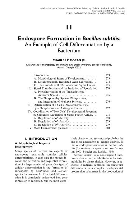
Modern Microbial Genetics, Second Edition. Edited by Uldis N. Streips, Ronald E. Yasb<strong>in</strong>Copyright # 2002 Wiley-Liss, Inc.ISBNs: 0-471-38665-0 Hardback); 0-471-22197-X Electronic)11<strong>Endospore</strong> <strong>Formation</strong> <strong>in</strong> <strong>Bacillus</strong> <strong>subtilis</strong>:<strong>An</strong> <strong>Example</strong> <strong>of</strong> <strong>Cell</strong> Differentiation by aBacteriumCHARLES P. MORAN JR.Department <strong>of</strong> Microbiology and Immunology, Emory University School <strong>of</strong> Medic<strong>in</strong>e,Atlanta, Georgia 30322I. Introduction ................................... 273A. Morphological Stages <strong>of</strong> Development. . ........ 273B. Developmentally Regulated Gene Expression .... 274C. The Cascade <strong>of</strong> RNA Polymerase Sigma Factor. . 275II. Signal Transduction and the Initiation <strong>of</strong> Sporulation 276A. Phosphorylation <strong>of</strong> the TranscriptionalActivator Spo0A ............................ 276B. The Phosphorelay System, Phosphatases,and Integration <strong>of</strong> Multiple Systems............ 276III. Determ<strong>in</strong>ation <strong>of</strong> a <strong>Cell</strong>'s Developmental Fateby a Phosphatase and <strong>An</strong>ti-sigma Factor . . . ........ 277IV. Coord<strong>in</strong>ation <strong>of</strong> Two <strong>Cell</strong>s' Developmental Programsby Crisscross Regulation <strong>of</strong> Sigma Factor Activity . . . 278A. Regulation <strong>of</strong> s E Activity..................... 278B. Regulation <strong>of</strong> s K Activity. .................... 279C. Regulation <strong>of</strong> s G Activity. .................... 279V. More Unanswered Questions ..................... 280I. INTRODUCTIONA. Morphological Stages <strong>of</strong>DevelopmentMany species <strong>of</strong> bacteria are capable <strong>of</strong>undergo<strong>in</strong>g remarkably complex cellulardifferentiations. In each case the process <strong>in</strong>volvesthe activation and sequential expression<strong>of</strong> a large number <strong>of</strong> genes. One type <strong>of</strong>cellular differentiation is the formation <strong>of</strong>endospores by Clostridium and <strong>Bacillus</strong>species. In no example <strong>of</strong> bacterial differentiationis it completely understood how geneexpression is regulated, but the most extensivelycharacterized system, and probably theone most ameanable to genetic analysis, isthat <strong>of</strong> endospore formation <strong>in</strong> <strong>Bacillus</strong> <strong>subtilis</strong>for reviews on sporulation, see Err<strong>in</strong>gton,1993; Stragier and Losick, 1996).<strong>Bacillus</strong> <strong>subtilis</strong> is a rod-shaped Grampositivebacterium, which like most bacteria,multiplies by b<strong>in</strong>ary fission. However, <strong>in</strong> responseto nutrient depletion, the bacteriumdifferentiates <strong>in</strong> a complex developmentalprocess that culm<strong>in</strong>ates <strong>in</strong> the production <strong>of</strong>
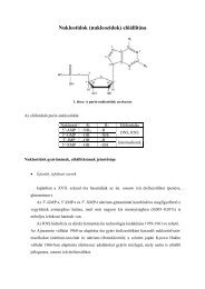
274 MORAN JR.a new cell type, the endospore. The endosporeis a dormant cell that can survivenumerous environmental <strong>in</strong>sults e.g., dessication,heat, chemical stresses, and nutrientdepletion). Although the endospore is a dormantcell, it responds to specific nutrientsignals by germ<strong>in</strong>at<strong>in</strong>g to produce a vegetativecell that resumes multiplication byb<strong>in</strong>ary fission.<strong>Endospore</strong> development is a morphologicallycomplex process that proceeds through awell-def<strong>in</strong>ed series <strong>of</strong> morphological stagesFig. 1). Multiplication <strong>of</strong> vegetative cellsby b<strong>in</strong>ary fission <strong>in</strong>volves a cell divisionthat produces two identical daughter cells.However, at the onset <strong>of</strong> sporulation thecell divides asymetrically to produce asmall cell and a large cell. These two cellshave different developmental fates. Thesmaller cell, forespore, develops <strong>in</strong>to themature endospore. The larger cell, knownas the mother cell, develops <strong>in</strong>to a term<strong>in</strong>allydifferentiated cell that nurtures the develop<strong>in</strong>gendospore, provid<strong>in</strong>g to it a largenumber <strong>of</strong> products. Ultimately the mothercell dies when the endospore is mature.In the next most conspicuous morphologicalevent after the asymmetric cell divisionthe peptidoglycan layer between theforespore and mother cells is hydrolyzed,and the mother cell engulfs the forespore <strong>in</strong>a phagocytic-like process <strong>in</strong> which themother cell cytoplasmic membrane surroundsthe membrane <strong>of</strong> the forespore. Theengulfed forespore protoplast floats with<strong>in</strong>the mother cell cytoplasm where it is surroundedby both its own orig<strong>in</strong>al membraneand a second membrane that orig<strong>in</strong>atedfrom the mother cell cytoplasmic membrane.This cell-with<strong>in</strong>-a-cell form <strong>of</strong> developmentis why this type <strong>of</strong> bacterial spore is called anendospore. At this stage both cells, foresporeand mother cell, are metabolically active.Therefore the develop<strong>in</strong>g endospore is builtfrom the <strong>in</strong>side and outside simultaneously,with some products synthesized with<strong>in</strong>the forespore where they accumulate e.g.,prote<strong>in</strong>s that b<strong>in</strong>d and protect the sporeDNA), while other products are synthesized<strong>in</strong> the mother cell cytoplasm from wherethey are donated to the endospore. In thelater stages <strong>of</strong> development a thick layer<strong>of</strong> peptidoglycan accumulates betweenthe two membranes <strong>of</strong> the forespore, and athick layer <strong>of</strong> prote<strong>in</strong>s forms a coat thatsurrounds and protects the peptidoglycanlayer.B. Developmentally Regulated GeneExpression<strong>Endospore</strong> formation requires the expression<strong>of</strong> over 100 genes that are not requiredFig. 1. Morphological stages <strong>of</strong> endospore formation <strong>in</strong> B. <strong>subtilis</strong>. The most conspicuous morphologicalchanges <strong>in</strong>clude an asymmetric cell division c), engulfment <strong>of</strong> the forespore by the mother cell membrane dand e), synthesis <strong>of</strong> the cortex f), assembly <strong>of</strong> the coat surround<strong>in</strong>g the forespore g), and lysis <strong>of</strong> the mothercell to release the dormant spore h). Also shown are the sigma factors s) active dur<strong>in</strong>g sporulation Thedashed arrows represent signals from one cell that signal activation <strong>of</strong> a s <strong>in</strong> the other cell. The symbol ?)<strong>in</strong>dicates the anti-sigma factor SpoIIAB that represses the activity <strong>of</strong> s F and s G .
ENDOSPORE FORMATION IN BACILLUS SUBTILIS 275for growth or survival <strong>of</strong> vegetative cells.These genes are regulated primarily atthe level <strong>of</strong> transcription, and are transcribed<strong>in</strong> a specific temporal sequence.Transcription <strong>of</strong> sporulation genes is alsoregulated <strong>in</strong> a cell-type specific manner;some genes are transcribed exclusively <strong>in</strong>the forespore, whereas other genes are transcribedexclusively <strong>in</strong> the mother cell.C. The Cascade <strong>of</strong> RNA PolymeraseSigma FactorsRegulation <strong>of</strong> both the temporal pattern<strong>of</strong> transcription and the cell-type specifictranscription <strong>of</strong> genes dur<strong>in</strong>g endosporedevelopment is primarily governed by afamily <strong>of</strong> transcription factors known asRNA polymerase sigma factors reviewed<strong>in</strong> Losick and Stragier, 1992; Kroos et al.,1999). RNA polymerase <strong>in</strong> bacteria ismade <strong>of</strong> several prote<strong>in</strong> subunits. Thecore enzyme is composed <strong>of</strong> two alpha, onebeta, and one beta 0 subunits. The sigma subunit,or sigma factor, associates with thecore RNA polymerase to form the holoenzyme.Association <strong>of</strong> the sigma withRNA polymerase confers on the polymeraseits ability to b<strong>in</strong>d to and utilize specificpromoters. All bacteria have a primarysigma factor called sigma A, s A ,<strong>in</strong>B. <strong>subtilis</strong>)that directs transcription <strong>of</strong> most housekeep<strong>in</strong>ggenes. RNA polymerase conta<strong>in</strong><strong>in</strong>gs A Es A ) recognizes specific promoters becausethe sigma <strong>in</strong>teracts with two specificsequences <strong>of</strong> DNA located 35 and 10 basepairs upstream from the start po<strong>in</strong>t <strong>of</strong> transcription,the 35 and 10 regions, respectively,<strong>of</strong> the promoter. Most bacteria conta<strong>in</strong>secondary sigma factors that enable RNApolymerase to utilize promoters that aredef<strong>in</strong>ed by different sequences <strong>of</strong> DNA.Five secondary sigma factors, each confer<strong>in</strong>ga different specificity for promoter recognitionon RNA polymerase, direct transcriptiondur<strong>in</strong>g endospore formation <strong>in</strong> B.<strong>subtilis</strong> see Helmann, this volume).One <strong>of</strong> the secondary sigma factors requiredfor endospore development, s H , ispresent <strong>in</strong> vegetative cells. However, theother secondary sigma factors required forendospore development are synthesized afterthe onset <strong>of</strong> sporulation <strong>in</strong> the order s F , s E ,s G , and s K . The sequential appearance <strong>of</strong>these sigma factors produces the temporallyregulated pattern <strong>of</strong> gene transcription.Therefore genes whose transcription isdirected by s F are transcribed before geneswhose transcription is dependent on s E , s G ,or s K . Although these secondary sigmafactors def<strong>in</strong>e the major temporal classes <strong>of</strong>gene transcription dur<strong>in</strong>g sporulation, eachclass is subdivided by ancillary transcriptionfactors that work with the sigma factors.These are DNA-b<strong>in</strong>d<strong>in</strong>g prote<strong>in</strong>s that repressor activate transcription <strong>of</strong> specficgenes. For example, s K is active dur<strong>in</strong>g thef<strong>in</strong>al stages <strong>of</strong> development. When s K becomesactive, it directs the transcription <strong>of</strong>several genes, one <strong>of</strong> which encodes a DNAb<strong>in</strong>d<strong>in</strong>gprote<strong>in</strong> called GerE. When GerEaccumulates, it b<strong>in</strong>ds to and represses transcriptionfrom a subset <strong>of</strong> s K dependent promoters.GerEalso b<strong>in</strong>ds to and activates adifferent subset <strong>of</strong> s K -dependent promotersthat are only active when both s K andGerEare present. Therefore GerEsubdividesthe s K regulon <strong>in</strong>to genes that are transcribedthroughout the period <strong>of</strong> s K activity,genes that are transcribed early but are shut<strong>of</strong>f when GerEaccumulates, and geneswhose transcription is activated only <strong>in</strong> thef<strong>in</strong>al stage <strong>of</strong> s K activity when GerEis present.The secondary sigma factors also play thecentral role <strong>in</strong> the cell-type specific transcription<strong>of</strong> genes. s F and s G are active exclusively<strong>in</strong> the forespore, whereas, s E and s Kare active exclusively <strong>in</strong> the mother cell.Therefore genes whose transcription is dependenton one <strong>of</strong> these sigma factors canonly occur <strong>in</strong> one cell type, either the foresporeor mother cell. Transcription <strong>of</strong> genesis coord<strong>in</strong>ated between the two cell types.The mechanisms that coord<strong>in</strong>ate the geneexpression <strong>in</strong> the two cells types <strong>in</strong>volveregulation <strong>of</strong> sigma factor activity. Thesemechanisms are discussed below <strong>in</strong> SectionIV.)
276 MORAN JR.II. SIGNAL TRANSDUCTIONAND THE INITIATION OFSPORULATIONA. Phosphorylation <strong>of</strong> theTranscriptionalActivator Spo0AMost <strong>of</strong> the developmental gene expressionrequired for endspore formation is the result<strong>of</strong> the production <strong>of</strong> RNA polymerase sigmafactors. But how is the synthesis <strong>of</strong> the firstsporulation-<strong>in</strong>duced sigma factors regulated?s F and s E are the first two sigma factorsproduced after the <strong>in</strong>itiation <strong>of</strong> sporulation.The structural genes encod<strong>in</strong>g these sigmafactors are part <strong>of</strong> the spoIIA and spoIIGoperons, respectively. Transcription <strong>of</strong> bothoperons is activated with<strong>in</strong> m<strong>in</strong>utes after the<strong>in</strong>itiation <strong>of</strong> sporulation. The promoter forthe spoIIG operon is used by RNA polymeraseconta<strong>in</strong><strong>in</strong>g s A , the primary sigma factor<strong>in</strong> vegetative cells. The promoter for thespoIIA operon is used by RNA polymeraseconta<strong>in</strong><strong>in</strong>g s H . s H , like s A , is present <strong>in</strong>vegetative cells; therefore these sigma factorscannot account for the activation <strong>of</strong> thespoIIA and spoIIG promoters at the onset<strong>of</strong> sporulation. Activation <strong>of</strong> these promotersrequires an additional factor, aDNA-b<strong>in</strong>d<strong>in</strong>g prote<strong>in</strong> known as Spo0A.Spo0A is similar to a large family <strong>of</strong> prote<strong>in</strong>sthat form the response regulator class <strong>of</strong>two-component regulatory systems. Likeother regulators <strong>of</strong> this class, Spo0A is activeonly when phosphorylated on a specific aspartateresidue. Once phosphorylated, itb<strong>in</strong>ds to specific sites on the spoIIA andspoIIG promoters where it <strong>in</strong>teracts withRNA polymerase to activate transcription.B<strong>in</strong>d<strong>in</strong>g <strong>of</strong> phosphorylated Spo0A to othersites on the chromosome activates, or somecases represses, transcription <strong>of</strong> other genes.B. The Phosphorelay System,Phosphatases, and Integration <strong>of</strong>Multiple SystemsSpo0A is the key to the <strong>in</strong>itiation <strong>of</strong> sporulation.Spo0A is present <strong>in</strong> grow<strong>in</strong>g cells, andits phosphorylation triggers the <strong>in</strong>itiation <strong>of</strong>sporulation. Phosphorylation <strong>of</strong> Spo0A iscontrolled by a phosphorelay system that ismore complex than most other two-componentsystems see the chapter by Bayles andFujimoto, this volume) Fig. 2) reviewed <strong>in</strong>Fabret et al., 1999; Hoch, 2000; Perego,1998). Unlike simple two-component systems<strong>in</strong> which a k<strong>in</strong>ase phosphorylates the responseregulator, the phosphoryl group thatis added to Spo0A is first passed from one <strong>of</strong>two k<strong>in</strong>ases to another response regulatorlikeprote<strong>in</strong> called Spo0F. The phosphorylgroup is then tranfered from Spo0FPto Spo0A by a phosphotransferase calledSpo0B. The concentration <strong>of</strong> Spo0AP isthe key determ<strong>in</strong>ant <strong>of</strong> whether the cell willsporulate or cont<strong>in</strong>ue to divide. The concentration<strong>of</strong> Spo0AP depends on the rate <strong>of</strong>flow <strong>of</strong> phosphoryl groups through the phosphorelay.The rate <strong>of</strong> flow <strong>of</strong> phosphorylgroups to Spo0A is modulated by the control<strong>of</strong> k<strong>in</strong>ase activity, and by phosphatases thatremove phosphoryl groups from the streamFig. 2).The complexity <strong>of</strong> the network <strong>of</strong> regulatorsthat control <strong>of</strong> Spo0AP concentrationallows the cell to <strong>in</strong>tegrate several signals, aFig. 2. The phosphorelay system that controls the<strong>in</strong>itiation <strong>of</strong> sporulation. K<strong>in</strong>ases K<strong>in</strong>A and K<strong>in</strong>B feedphosphate <strong>in</strong>to the phosphorelay. The flow <strong>of</strong> phosphorylgroups to Spo0A is contolled by KipI, whichcontols K<strong>in</strong>A activity, and by phosphatases RapB andSpo0E, which remove phosphoryl groups fromSpo0F and Spo0A, respectively. PhosphorylatedSpo0A Spo0AP) acts as a DNA-b<strong>in</strong>d<strong>in</strong>g prote<strong>in</strong>to directly activate some promoters and represstranscription from other promoters.
ENDOSPORE FORMATION IN BACILLUS SUBTILIS 277way <strong>of</strong> consider<strong>in</strong>g other responses to nutrientdepletion, before committ<strong>in</strong>g to the mostdrastic response <strong>of</strong> produc<strong>in</strong>g a dormant cell.Other responses, such as chemotaxis and thedevelopment <strong>of</strong> the competent state forDNA uptake, are discussed <strong>in</strong> other chapters.Nutrient depletion may affect phosphateflow to Spo0A by modulat<strong>in</strong>g k<strong>in</strong>aseactivity. KipI, an <strong>in</strong>hibitor <strong>of</strong> K<strong>in</strong>A, themajor k<strong>in</strong>ase <strong>of</strong> the phosphorelay, andKipA, an anti-k<strong>in</strong>ase <strong>in</strong>hibitor, are encoded<strong>in</strong> an operon that is responsive to glucoseand nitrogen levels. Recently, A. L. Sonenshe<strong>in</strong>and his colleagues found that CodY, aDNA-b<strong>in</strong>d<strong>in</strong>g prote<strong>in</strong>, is also a GTP-b<strong>in</strong>d<strong>in</strong>gprote<strong>in</strong>, and CodY acts as an <strong>in</strong>tracellularsensor <strong>of</strong> GTP levels Ratnayake-Lecamwasamet al., 2001). Under conditions <strong>of</strong> nutrientexcess the <strong>in</strong>tracellular level <strong>of</strong> GTP ishigh so CodY b<strong>in</strong>ds GTP and acts as a transcriptionalrepressor <strong>of</strong> several genes. Underconditions <strong>of</strong> nutrient depletion the <strong>in</strong>tracellularlevel <strong>of</strong> GTP decreases. In the absence<strong>of</strong> bound GTP CodY fails to acts as a transcriptionalrepressor. CodY may regulate the<strong>in</strong>itiation <strong>of</strong> sporulation <strong>in</strong> response to nutrientconditions by regulat<strong>in</strong>g the transcription<strong>of</strong> one <strong>of</strong> more structural genes forcomponents <strong>of</strong> the phosphorelay.Other responses to nutrient depletion andother physiological signals compete with thesporulation response by affect<strong>in</strong>g the flow <strong>of</strong>phosphate through the phosphorelay. Thedevelopment <strong>of</strong> competence affects the activity<strong>of</strong> a phosphatase, RapA, which dephosphorylatesSpo0F. A second Spo0FPphosphatase, RapB, is expressed <strong>in</strong> grow<strong>in</strong>gcells. <strong>An</strong>other phosphatase Spo0EdephophorylatesSpo0AP directly, but it isunknown how its activity is regulated. PresumablySpo0Ephosphatase activity is usedto <strong>in</strong>tegrate another, as yet unknown, <strong>in</strong>put<strong>in</strong>to the decision to sporulate. <strong>An</strong>other level<strong>of</strong> complexity was revealed by the discovery<strong>of</strong> peptides that regulate phosphatase activity.PhrA is a 44 am<strong>in</strong>o acid peptide that<strong>in</strong>hibits RapA activity. This peptide is secretedand then a peptide consist<strong>in</strong>g <strong>of</strong> atleast the five am<strong>in</strong>o acyl residues at the carboxyterm<strong>in</strong>al end PhrA is <strong>in</strong>ternalizedwhere it may act directly on the phosphatase.It is not known whether the ma<strong>in</strong> role <strong>of</strong>PhrA is to serve as a tim<strong>in</strong>g device or as an<strong>in</strong>tercellular signal. However, it is evidentthat PhrA is essential for <strong>in</strong>hibition <strong>of</strong> theRapA phosphatase and thereby for the <strong>in</strong>itiation<strong>of</strong> sporulation.III. DETERMINATION OF ACELL'S DEVELOPMENTAL FATEBY A PHOSPHATASE ANDANTI-SIGMA FACTOREarly dur<strong>in</strong>g endospore formation the asymetriccell division produces two cells thathave different developmental fates. Differentsets <strong>of</strong> genes are transcribed <strong>in</strong> each <strong>of</strong> thesetwo cells because the activities <strong>of</strong> specificsigma factors are restricted to specific cellsfor review on the control <strong>of</strong> sigma activitydur<strong>in</strong>g sporulation, see Kroos et al., 1999;Losick, 1994; Losick and Stragier, 1992).Specific genes are transcribed exclusively <strong>in</strong>the forespore with<strong>in</strong> m<strong>in</strong>utes after the asymmetricdivision. Transcription <strong>of</strong> these genes<strong>in</strong> the forespore is directed by s F , one <strong>of</strong> thesigma factors synthesized early after the <strong>in</strong>itiation<strong>of</strong> sporulation. Although s F is produced<strong>in</strong> the predivisional cell with<strong>in</strong> m<strong>in</strong>utesafter the onset <strong>of</strong> sporulation, s F is notactive <strong>in</strong> the predivisional cell. After theasymmetric cell division, s F rema<strong>in</strong>s <strong>in</strong>active<strong>in</strong> the mother cell but becomes active <strong>in</strong> theforespore. It is the activity <strong>of</strong> s F <strong>in</strong> the foresporethat leads to expression <strong>of</strong> foresporespecific genes, and as is discussed <strong>in</strong> the nextsection, s F activity <strong>in</strong> the forespore also signalsthe activation <strong>of</strong> mother cell specificgene expression. S<strong>in</strong>ce activation <strong>of</strong> s F <strong>in</strong>the forespore determ<strong>in</strong>es its developmentalfate, it is important to understand how s Factivity is restricted to the forespore.In order to discuss the regulation <strong>of</strong> s Factivity three additional prote<strong>in</strong>s must be<strong>in</strong>troduced Fig. 3). The first is SpoIIAB,an anti-sigma factor that is capable <strong>of</strong> b<strong>in</strong>d<strong>in</strong>gto s F and keep<strong>in</strong>g it <strong>in</strong>active. SpoIIAB isproduced <strong>in</strong> the predivisional cell at the same
278 MORAN JR.Fig. 3. Regulation <strong>of</strong> s F activity by the anti-sigmafactor SpoIIAB. SpoIIAB b<strong>in</strong>ds to s F <strong>in</strong>hibit<strong>in</strong>g itsfunction. SpoIIAB can also b<strong>in</strong>d to SpoIIAA but cannotstably b<strong>in</strong>d s F and SpoIIAA simultaneously. SpoIIABalso is a k<strong>in</strong>ase that phosphorylates SpoIIAA. However,SpoIIAAP does not b<strong>in</strong>d SpoIIAB. In the predivisionalcell Fig. 1, panel b) almost all <strong>of</strong> SpoIIAA isfound <strong>in</strong> its phosphorlated form SpoIIAAP). ThereforeSpoIIAB is free to b<strong>in</strong>d and <strong>in</strong>hibit s F . After theasymmetric septation Fig. 1, panel c) SpoIIAAP isdephosphorylated by the phosphatase SpoIIE exclusively<strong>in</strong> the forespore compartment. The dephosphorylatedSpoIIAA b<strong>in</strong>ds SpoIIAB releas<strong>in</strong>g s F ,which is therefore active exclusively <strong>in</strong> the forespore.time that s F is produced. SpoIIAB b<strong>in</strong>ds tos F <strong>in</strong> the predivisional cell antagoniz<strong>in</strong>g itsactivity. SpoIIAB also b<strong>in</strong>ds and antagonizess F <strong>in</strong> the mother cell after cell division.SpoIIAB releases s F <strong>in</strong> the forespore because<strong>of</strong> the action <strong>of</strong> an anti-anti-sigma,SpoIIAA. SpoIIAA b<strong>in</strong>ds to SpoIIAB forc<strong>in</strong>git to release s F . The SpoIIAA prote<strong>in</strong> ispresent <strong>in</strong> the predivisional cell, and after theasymmetric cell division it is present <strong>in</strong> boththe mother cell and forespore. However,SpoIIAA disrupts the SpoIIAB-s F complexexclusively <strong>in</strong> the forespore because <strong>in</strong>the predivisional cell and mother cellSpoIIAA rema<strong>in</strong>s phosphorylated. SpoIIABis the k<strong>in</strong>ase that phosphorylates SpoIIAA.The phosphorylated form <strong>of</strong> SpoIIAASpoIIAAP) does not b<strong>in</strong>d to SpoIIAB.Therefore SpoIIAAP leaves SpoIIAB freeto b<strong>in</strong>d and <strong>in</strong>hibit s F . SpoIIAAP is dephosphorylated<strong>in</strong> the forespore by a membranebound phosphatase called SpoIIE. SpoIIE issynthesized <strong>in</strong> the predivisional cell, but afterthe assymmetric cell division SpoIIEis located<strong>in</strong> the septum that divides the foresporeand mother cell. The reason that SpoIIEisactive exclusively <strong>in</strong> the forespore is a topicthat is currently be<strong>in</strong>g <strong>in</strong>vestigated. One hypothesisstates that SpoIIEmay be locatedprimarily on the forespore side <strong>of</strong> the septum.However, even if SpoIIEis located on bothsides <strong>of</strong> the septum, its concentration wouldbe much higher <strong>in</strong> the forespore because thevolume <strong>of</strong> the forespore is much smaller thanthe mother cell. One hypothesis suggests thatthis difference <strong>in</strong> volume may be sufficient toaccount for the forespore-specific activation<strong>of</strong> s F through the action <strong>of</strong> the SpoIIEphosphatase.In summary, s F activation <strong>in</strong> the foresporedeterm<strong>in</strong>es the developmental fate <strong>of</strong>this cell, and as described below, dictates thefate <strong>of</strong> the mother cell. s F activity is regulatedby an anti-sigma factor SpoIIAB) andan anti-anti-sigma factor SpoIIAA), whosephosphorylation state is controlled by aphosphatase SpoIIE) whose activity is restrictedto the forespore.IV. COORDINATION OF TWOCELLS' DEVELOPMENTALPROGRAMS BY CRISSCROSSREGULATION OF SIGMAFACTOR ACTIVITYA. Regulation <strong>of</strong> s E ActivityLike s F , s E is synthesized <strong>in</strong> the predivisionalcell but is not active until after theasymmetric cell division dur<strong>in</strong>g endspore development.However, the mechanism controll<strong>in</strong>gs E activity is very different fromthat controll<strong>in</strong>g s F . s E , is <strong>in</strong>itially producedas an <strong>in</strong>active precursor pro-s E ) that conta<strong>in</strong>s27 am<strong>in</strong>o acids at its am<strong>in</strong>o term<strong>in</strong>us,-which must be removed to produce the activeform <strong>of</strong> s E . The protease SpoIIGA) thatprocesses pro-s E to its mature form is membranebound and located <strong>in</strong> the septum thatseparates the mother cell and forespore. Activation<strong>of</strong> the protease requires the product<strong>of</strong> the spoIIR locus, which is transcribed <strong>in</strong>the forespore under the direction <strong>of</strong> s F . ThespoIIR product is a small peptide that issecreted through the forespore membraneso that it can <strong>in</strong>teract with the membranebound SpoIIGA protease to activate process<strong>in</strong>g<strong>of</strong> pro-s E to form active s E . s Eis active exclusively <strong>in</strong> the mother cell. It isclear that its activation depends on process<strong>in</strong>gby SpoIIGA, but it is not known whether
ENDOSPORE FORMATION IN BACILLUS SUBTILIS 279this is the mechanism <strong>of</strong> control is responsiblefor restrict<strong>in</strong>g s E activity to the mothercell. Some evidence supports a model <strong>in</strong>which s E is degraded <strong>in</strong> the forespore by anunidentified protease.Activation <strong>of</strong> s E requires spoIIR expressionthat is dependent on s F ; therefore s Eactivation is dependent on s F activation,which <strong>in</strong> turn is coupled to the asymmetriccell division. In this way both s F and s Eactivation is coupled to completion <strong>of</strong> thedivision septum. Coupl<strong>in</strong>g <strong>of</strong> sigma activationto a morphological landmark <strong>in</strong> thiscase, the completion <strong>of</strong> the division septum)appears to be a reoccurr<strong>in</strong>g theme throughoutdevelopment <strong>of</strong> the endospore seebelow). A second theme is illustrated by thefact that activation <strong>of</strong> s E <strong>in</strong> the mother cell isregulated by events <strong>in</strong> the forespore the s F -dependent expression <strong>of</strong> spoIIR). Gene expression<strong>in</strong> the two cells may be coord<strong>in</strong>atedto ensure the proper assembly <strong>of</strong> spore componentsfrom the <strong>in</strong>side and outside <strong>of</strong> theforespore dur<strong>in</strong>g development. The coord<strong>in</strong>ation<strong>of</strong> gene expression <strong>in</strong> the two cells isregulated by a crisscross pattern <strong>of</strong> regulation<strong>of</strong> sigma factor activity Fig. 1). s F <strong>in</strong>the forespore is an essential prerequisite foractivation <strong>of</strong> s E <strong>in</strong> the mother cell. s E activity<strong>in</strong> the mother cell is required for activation<strong>of</strong> s G <strong>in</strong> the forespore. F<strong>in</strong>ally, s Gactivity <strong>in</strong> the forespore is required for theactivation <strong>of</strong> s K <strong>in</strong> the mother cell. Themechanisms controll<strong>in</strong>g these sigma factorsare described below.B. Regulation <strong>of</strong> s K ActivityWe will exam<strong>in</strong>e regulation <strong>of</strong> s K activityfirst because its mechanism seems superficallysimilar to that regulat<strong>in</strong>g s E activity.The structural gene encod<strong>in</strong>g s K is transcribed<strong>in</strong> the mother cell by RNA polymeraseconta<strong>in</strong><strong>in</strong>g s E . The primary product prosK conta<strong>in</strong>s 22 am<strong>in</strong>o acids at its am<strong>in</strong>oterm<strong>in</strong>al end, which must be proteolyticallyremoved to produce the active s K . However,here the similarity ends, s<strong>in</strong>ce the mechanisms<strong>of</strong> process<strong>in</strong>g for these two mother cellsigma factors are not homologous. The proteasethat activates s E appears to be an aspartateprotease, whereas the protease thatactivates s K is a member <strong>of</strong> a subset <strong>of</strong> z<strong>in</strong>cmetalloproteases. Process<strong>in</strong>g <strong>of</strong> s K <strong>in</strong> themother cell is dependent on events <strong>in</strong>the forespore, but the mechanism that transducesthe signal from forespore to the proteaseis not homologous to the SpoIIRpathway that signals process<strong>in</strong>g <strong>of</strong> s E .In its default state the SpoIIGA proteasethat processes pro-s E is <strong>in</strong>active. It is activatedby the spoIIR product, which is produced<strong>in</strong> the forespore. In its default state theprotease that processes pro-s K SpoIVFB) isactive. It resides <strong>in</strong> the forespore membranewhere it is held <strong>in</strong>active by two other prote<strong>in</strong>s.The forespore-expressed gene productthat signals pro-s K process<strong>in</strong>g spoIVB) isnot homologous to SpoIIR, and it acts byantagoniz<strong>in</strong>g the <strong>in</strong>hibitors <strong>of</strong> the SpoIVFBprotease, thus activat<strong>in</strong>g the proteolytic activation<strong>of</strong> s K . Transcription <strong>of</strong> the spoIVBgene <strong>in</strong> the forespore, which signals pro-s Kprocess<strong>in</strong>g, is directed by s G . Therefore s Kactivation <strong>in</strong> the mother cell is tied to theactivation <strong>of</strong> s G activation <strong>in</strong> the forespore,which, as discussed below, may be coupledto another morphological landmark.C. Regulation <strong>of</strong> s G Activitys G activity also is regulated. s F directs transcription<strong>of</strong> spoIIIG, the structural gene fors G . However, s G does not become activeuntil after the forespore protoplast isengulfed by the mother cell Fig. 1). Activation<strong>of</strong> s G <strong>in</strong> the forespore is dependenton the activity <strong>of</strong> s E <strong>in</strong> the mother cell. It isnot known how s G activity is regulated <strong>in</strong>the forespore, or how s E dependent events <strong>in</strong>the mother cell affect this regulation.Accord<strong>in</strong>g to one hypothesis, the anti-sigmafactor SpoIIAB may regulate s G activity.However, this model raises a number <strong>of</strong> unansweredquestions. For example, <strong>in</strong> orderfor SpoIIAB to sequentially regulate both s Fand s G , the SpoIIAA-dependent <strong>in</strong>hibition<strong>of</strong> SpoIIAB's anti-s activity must be temporaryso that, after s F directs expression <strong>of</strong> s G ,SpoIIAB will be active to <strong>in</strong>hibit the newly
280 MORAN JR.Fig. 4. <strong>Endospore</strong> structure. Shown is an electronmicrograph<strong>of</strong> a th<strong>in</strong> cross section <strong>of</strong> a B. <strong>subtilis</strong>endospore. The prote<strong>in</strong>aeous coat forms threelayers: an straited electron-dense outer coat Oc),a lamellar <strong>in</strong>ner coat Ic), and diffuse undercoat Uc).A thick layer <strong>of</strong> peptidoglycan known as the cortexCx) lies beneath the coat. The core <strong>of</strong> the spore isalso <strong>in</strong>dicated Cr). The radius <strong>of</strong> this spore is approximately0.5 mm.synthesized s G . It is known that the unphosphorylatedform <strong>of</strong> SpoIIAA disappearsafter s F is activated; however, it is notknown how SpoIIAB is <strong>in</strong>activated dur<strong>in</strong>gthis later stage to release s G activity.V. MORE UNANSWEREDQUESTIONSThe assembly <strong>of</strong> structures <strong>in</strong> bacteria requiresthat prote<strong>in</strong>s be targeted to specificsubcellular locations. For example, prote<strong>in</strong>sare targeted to the division septum dur<strong>in</strong>gbacterial cell division. Morphogenesis <strong>of</strong> theendospore is a complex process <strong>in</strong> whichmany prote<strong>in</strong>s are targeted to specific locations.For example, the mature spore is surroundedby coat composed <strong>of</strong> over twodozen prote<strong>in</strong>s. These prote<strong>in</strong>s are arranged<strong>in</strong>to at least three layers around the foresporemembrane Fig. 4). Most <strong>of</strong> the sporecoat prote<strong>in</strong>s are synthesized <strong>in</strong> the mothercell. However, it is not known how theyare targeted to their f<strong>in</strong>al locations. Futurestudies <strong>of</strong> spore coat assembly may lead toimportant <strong>in</strong>sights <strong>in</strong>to how bacteria buildcomplex cellular structures for reviews onthe the spore coat, see Driks, 1999; Henriquesand Moran, 2000).REFERENCESDriks A 1999): <strong>Bacillus</strong> <strong>subtilis</strong> spore coat. MicrobiolMol Biol Rev 63:1±20.Err<strong>in</strong>gton J 1993): <strong>Bacillus</strong> <strong>subtilis</strong> sporulation: Regulation<strong>of</strong> gene expression and control <strong>of</strong> morphogenesis.Microbiol Rev 57:1±33.Fabret C, Feher VA, Hoch JA 1999): Two-componentsignal transduction <strong>in</strong> <strong>Bacillus</strong> <strong>subtilis</strong>: how one organismsees its world. J Bacteriol 181:1975±1983.Henriques AO, Moran CP, Jr 2000): Structure andassembly <strong>of</strong> the bacterial endospore coat. Methods20:95±110.Hoch JA 2000): Two-component and phosphorelaysignal transduction. Curr Op<strong>in</strong> Microbiol 3:165±170.Kroos L, Zhang B, Ichikawa H, Yu YT 1999): Control<strong>of</strong> sigma factor activity dur<strong>in</strong>g <strong>Bacillus</strong> <strong>subtilis</strong> sporulation.Mol Microbiol 31:1285±1294.Losick R 1994): RNA polymerase sigma factors andcell-specific gene transcription <strong>in</strong> a simple develop<strong>in</strong>gorganism. Harvey Lect 90:1±17.Losick R, Stragier P 1992): Crisscross regulation <strong>of</strong> celltype-specificgene expression dur<strong>in</strong>g development <strong>in</strong>B. <strong>subtilis</strong>. Nature 355:601±604.Perego M 1998): K<strong>in</strong>ase-phosphatase competition regulates<strong>Bacillus</strong> <strong>subtilis</strong> development. Trends Microbiol6:366±370.Ratnayake-Lecamwasam M, Serror P, Wong KW, Sonenshe<strong>in</strong>AL 2001): <strong>Bacillus</strong> <strong>subtilis</strong> CodY repressesearly-stationary-phase genes by sens<strong>in</strong>g GTP levels.Genes Dev 15:1093±1003.Stragier P, Losick R 1996): Molecular genetics <strong>of</strong> sporulation<strong>in</strong> <strong>Bacillus</strong> <strong>subtilis</strong>. <strong>An</strong>nu Rev Genet 30:297±341.