APPROACHES TO MESSENGER RNA DETECTION - ResearchGate
APPROACHES TO MESSENGER RNA DETECTION - ResearchGate
APPROACHES TO MESSENGER RNA DETECTION - ResearchGate

Create successful ePaper yourself
Turn your PDF publications into a flip-book with our unique Google optimized e-Paper software.
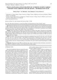
132 Z. Dvořák, J.-M. Pascussi, M. Modrianskýhowever, is accompanied by a higher risk of ribonucleasecontamination.Since <strong>RNA</strong> is extremely sensitive to the action ofribonucleases, it is essential to use disposable laboratoryware (eppendorfs, tubes, tips) autoclaved beforeuse. Plastic gloves are strictly recommended for workingwith <strong>RNA</strong> and it is useful and convenient to designate“<strong>RNA</strong>se free zone” in the laboratory. Following isolation,the concentration of <strong>RNA</strong> in samples is determined byUV-spectrometry at 260 nm. It is recommended to evaluateabsorbance at 280 nm as well to have an estimateof proteins impurities, with the ratio A 260/A 280> 1.8 beingsatisfactory for further use of a particular sample. Highabsorbance at λ < 260 nm (absence of a clear peak at260 nm) suggests traces of phenol contaminating the sample.Residual phenol may be removed by an additionalwash step and it is needed particularly for RT-PCR. Owingto thermal instability of m<strong>RNA</strong>, the samples are stored(in formamide; FA or water; W) in –80 °C freezer and onice when in use.NORTHERN BLOTEssentially a variation of a method developed forDNA detection, it serves two main purposes: i) detectionof m<strong>RNA</strong> presence in a cell as evidence for genetranscription, and ii) estimation of m<strong>RNA</strong> size. Becauseof lower sensitivity, it is limited to high abundance m<strong>RNA</strong>.On the other hand, shift from low to high abundance, i.e.inducibility, of a gene can be easily demonstrated becauseof this. Different size m<strong>RNA</strong>s of a specific gene detectedusing this technique suggest presence of a pseudogeneor a splice variant which can be utilized gene expressionregulation studies.Method description: Samples dissolved in either FA orW can be analysed by this, let us say, classic technique 3 .Depending on the gene of interest expression level analiquot of 10–50 µg of total <strong>RNA</strong> is mixed with a formaldehyde-basedloading buffer and denatured by heat. Then,electrophoretic separation of <strong>RNA</strong>s is performed on anagarose gel under denaturing conditions. Formaldehyde ispresent both in the gel and the migration buffer ensuringthe inhibition of ribonuclease activity. Formaldehyde canbe substituted by other substances, e.g. glyoxal, but witha bit higher risk of ribonuclease contamination. Procedureis performed in a horizontal arrangement, with constantstirring of the migration buffer and using constantcurrent. The time of analysis varies between 1–2 h and5–6 h for small and large gels, respectively. Separation isfollowed by transfer of <strong>RNA</strong>s onto the nylon membrane,driven by capillary forces, the so-called Northern blot.The procedure usually runs overnight. Quality of transferis quickly checked under UV light (254 nm) when twomajor bands of ribosomal 18S and 28S <strong>RNA</strong> fraction arevisible. According to the time schedule/equipment, themembrane is: a) exposed to an intense UV light to inducem<strong>RNA</strong> cross-link (for immediate use); or b) dried undervacuum. Following the latter procedure, the membranecan be stored in plastic wrap for months.Since m<strong>RNA</strong> (+, sense) is being synthesised by transcriptionof a template DNA strand, it has the nucleotidesequence of the coding DNA strand (note that uridineis present instead of thymidine). Accordingly, an appropriateplasmid with the cloned cDNA fragment of desirablegene is used for the synthesis of the probe, whichhas then sequence of the original template DNA strand.Commercial kits are used, employing DNA-dependentDNA polymerase (Klenow) and microcolumns forprobe purification. The probe is most often radio-labelledusing 32 P-α-dCTP or by biotin. The principle of detectionis the hybridization of m<strong>RNA</strong> fixed in membrane withthe labelled cDNA probe (sequence complementaryto the m<strong>RNA</strong>) forming a double stranded <strong>RNA</strong>-DNAstructure. Hybridization is usually carried out overnightin a rotating oven and detected by: a) autoradiographyor Phosphoimager apparatus for 32 P labelled probe; orb) immuno-chemiluminiscence detection for biotinylatedprobe.A representative example of NB is shown in Fig 1.3A43.0 kb2.2 kbGAPDHFig. 1. Example of Northern blot analysis. Northern blot oftotal <strong>RNA</strong> (20 µg) obtained from primary humanhepatocytes using Trizol isolation procedure. kb == kilobase; GAPDH = glyceraldehyde-3-phosphatedehydrogenase; 3A4 = cytochrome P450 isoform3A4 (band at 2.2 kb); C = control cells; I = cellstreated with 3A4 inducer rifampicin. Band at 3.0 kbcorresponds to 3A4 pseudogene.SWOT: Strength of the method is its simplicity. Timeschedule allows spreading of the work into two stages ofsample-separation and hybridization-detection possiblyperformed weeks apart. Specificity is relatively high. Financialdemands are the lowest of all described methodswith added cost if Phosphoimager apparatus is being used.Weakness is linked to the work and environmental riskswhen high doses of radioactivity (on the order of 10 7 cpm)and formaldehyde are used. The sensitivity of Northernblot technique is the lowest of all the described methods.Opportunity emerges in the possibility to detect two ormore genes in one hybridization step (with respect toCI