APPROACHES TO MESSENGER RNA DETECTION - ResearchGate
APPROACHES TO MESSENGER RNA DETECTION - ResearchGate
APPROACHES TO MESSENGER RNA DETECTION - ResearchGate

You also want an ePaper? Increase the reach of your titles
YUMPU automatically turns print PDFs into web optimized ePapers that Google loves.
Biomed. Papers 147(2), 131–135 (2003)© Z. Dvořák, J.-M. Pascussi, M. Modrianský131<strong>APPROACHES</strong> <strong>TO</strong> <strong>MESSENGER</strong> <strong>RNA</strong> <strong>DETECTION</strong> – COMPARISON OF METHODSZdeněk Dvořák a *, Jean-Marc Pascussi b , Martin Modrianský aaInstitute of Medical Chemistry and Biochemistry, Faculty of Medicine, Palacký University Olomouc, Hněvotínská 3,775 15 Olomouc, Czech Republic.bCNRS – INSERM – U128, 1919 Route de Mende, 34293 Montpellier, France.Received: September 23, 2003; Accepted: October 15, 2003Key words: Messenger <strong>RNA</strong> / Northern blot / Ribonuclease protection assay / Real time polymerase chain reactionDetection of messenger <strong>RNA</strong> is an important part of current biomedical research, although utilized for decades.This communication endeavors to compare three most commonly used techniques of m<strong>RNA</strong> detection, i.e. Northernblot, ribonuclease protection assay (RPA), and real-time polymerase chain reaction (RT-PCR). Principles and generalprocedures of these methods are described, and advantages and weaknesses of each are discussed in terms of theirspecificity, sensitivity, difficulty, time and material demands as well as health and environmental risks. We concludethat choice of any method discussed depends on particular purpose, experience of the researcher, and on laboratoryequipment and organization.INTRODUCTIONMessenger ribonucleic acid (m<strong>RNA</strong>) is an essentialfunctional constituent of a DNA-containing cell in anyliving organism. It is a polymer of four nucleotides, theorder of which gives primary structure unique to anindividual functional gene or a pseudogene. During theprocess of gene expression, the functional single strandstructure of m<strong>RNA</strong> is synthesized and serves thereafteras an intermediate template for translation process inproteosynthesis. In many fields of current research, includingmolecular biology, toxicology, physiology, pharmacology,biochemistry etc., the detection of individualm<strong>RNA</strong>s is an indispensable tool. From the point of viewof analytical chemistry, the detection/determination ofm<strong>RNA</strong> represents the following: 1) The nature of m<strong>RNA</strong>as a macromolecule; 2) Identical physico-chemical propertiesof m<strong>RNA</strong>s representing different genes; 3) Verylow level/concentration of m<strong>RNA</strong> of interest; 4) Lowstability at room temperature together with extremesensitivity to destructive action of ribonucleases and pHchange in aqueous milieu. Taken together, any of methodsemployed for m<strong>RNA</strong> detection face the problems of sensitivity,specificity, and stability.Hereby we attempt to describe in brief and to comparethe three most commonly used techniques of m<strong>RNA</strong> detection,i.e. Northern blot (NB), ribonuclease protectionassay (RPA) and real-time polymerase chain reaction(RT-PCR), with respect to the abovementioned featuresof m<strong>RNA</strong> analysis. Each technique is first described andthan its advantages and weak spots are emphasised. Forthe “rating” of the methods, we used literature data aswell as our own experimental experience. We chose anapproach similar to “SWOT” analysis (Strength vs Weaknessand Opportunity vs Threats) in economy.We do not claim that the presented discourse is exhaustive,it is intended to be rather a simplified guide forresearchers starting their work in the m<strong>RNA</strong> field.SAMPLE PREPARATIONm<strong>RNA</strong> obviously comes from biological samples,i.e. tissues or cells, consisting of vast number of variousmicro- and macromolecules, it is therefore primordialto isolate it properly. The widespread technique usedfor years employs commercial reagent Trizol ® . The principleis based on cell/tissue lysis by this reagent, followedby extraction (phenol/chloroform/water), precipitation(isopropyl alcohol), and washing (75 % ethanol) steps 1 .Resulting pellet of <strong>RNA</strong> is then dissolved either in formamide(for NB and RPA) or water (for RT-PCR). Notethat using this method the total <strong>RNA</strong> is obtained, i.e.a mixture of all m<strong>RNA</strong>, t<strong>RNA</strong>, and r<strong>RNA</strong>. One mustbear in mind that m<strong>RNA</strong> represents only about 1 % ofthat total amount. Depending on a particular case, theyield is around 50–250 µg of total <strong>RNA</strong> per 10 millionsof mammalian cells. Advantage of Trizol ® technique isthe possibility to obtain genomic DNA as well as totalcellular protein from the same sample by an extendedextraction procedure.It is possible, and in case of RT-PCR highly recommended,to isolate m<strong>RNA</strong> directly from tissue homogenate,cell culture, or total <strong>RNA</strong>. Commercial kits for thispurpose are available which utilize adsorption of m<strong>RNA</strong>to sorbents with anchored oligo(dT) probe due to interactionwith the poly-A end of m<strong>RNA</strong>s 2 . Following extensivewashing the m<strong>RNA</strong> is eluted and used for further applications.Advantage is the purity of obtained m<strong>RNA</strong>. This,
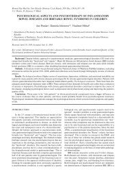
132 Z. Dvořák, J.-M. Pascussi, M. Modrianskýhowever, is accompanied by a higher risk of ribonucleasecontamination.Since <strong>RNA</strong> is extremely sensitive to the action ofribonucleases, it is essential to use disposable laboratoryware (eppendorfs, tubes, tips) autoclaved beforeuse. Plastic gloves are strictly recommended for workingwith <strong>RNA</strong> and it is useful and convenient to designate“<strong>RNA</strong>se free zone” in the laboratory. Following isolation,the concentration of <strong>RNA</strong> in samples is determined byUV-spectrometry at 260 nm. It is recommended to evaluateabsorbance at 280 nm as well to have an estimateof proteins impurities, with the ratio A 260/A 280> 1.8 beingsatisfactory for further use of a particular sample. Highabsorbance at λ < 260 nm (absence of a clear peak at260 nm) suggests traces of phenol contaminating the sample.Residual phenol may be removed by an additionalwash step and it is needed particularly for RT-PCR. Owingto thermal instability of m<strong>RNA</strong>, the samples are stored(in formamide; FA or water; W) in –80 °C freezer and onice when in use.NORTHERN BLOTEssentially a variation of a method developed forDNA detection, it serves two main purposes: i) detectionof m<strong>RNA</strong> presence in a cell as evidence for genetranscription, and ii) estimation of m<strong>RNA</strong> size. Becauseof lower sensitivity, it is limited to high abundance m<strong>RNA</strong>.On the other hand, shift from low to high abundance, i.e.inducibility, of a gene can be easily demonstrated becauseof this. Different size m<strong>RNA</strong>s of a specific gene detectedusing this technique suggest presence of a pseudogeneor a splice variant which can be utilized gene expressionregulation studies.Method description: Samples dissolved in either FA orW can be analysed by this, let us say, classic technique 3 .Depending on the gene of interest expression level analiquot of 10–50 µg of total <strong>RNA</strong> is mixed with a formaldehyde-basedloading buffer and denatured by heat. Then,electrophoretic separation of <strong>RNA</strong>s is performed on anagarose gel under denaturing conditions. Formaldehyde ispresent both in the gel and the migration buffer ensuringthe inhibition of ribonuclease activity. Formaldehyde canbe substituted by other substances, e.g. glyoxal, but witha bit higher risk of ribonuclease contamination. Procedureis performed in a horizontal arrangement, with constantstirring of the migration buffer and using constantcurrent. The time of analysis varies between 1–2 h and5–6 h for small and large gels, respectively. Separation isfollowed by transfer of <strong>RNA</strong>s onto the nylon membrane,driven by capillary forces, the so-called Northern blot.The procedure usually runs overnight. Quality of transferis quickly checked under UV light (254 nm) when twomajor bands of ribosomal 18S and 28S <strong>RNA</strong> fraction arevisible. According to the time schedule/equipment, themembrane is: a) exposed to an intense UV light to inducem<strong>RNA</strong> cross-link (for immediate use); or b) dried undervacuum. Following the latter procedure, the membranecan be stored in plastic wrap for months.Since m<strong>RNA</strong> (+, sense) is being synthesised by transcriptionof a template DNA strand, it has the nucleotidesequence of the coding DNA strand (note that uridineis present instead of thymidine). Accordingly, an appropriateplasmid with the cloned cDNA fragment of desirablegene is used for the synthesis of the probe, whichhas then sequence of the original template DNA strand.Commercial kits are used, employing DNA-dependentDNA polymerase (Klenow) and microcolumns forprobe purification. The probe is most often radio-labelledusing 32 P-α-dCTP or by biotin. The principle of detectionis the hybridization of m<strong>RNA</strong> fixed in membrane withthe labelled cDNA probe (sequence complementaryto the m<strong>RNA</strong>) forming a double stranded <strong>RNA</strong>-DNAstructure. Hybridization is usually carried out overnightin a rotating oven and detected by: a) autoradiographyor Phosphoimager apparatus for 32 P labelled probe; orb) immuno-chemiluminiscence detection for biotinylatedprobe.A representative example of NB is shown in Fig 1.3A43.0 kb2.2 kbGAPDHFig. 1. Example of Northern blot analysis. Northern blot oftotal <strong>RNA</strong> (20 µg) obtained from primary humanhepatocytes using Trizol isolation procedure. kb == kilobase; GAPDH = glyceraldehyde-3-phosphatedehydrogenase; 3A4 = cytochrome P450 isoform3A4 (band at 2.2 kb); C = control cells; I = cellstreated with 3A4 inducer rifampicin. Band at 3.0 kbcorresponds to 3A4 pseudogene.SWOT: Strength of the method is its simplicity. Timeschedule allows spreading of the work into two stages ofsample-separation and hybridization-detection possiblyperformed weeks apart. Specificity is relatively high. Financialdemands are the lowest of all described methodswith added cost if Phosphoimager apparatus is being used.Weakness is linked to the work and environmental riskswhen high doses of radioactivity (on the order of 10 7 cpm)and formaldehyde are used. The sensitivity of Northernblot technique is the lowest of all the described methods.Opportunity emerges in the possibility to detect two ormore genes in one hybridization step (with respect toCI
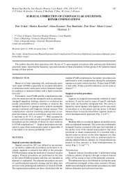
Approaches to messenger <strong>RNA</strong> detection – comparison of methods133separation efficiency and properties of the probe) and/orto perform additional detection after former <strong>RNA</strong>-DNAcomplex deshybridization. Again the time schedule ofthe method brings the chance to optimise the work or totransport the samples which is frequently important forco-operation between laboratories. Threat is representedby the risk of m<strong>RNA</strong> degradation during electrophoresis(pH change).RIBONUCLEASE PROTECTION ASSAYThis method can be used for detection and, to someextent, to quantification of m<strong>RNA</strong>. Its full potential,however, lies in the possibility to map <strong>RNA</strong> termini, todetermine alternative splicing and even single base mutations.Positions of introns within a corresponding genecan be determined as well.Method description: In this case, m<strong>RNA</strong> complementaryto the detected, i.e. antisense (–) m<strong>RNA</strong>, servesas the probe 4, 5 . In contrast to the NB principle, probepreparation and hybridisation precedes the separationstep. A sequence of usually 200–450 bp correspondingto the desired m<strong>RNA</strong> is cloned into a plasmid betweenSP6, T7 or T3 promoter and a stop codon. For ribo-probepreparation, the plasmid is linearised such that it can betranscribed in the 3’–5’ direction, contrary to the complementarystrand which is transcribed in the 5’–3’ direction.Then using corresponding DNA-dependent<strong>RNA</strong> polymerase (SP6, T7, T3), mixture of nucleotidetriphosphates (ATP, GTP, TTP, UTP) and radioactivelylabelled 32 P-α-UTP, the radio-labelled probe is syntesisedas the anti-sense (–) m<strong>RNA</strong>. When the original plasmidis linearised to allow the correct transcription, the sense(+) m<strong>RNA</strong> identical to the detected one is synthesisedand can serve as a standard (positive control). Followingthe transcription, the radio-labelled riboprobe is sequentiallypurified by 1) extraction/precipitation (phenol/chloroform reagent); 2) electrophoresis on acrylamidegel containing urea; 3) extraction from acrylamide geland precipitation. A riboprobe is then dissolved in formamideand stored in –20 °C. Presence of 32 P is limitingfactor for storage because of half-life of approx. 15 days.As an alternative to radiolabeling, it is possible to usenucleotides labeled with e.g. biotin or digoxigenin, usuallyfollowed by indirect detection by chemiluminescence orfluorescence. However, nucleotides labeled in this fashionare not efficiently used by <strong>RNA</strong> polymerases necessaryfor riboprobe synthesis hence lowering the probe yield.Sensitivity of detection may also be lower than in case ofradiolabeled probes making detection of low abundancem<strong>RNA</strong> difficult if not impossible.Analytic procedure begins by hybridization, usuallycarried out overnight, when aliquot of 10–50 µg of total<strong>RNA</strong> in FA is incubated with riboprobe, and a doublestrand m<strong>RNA</strong> structure is formed. Mixture is then incubatedwith ribonucleases A and T1 which attack singlestrand <strong>RNA</strong> only. Double strand <strong>RNA</strong> structures areprotected – hence the name of the method “ribonucleaseprotection assay”. Since <strong>RNA</strong>ses digest <strong>RNA</strong> even if a singlebase pair mismatch is present, the method providesalmost absolute specificity. In parallel, riboprobe itself istreated to reveal the quality of <strong>RNA</strong> digestion (negativecontrol, digested probe). Following <strong>RNA</strong> digestion the<strong>RNA</strong>ses are eliminated by protease K cleavage. Remainingdouble strand <strong>RNA</strong> is extracted by phenol/chloroform,precipitated, and dissolved in a loading buffer basedon FA. Prior to loading on a gel, double strand structureof <strong>RNA</strong> is disrupted by heat. Electrophoretic separationis performed in vertical arrangement on polyacrylamidegel containing urea. Constant current is applied and thetime of analysis is between 1.5–2.5 hours. The gel is thensoaked in a fixation solution and dried under vacuum.Detection, as for NB, is performed by autoradiographyor Phosphoimager apparatus.Representative example of RPA is shown in Fig 2.480 bp440 bp240 bp2C9MW NP DP C IFig. 2. Example of Ribonuclease protection assay analysis. RPA of total <strong>RNA</strong> (20 µg) isolated from primary human hepatocytesusing Trizol isolation procedure. bp = base pair; 2C9 = cytochrome P450 isoform 2C9; MW = molecular weightmarker; NP = native probe; DP = digested probe; C = control cells; I = cells treated with 2C9 inducer rifampicin.
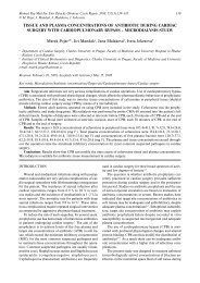
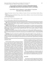
134 Z. Dvořák, J.-M. Pascussi, M. ModrianskýSWOT: Strength: Owing to its unique principle, themethod gives nearly absolute specificity. It is therefore themost specific from the three methods under review 6 . Consequently,the design of the probe is intended to recognizea specific sequence like in other techniques. Compared tothe NB, RPA is approximately ten times more sensitive.Weakness: The method is time consuming and requiresa highly skilled operator. The work organisation is verytiresome for the operator because there is no chance forpostponing any of the steps after hybridization. Furthermore,economical expenses are higher as compared toNB. Opportunity: Due to its high specificity, two or severalgenes with high homology in primary structure can be distinguished.Recall again that only one base pair mismatchleads to the cleavage by <strong>RNA</strong>ses. Threat: Since the procedureconsists of many steps, the risk of <strong>RNA</strong> degradationin sample during the analysis is very high and occursquite frequently. The risk of radioactivity contaminationwhen handling samples and particularly during riboprobepreparation is also considerable, even though the activitiesused are much lower than in the NB method.REAL TIME POLYMERASE CHAIN REACTIONThanks to its sensitivity even very low abundancem<strong>RNA</strong> can be detected using this technique. Other frequentuses include: primary validation of gene expressionand hence confirmation of microarray data, splice variantanalysis, allelic discrimination, viral titre (e.g. HCV),bacterial species identification, and single nucleotidepolymorphism genotyping.Method description: This method takes an advantagefrom well known polymerase chain reaction (PCR)which is used for DNA detection 7 . The abbreviation RT-is somewhat ambiguous standing on one hand for reversetranscription reaction, a key step in the procedure, and onthe other hand for real time format of the analysis whenLightcycler apparatus is used 8 .First of all, the reverse transcription reaction is performedusing <strong>RNA</strong>-dependent DNA polymerase (reversetranscriptase), synthesising the cDNAs complementaryto the m<strong>RNA</strong>s present in the sample. It is sine qua nonto use sample of <strong>RNA</strong> in water free of organic solventsresidues, e.g. phenol, isopropanol, chloroform, ethanoletc., to avoid inhibition of reverse transcriptase. Forthe starting material it is possible to use total <strong>RNA</strong>, atleast 1 µg, but isolated m<strong>RNA</strong> is more appropriate givinghigher probability of low copy m<strong>RNA</strong> transcription.The synthesised cDNA is mixed with DNA-dependentDNA polymerase, sufficient amount of deoxynucleotidetriphosphates (dATP, dCTP, dGTP, and dTTP), andfluorescently labeled oligonucleotide primers. As in thecase of “classical” PCR, one cycle of a PCR reactionconsists of strand separation, annealing, and elongationsteps. Duration of and temperature during each of thethree steps are set on the thermocycler apparatus in use.Fluorescence is monitored in each sample following everycycle of the PCR reaction. When threshold fluorescenceintensity is reached due to increased number of copies ofthe cDNA of interest, the fluorescence linearly increasesin each Asubsequent cycle eventually reaching a maximumplateau. The threshold RIF (M) is so-called Actin“crossing CYP3A4 point” whichis essential for comparison between samples. Computersoftware provided0with the Lightcycler14.69usually27.84calculatesthe cycle number 1corresponding 14.86to the crossing 22.94 point usingsecond derivative 5 maximum. 15.07If calibrated 22.24for knownnumber of cDNA copies present in a sample, RT-PCR canbe used for very precise 10 quantification 14.78 of 21.56 original m<strong>RNA</strong>copies per number 20 of cells 8, 15.319 . Representative 21.52computeroutput from a Lightcycler apparatus is shown in Fig 3.ARIF (M)0151020Actin14.6914.8615.0714.7815.31CYP3A427.8422.9422.2421.5621.52BFlod inductionnormalized per actinInduction of CYP3A4 by rifampicin1501005000 1 5 10 20Rifampicin (M)Fig. 3. Example of Real-Time Polymerase Chain Reaction analysis. RT-PCR computer data from total <strong>RNA</strong> isolated fromB primary Induction human hepatocytes of CYP3A4 by using rifampicin Trizol procedure. Panel A) shows cycle thresholds for CYP3A4 and actin m<strong>RNA</strong>.Panel B) 150shows fold induction of CYP3A4 gene by rifampicin. Data are normalized per actin value.Flod inductionnormalized per actin1005000 1 5 10 20Rifampicin (M)
Approaches to messenger <strong>RNA</strong> detection – comparison of methods135SWOT: Strength: Method is elegant, safe, simple touse, and requires only minimum experience and skills.It provides maximum sensitivity of all three discussedmethods (approx. 10 000 times more sensitive than NB)and among them it is the only one allowing quantification.Weakness: This method is the most expensive: Lightcyclerapparatus, fluorescently labeled primers, and m<strong>RNA</strong>isolation kits are the principal costs. There is a need ofhighly pure samples to be used, free of organic solvents,often present after Trizol isolation increasing the needfor alternative isolation procedures. Opportunity: Owingto its high sensitivity, a large number of genes can beanalysed from a single m<strong>RNA</strong> preparation. Threat: Theselection and design of the primers must be done carefullybecause of the possibility, albeit minuscule, thatother homologous genes may be detected. The risk of<strong>RNA</strong> degradation, until the reverse transcription stepis performed, is high because of water used as solvent.Contamination of <strong>RNA</strong> by fragments of genomic DNAcan occur if Trizol is used for extraction. Consequently, itcannot be distinguished whether genomic DNA or reversetranscript are detected.CONCLUSIONWith the human genome now available a principalquestion was raised: what do all those genes do? In view ofmany a researcher this question translates primarily intothe problem of: Are all those genes transcribed? It meansto look for m<strong>RNA</strong> occurrence within a cell as many ofthe genes may be cell type specific. Area of genomics isnot the only one with craving for m<strong>RNA</strong> data. The threemethods described herein are the tools currently availablefor a researcher to deal with a particular problem involvingm<strong>RNA</strong>.We offer a SWOT analysis in attempt to help othersduring pros and cons consideration when selectinga method for their purpose. Things to consider areobviously intertwined as specificity of each method islinked to financial demands together with health and environmentalrisks. An ideal of a highly specific, safe, andinexpensive method for m<strong>RNA</strong> detection does not exist.Quite often combination of two, out of the three methodsdiscussed, is utilized in many a lab throughout the world.This is important especially in pilot studies in order toconfirm data obtained. Therefore we recommend to haveone of these methods for routine use as the core methodand then to select another one not as an alternative butas a confirmation tool.ACKNOWLEDGEMENTThe authors gratefully acknowledge financial supportfrom the Ministry of Education of the Czech Republic grantMSM 151100003.REFERENCES1. Chomczynski P, Sacchi N. (1993) Single-step method of <strong>RNA</strong>isolation by acid guanidinium thiocyanate-phenol-chloroformextraction. Anal Biochem 162, 156–9.2. Kingston RE. (1993) Preparation of Poly(A) + <strong>RNA</strong>. In: CurrentProtocols in Molecular Biology (Eds.: Ausubel EM, Brent R,Kingston RE, Moore DD, Siedman JG, Smith JA, Struhl K),p 4.5.1.–4.5.3. John Wiley and Sons, New York, NY, 1993.3. Brown T. (1993) Analysis of <strong>RNA</strong> by Northern and Slot BlotHybridization. In: Current Protocols in Molecular Biology (Eds.:Ausubel EM, Brent R, Kingston RE, Moore DD, Siedman JG,Smith JA, Struhl K), p 4.9.1.–4.9.14. John Wiley and Sons, NewYork, NY, 1993.4. Gilman M. (1993) Ribonuclease protection assay. In: CurrentProtocols in Molecular Biology (Eds.: Ausubel EM, Brent R,Kingston RE, Moore DD, Siedman JG, Smith JA, Struhl K),p 4.7.1.–4.7.8. John Wiley and Sons, New York, NY, 1993.5. Melton DA, Krieg PA, Rebagliati MR, Maniatis T, Zinn K, GreenMR. (1984) Efficient in vitro synthesis of biologically active<strong>RNA</strong> and <strong>RNA</strong> hybridization probes from plasmids containinga bacteriophage SP6 promoter. Nucleic Acids Res 12, 7035–56.6. Rottman JB. (2002) The ribonuclease protection assay: a powerfultool for the veterinary pathologist. Vet Pathol 39, 2–9.7. Erlich HA. (ed.) (1989) PCR Technology: Principles and Applicationsfor DNA amplification. Stockton Press, London.8. Wilkening S, Bader A. (2003) Influence of culture time on theexpression of drug-metabolizing enzymes in primary human hepatocytesand hepatoma cell line HepG2. J Biochem Mol Toxicol 17,207–13.9. Wilkening S, Stahl F, Bader A. (2003) Comparison of primaryhuman hepatocytes and hepatoma cell line HepG2 with regardto their biotransformation properties. Drug Metab Dispos 31,1035–42.