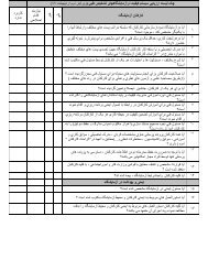
Original Article Immunophenotyping of Leukemia in Children ...
Original Article Immunophenotyping of Leukemia in Children ...
Original Article Immunophenotyping of Leukemia in Children ...
- No tags were found...

You also want an ePaper? Increase the reach of your titles
YUMPU automatically turns print PDFs into web optimized ePapers that Google loves.
IntroductionOf all cancers <strong>in</strong> childhood, leukemias are one <strong>of</strong> the most important that are prevalent <strong>in</strong>children with the <strong>in</strong>cidence <strong>of</strong> 25-30% <strong>of</strong> all childhood cancers (1-3).Generally, leukemia is divided <strong>in</strong>to two categories, acute and chronic leukemia. Acuteleukemia is a heterogeneous group <strong>of</strong> neoplastic diseases and is categorized <strong>in</strong>to two ma<strong>in</strong>subgroups: acute lymphoid leukemia (ALL) and acute myeloid leukemia (AML) (4). ALL is aheterogeneous disease with abnormal proliferation and accumulation <strong>of</strong> immature lymphoidcells with<strong>in</strong> the bone marrow, peripheral blood and lymphoid tissues. ALL patients aresubdivided <strong>in</strong>to three morphological subsets <strong>in</strong>clud<strong>in</strong>g L1, L2 and L3 (5, 6). AML is anaggressive malignancy same as ALL and it is characterized by accumulation <strong>of</strong> immaturemyeloid progenitors <strong>in</strong> the bone marrow (7).<strong>Immunophenotyp<strong>in</strong>g</strong> is important not only <strong>in</strong> the classification and diagnosis <strong>of</strong> leukemia, butalso <strong>in</strong> the prediction and prognosis <strong>of</strong> these disorders (8). It makes complete morphological,cytochemical, cytogenetic and molecular studies for def<strong>in</strong><strong>in</strong>g diagnosis and prognosis. Thenvarious <strong>in</strong>vestigations have been conducted <strong>in</strong> this field.<strong>Immunophenotyp<strong>in</strong>g</strong> <strong>in</strong> patients with ALL <strong>in</strong> Ch<strong>in</strong>a beside cl<strong>in</strong>ical and cytogenetic featuresreveal the importance <strong>of</strong> <strong>Immunophenotyp<strong>in</strong>g</strong> <strong>in</strong> diagnosis and determ<strong>in</strong><strong>in</strong>g ALL type (9).<strong>Immunophenotyp<strong>in</strong>g</strong> CD antigens were studied <strong>in</strong> patients with acute leukemia <strong>in</strong> Iran (10).<strong>Immunophenotyp<strong>in</strong>g</strong> <strong>in</strong> children is so important, which present study for the first time wasdone among leukemic patients <strong>in</strong> Golestan.Materials and MethodsCasesThis was a descriptive and analytical study and 62 patients with leukemia were exam<strong>in</strong>eddur<strong>in</strong>g 2004-2009.Sample prepar<strong>in</strong>g and immunophenotype detectionPatients suspected to leukemia that were admitted <strong>in</strong> Talghani hospital <strong>of</strong> Gorgan werechecked by CBC test and then if their test were suspicious bone marrow aspiration would bedone. After confirm<strong>in</strong>g for leukemia, results <strong>of</strong> hematological, precl<strong>in</strong>ical and cl<strong>in</strong>ical studiesand morphological tests were recorded <strong>in</strong> patients’ pr<strong>of</strong>iles. Bone marrow aspiration sampleswere collected <strong>in</strong> tubes conta<strong>in</strong><strong>in</strong>g EDTA and sent to pathology center <strong>of</strong> Baghiatallahhospital <strong>of</strong> Tehran. The steps <strong>of</strong> <strong>Immunophenotyp<strong>in</strong>g</strong> were <strong>in</strong>cluded: 1) prepar<strong>in</strong>g 4 glassslides <strong>of</strong> bone marrow and 1 glass slide <strong>of</strong> peripheral blood; 2) sta<strong>in</strong><strong>in</strong>g one glass slide <strong>of</strong> eachsample by Wright method. Then sta<strong>in</strong>ed glass slides were checked by a pathologist.Accord<strong>in</strong>g to flow cytometry <strong>in</strong>struction, it is determ<strong>in</strong>ed which markers should becharacterized for each patient; 3) dilut<strong>in</strong>g samples accord<strong>in</strong>g to required marker (50µlsample+ 5 µl monoclonal antibody); 4) <strong>in</strong>cubation all <strong>of</strong> samples for 20 m<strong>in</strong>utes <strong>in</strong>refrigerator; 5) putt<strong>in</strong>g the samples <strong>in</strong> Q-Prep. In this device 3 solutions are added to samplesdur<strong>in</strong>g 35 seconds. Solutions <strong>in</strong>clude A (lyses buffer), B (buffer), C (fixative buffer); 6)f<strong>in</strong>ally all <strong>of</strong> sample tubes were transferred to flow cytometry device and then the results wererecorded.Statistical analysisStatistical analysis was performed by SPSS 18 and Mann-Whitney test, Leven test, relativerisk and Shopiro-Wilk test were used to compare the groups.116 Iranian Journal <strong>of</strong> Pediatric Hematology Oncology Vol 1. No 4.