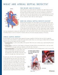
Fibroid Torsion.pdf - Henry Ford Health System
Fibroid Torsion.pdf - Henry Ford Health System
Fibroid Torsion.pdf - Henry Ford Health System

Create successful ePaper yourself
Turn your PDF publications into a flip-book with our unique Google optimized e-Paper software.
Department of Radiology<strong>Henry</strong> <strong>Ford</strong> <strong>Health</strong> <strong>System</strong>Detroit, MichiganCase Report: Abdominal PainAndre’ PorchiaWayne State SOMFebruary 19, 2010Fellow: Dr. Merchant
• 48 y/o AAFHPI• Two day h/o abdominal pain• Sharp• RUQ and LLQ• No radiation• 8/10• Relieved briefly after emesis• 10 episodes of emesis
PMH• HTN• Chronic pancreatitis• Uterine <strong>Fibroid</strong>sPSH• Cholecystectomy
RationaleOnly leukocytosis was found and the pelvic examwas unimpressive thus a CT was done to r/ocolitis/diverticulitis
CT Image
Pelvic Findings• Enlarged uterus with multiple masses and fibroids• A large subserosal fibroid on that was previously observed with aleft axis is now directed right and there is hypoenhancement ofthe area with fat stranding present.• No adenopathyPelvic Impressions• Findings highly suspicious for interval torsion of a left-sidedsubserosal uterine fibroid.
Differential Diagnosis• <strong>Fibroid</strong> <strong>Torsion</strong>• Ovarian <strong>Torsion</strong>• Endometriosis• Adenomyosis• Ovarian Mass• Cyst, dermoid, tumor, etc• Other diseases to be considered:• Hydrometra• Leiomyosarcoma• Ovary Malignant Tumor
Twisted <strong>Fibroid</strong>initial1 month later
CT: Degenerated <strong>Fibroid</strong>• Enlargement of the uterus with nodularappearance due to the possible fibroid locations:submucosal, intramural, subserosal• Decreased contrast enhancement with thepossibility of a ring enhancement• Possibly cystic in appearance
Use of MR• Depending on the characteristics of the fibroid,they can be difficult to diagnose, often gettingconfused with a different structure.• To clear up that ambiguity, a MR is oftenrecommended because there is a characteristiclow signal intensity or perhaps a blood vessel isnoted on a T2 image.
Inappropriate placement ofendometrial tissue outside ofthe endometrium• On CT• Variable appearance: can rangefrom mostly solid appearanceto a cystic mass• On MR• “Shading”• Lesion has high signalintensity on T1-weightedimages but loss of intensity onT2Endometriosis
MR: EndometriosisT1T2
AdenomyosisInappropriate presence ofendometrial tissue within themyomentrium• MR• Focal form of adenomyosis:often have ill-defined borderswith low signal intensity• <strong>Fibroid</strong>s often wellcircumscribed• Significant difference• Lesion is diffuse
MR: Adenomyosis
Ovarian <strong>Torsion</strong>• Appearance on CT• Often occurs in the presenceof an ovarian cyst or mass• Can lead to the uterusdeviated toward the twistedside• Ascities• Loss of fat planes,• Enlarged and displaced ovary
MR: Ovarian <strong>Torsion</strong>T2
HydrometraFluid filled endometriumDifferences on CT• Symmetrically enlargeduterus• Central mass does notenhance• Usually secondary tocervical os obstruction.
<strong>Fibroid</strong> Treatment• Uterine artery embolization• Surgery• Hysterectomy• Myomectomy
References• Ahmad, A., Qadan, L., Hassan, N., Najarian, K. Uterine Artery Embolizationfor Treatment of Uterine <strong>Fibroid</strong>s: Effect on ovarian function in youngerwomen. Journal of Vascular and Interventional Radiology. 13 (10), 1017-1020.• Bennett, G. Slywotzky, C, Giovanniello, G. Gynecologic causes of acutepelvic pain: Spectrum of CT findings. Radiographics, 22 (4), 785-800.• Murase, E., Siegelman, E., Outwater, E., Perez-Jaffe, L., Tureck, R. UterineLeiomyomas: Histopathologic features, MR imaging findings, differentialdiagnosis, and treatment. Radiographics, 19 (5), p 1179-1197.• Pedrosa, I., Zeikus, E., Levine, D., Rofsky, N. MR Imaging of acute rightlower quadrant pain in pregnant and nonpregnant patients. Radiographics.27 (3). 721-743.• Tempany, C. Image-guided thermal therapy of uterine fibroids.Radiographics. 27, 1819-1826.