1 The questions for the final examination in anatomy for students of ...
1 The questions for the final examination in anatomy for students of ...
1 The questions for the final examination in anatomy for students of ...
You also want an ePaper? Increase the reach of your titles
YUMPU automatically turns print PDFs into web optimized ePapers that Google loves.
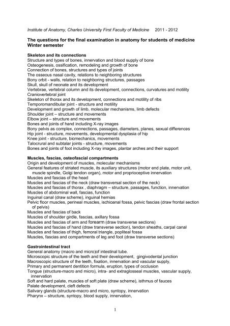
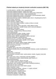
Institute <strong>of</strong> Anatomy, Charles University First Faculty <strong>of</strong> Medic<strong>in</strong>e 2011 - 2012<strong>The</strong> <strong>questions</strong> <strong>for</strong> <strong>the</strong> <strong>f<strong>in</strong>al</strong> <strong>exam<strong>in</strong>ation</strong> <strong>in</strong> <strong>anatomy</strong> <strong>for</strong> <strong>students</strong> <strong>of</strong> medic<strong>in</strong>eW<strong>in</strong>ter semesterSkeleton and its connectionsStructure and types <strong>of</strong> bones, <strong>in</strong>nervation and blood supply <strong>of</strong> boneOsteogenesis, ossification, remodel<strong>in</strong>g and growth <strong>of</strong> boneConnection <strong>of</strong> bones, structures and types <strong>of</strong> jo<strong>in</strong>ts<strong>The</strong> osseous nasal cavity, relations to neighbor<strong>in</strong>g structuresBony orbit - walls, relation to neighbor<strong>in</strong>g structures, passagesSkull, skull <strong>of</strong> neonate and its developmentVertebrae, vertebral column and its development, connections, curvatures and motilityCraniovertebral jo<strong>in</strong>tSkeleton <strong>of</strong> thorax and its development, connections and motility <strong>of</strong> ribsTemporomandibular jo<strong>in</strong>t - structure and motilityDevelopment and growth <strong>of</strong> limb, molecular mechanisms, limb defectsShoulder jo<strong>in</strong>t – structure and movementsElbow jo<strong>in</strong>t – structure and movementsBones and jo<strong>in</strong>ts <strong>of</strong> hand <strong>in</strong>clud<strong>in</strong>g X-ray imagesBony pelvis as complex, connections, passages, diameters, planes, sexual differencesHip jo<strong>in</strong>t - structure, movements, developmental dysplasia <strong>of</strong> hipKnee jo<strong>in</strong>t - structure, biomechanics, movementsTalocrural and subtalar jo<strong>in</strong>ts - structure, movementsBones and jo<strong>in</strong>ts <strong>of</strong> foot <strong>in</strong>clud<strong>in</strong>g X-ray images, plantar arches and <strong>the</strong>ir supportMuscles, fascias, oste<strong>of</strong>ascial compartmentsOrig<strong>in</strong> and development <strong>of</strong> muscles, molecular mechanismsGeneral features <strong>of</strong> striated muscle, its auxiliary structures (motor end plate, motor unit,muscle sp<strong>in</strong>dle, Golgi tendon organ), motor and proprioceptive <strong>in</strong>nervationMuscles and fascias <strong>of</strong> <strong>the</strong> headMuscles and fascias <strong>of</strong> <strong>the</strong> neck (draw transversal section <strong>of</strong> <strong>the</strong> neck)Muscles and fascias <strong>of</strong> thorax , diaphragm – structure, passages, function, <strong>in</strong>nervationMuscles <strong>of</strong> abdom<strong>in</strong>al wall, fascias, functionIngu<strong>in</strong>al canal (draw scheme), <strong>in</strong>gu<strong>in</strong>al herniasPelvic floor muscles, per<strong>in</strong>eal muscles, ischioanal fossa, pelvic fascias (draw frontal section<strong>of</strong> pelvis)Muscles and fascias <strong>of</strong> backMuscles <strong>of</strong> shoulder girdle, fascias, axillary fossaMuscles and fascias <strong>of</strong> arm and <strong>for</strong>earm (draw transverse sections)Muscles and fascias <strong>of</strong> hand (draw transverse section), tendon sheaths, carpal canalMuscles and fascias <strong>of</strong> thigh, femoral triangle, popliteal fossaMuscles, fascias and compartments <strong>of</strong> leg and foot (draw transverse sections)Gastro<strong>in</strong>test<strong>in</strong>al tractGeneral <strong>anatomy</strong> (macro and micro)<strong>of</strong> <strong>in</strong>test<strong>in</strong>al tube.Microscopic structure <strong>of</strong> <strong>the</strong> teeth and <strong>the</strong>ir development, g<strong>in</strong>givodental junctionMacroscopic structure <strong>of</strong> <strong>the</strong> teeth, fixation, <strong>in</strong>nervation and vascular supply,Primary and permanent dentition <strong>for</strong>mula, eruption, types <strong>of</strong> occlusionTongue (structure-macro and micro), <strong>in</strong>tra- and extraglosseal muscles, vascular supply,<strong>in</strong>nervationS<strong>of</strong>t and hard palate, muscles <strong>of</strong> s<strong>of</strong>t plate (draw scheme), isthmus <strong>of</strong> faucesPalate development, cleft defectsSalivary glands (structure-macro and micro, syntopy, <strong>in</strong>nervationPharynx – structure, syntopy, blood supply, <strong>in</strong>nervation,1
Nasal, palat<strong>in</strong>e and l<strong>in</strong>gual tonsills (structure-macro and micro) (Waldeyer circle)Oesophagus – structure (macro and micro), syntopyStomach – shape, position, syntopy, projectionsStomach – structure <strong>of</strong> <strong>the</strong> wall, divisions, vascular supply, <strong>in</strong>nervation, lymphatic dra<strong>in</strong>ageDevelopment <strong>of</strong> oesophagus, stomach and duodeumSmall <strong>in</strong>est<strong>in</strong>e – structure (macro and micro), divisions, vascular supply, <strong>in</strong>nervation,lymphatic dra<strong>in</strong>ageDuodenum – divisions, positions, syntopy (draw scheme), blood supplyLarge <strong>in</strong>test<strong>in</strong>e, structure (macro and micro), divisions (draw scheme), syntopy, vascularsupply, <strong>in</strong>nervation, positions <strong>of</strong> vermi<strong>for</strong>m appendixDevelopment <strong>of</strong> small and large <strong>in</strong>test<strong>in</strong>e, <strong>in</strong>test<strong>in</strong>al rotationPancreas – structure (macro and micro), , Langerhans islets, syntopy,Liver – segments, syntopy (draw scheme <strong>of</strong> visceral surface)Liver – structure (macro and micro), nutritional and portal vascular bed,<strong>in</strong>trahepatic bile ductsGallbladder and extrahepatic bile ducts (draw scheme)Development <strong>of</strong> pancreas and liverRectum and anal canal - structure (macro and micro), syntopy (draw frontal and sagittalsections), vascular supply, sph<strong>in</strong>cters and <strong>the</strong>ir <strong>in</strong>nervationPeritoneum - parietal and visceral, greater and lesser omentumLesser sac (omental bursa), its recessesDevelopment <strong>of</strong> visceral situs and mesenteryRegional AnatomyInfratemporal fossa and parapharyngeal spaceExternal and <strong>in</strong>ternal cranial base - open<strong>in</strong>gs <strong>for</strong> vessels and nervesSubmandibular triangle, carotid triangle (draw scheme)Lateral neck region, scalenic fissureAxilla – boundaries, contentAnterior and posterior regions <strong>of</strong> arm (draw transverse section)Cubital fossa, elbow jo<strong>in</strong>tTopographic <strong>anatomy</strong> <strong>of</strong> <strong>the</strong> hand and f<strong>in</strong>gersGluteal region, supra- and <strong>in</strong>frapiri<strong>for</strong>m <strong>for</strong>amensAnterior thigh region, vascular and muscular lacuna, iliopect<strong>in</strong>eal fossa, femoral triangle(draw schema), femoral herniasPopliteal fossa, adductor canalRegions <strong>of</strong> lower leg (draw transverse section)Retromalleolar regionsTopography <strong>of</strong> foot (draw transverse section)Topography <strong>of</strong> chest wall, surface projections <strong>of</strong> heart, lungs and pleuraTopography <strong>of</strong> abdom<strong>in</strong>al wall, blood supply, <strong>in</strong>nervation and surface projections <strong>of</strong>abdom<strong>in</strong>al organsIngu<strong>in</strong>al region, <strong>in</strong>gu<strong>in</strong>al canal, hernias (draw schema <strong>of</strong> <strong>in</strong>gu<strong>in</strong>al canal)Topography <strong>of</strong> supramesocolic part <strong>of</strong> peritoneal cavity (draw transverse section throughlesser sac), <strong>in</strong>framesocolic part <strong>of</strong> peritoneal cavityTopography <strong>of</strong> duodenum and pancreas (draw schema)2


