Pierre Gloor (1923–2003) - International League Against Epilepsy
Pierre Gloor (1923–2003) - International League Against Epilepsy
Pierre Gloor (1923–2003) - International League Against Epilepsy

You also want an ePaper? Increase the reach of your titles
YUMPU automatically turns print PDFs into web optimized ePapers that Google loves.
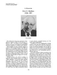
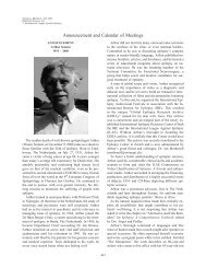
Epilepsia, 45(7):882–886, 2004Blackwell Publishing, Inc.C○ 2004 <strong>International</strong> <strong>League</strong> <strong>Against</strong> <strong>Epilepsy</strong>In Memoriam<strong>Pierre</strong> <strong>Gloor</strong> (<strong>1923–2003</strong>): An AppreciationMassimo AvoliOn October 24, 2003, the world’s neuroscience communitylost <strong>Pierre</strong> <strong>Gloor</strong>, one of its most influential leadersin epilepsy research. <strong>Gloor</strong> passed away peacefullyat the Montreal Neurological Institute and Hospital (the“Neuro”) of McGill University, where he had worked relentlesslybetween his arrival to Canada in 1952 and 1994,when he was felled by a severe stroke. A native of Basel(Switzerland), <strong>Gloor</strong> received his medical education at theUniversity of Basel and did his resident training in Neurologyand Neurosurgery at l’Hôpital Louis Pasteur in Colmar,France. In 1952, he joined the Montreal NeurologicalInstitute as a fellow in the Neurophysiology Laboratoryand worked with W. Penfield and H. Jasper. At that time,the Neurophysiology Laboratory, located in a relativelysmall area on the 7th floor of the Neuro, was crammedwith research fellows who would shape our knowledge onbrain function under normal and pathological conditionsin the years to come (Fig. 1).In this early period (1952–1957), <strong>Gloor</strong> explored byelectrophysiological means the connections of the amygdala.He found that this limbic area projected intensely toa large brain region comprising the basal core of the forebrainand midbrain extending to the hypothalamus andthe brainstem tegmentum. Data originating from these experimentshave, indeed, stood the test of time. They wereincluded in <strong>Gloor</strong>’s Ph.D. thesis, submitted in 1957, andbecame a chapter in the first edition of the Handbook ofPhysiology published by the American Physiological Society(1). This chapter has been much quoted over the lastfour decades, even though our knowledge on the anatomyand function of the amygdala has enormously increasedsince 1960, when it was published.The link of the amygdala with a disparate number ofphysiological and pathological conditions was probablycongenial with <strong>Gloor</strong>’s ability to explore the intimate functionof different brain systems such as the hippocampusand the hypothalamus. During the 1960s, he undertooka series of pioneering studies aimed at characterizing theresponses of hippocampal and dentate neurons to stimulationof amygdala and entorhinal cortex (2,3). He demonstratedthat these limbic structures contacted the apicaldendrites of hippocampal pyramidal neurons and dentategranule cells through excitatory synapses, whereas stimulationof the fimbria engaged mostly the basal dendrites.The laminar profile of these responses demonstrated thathippocampal cells behave as dipoles. Similar conclusionswere drawn at the same time by Per Andersen in Oslo(Fig. 2). <strong>Gloor</strong>’s studies also identified some intriguingphenomena that accompany hippocampal electrographicseizures; he found that epileptiform discharges were associatedwith DC shifts that may play a significant rolein seizure maintenance (4). These events are presently explained,at least in part, as reflecting extracellular potassiumaccumulation.During these years, <strong>Gloor</strong> remained focused on thestudy of amygdaloid connections. He demonstrated thatventromedial hypothalamic neurons receive excitatoryand inhibitory inputs from the amygdala basolateral andthe corticomedial nucleus, respectively (5,6). Hence theseexperiments led to the identification of the ability of amygdaloidoutputs to exert a dual influence on hypothalamicactivity. These studies also clarified how epileptic dischargesoriginating from limbic areas can elicit a varietyof autonomic and behavioral responses.A fundamental characteristic of his investigative approachwas the careful analysis of the clinical and neurophysiologicalfeatures of epileptic syndromes. <strong>Gloor</strong> usedthis clinical information to formulate specific hypothesesto be tested in the experimental laboratory. In turn he carefullyused the latest experimental results to bear on clinicaldecisions. An example of this approach can be found inhis identification of primary generalized epileptic disorderswith spike-and-wave discharges as “corticoreticularepilepsies.” The rationale behind this choice is detailed ina review chapter that was written in 1978 (7). I quote afew passages:About 10 years ago I received an invitation from Dr.Gastaut to go to Marseille to present a review of theneurophysiological basis of generalized seizures termed882
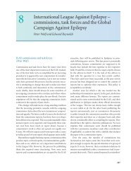
IN MEMORIAM: DR. PIERRE GLOOR 883FIG. 1. Fellows of the Neurophysiology Laboratory of the Neuro in 1952. Front row: Annie Courtois, Doris Oeconomos, William Feindel;middle row: Guss Stoll, Herbert Jasper, Cosimo Ajmone Marsan; Back row: Guy Courtois, <strong>Pierre</strong> <strong>Gloor</strong>, John Hunter, Kenan Tukel, DavidIngvar, Maida Tukel, Jake Hanbery.“centrencephalic.” I suspect he selected me for this assignmentbecause I came from the Montreal Neurological Instituteand he expected that I would review the clinical andexperimental data which supported the centrencephalic hypothesisof generalized seizures. Originally that was exactlywhat I intended to do. After reviewing many experimentaland clinical studies ..., I came to the conclusion that it wasno longer tenable to conceive of the mechanism of generalizedseizures in terms of either a purely centrencephalicor purely cortical mechanism .... Rather than to look uponthe “centrencephalic” and upon the “cortical” hypothesisas being mutually exclusive, I began to explore the notionthat generalized epilepsy was dependent upon more complexmechanisms which involved both cortical and subcorticalstructures .... Some abnormal interaction between thesetwo levels was thought to be responsible for the conditions,but at that time it was not very clear to me how this abnormalinteraction between the two levels could take place. In thelight of this rather vague hypothesis, I proposed the term generalizedcortico-reticular epilepsies for generalized epileptogenicconditions associated with bilaterally synchronousspike and wave activity (8,9).It soon became imperative to search for an experimentalmodel of which this new concept could be tested.The model selected by <strong>Gloor</strong> was generalized penicillinepilepsy in the cat. This model was the focus of his experimentalstudies for more than two decades, and I wasfortunate to work with him on many of the experimentsthat were aimed at identifying the relative contributionsmade by thalamic and neocortical networks to generalizedspike-and-wave activity in cats. Most of the originalfindings obtained in penicillin-treated animals have beenrecently confirmed by other researchers. Once more, thesestudies have demonstrated the correctness of his clinicallybased intuitions.Proof of <strong>Gloor</strong>’s unique ability to synthesize clinical andexperimental evidence and to pose straight questions alsois found in a letter he sent to Dan McIntyre to summarizean invited talk to be given at the annual meeting of theAmerican <strong>Epilepsy</strong> Society in 1994. This letter, whichwas written just a few days before his debilitating stroke,identifies questions on temporal lobe epilepsy that are asyet to be answered. I quote:I believe what I shall do is to stress that even though we knowthat most temporal lobe seizures in humans originate fromEpilepsia, Vol. 45, No. 7, 2004
884 M. AVOLIFIG. 2. <strong>Pierre</strong> <strong>Gloor</strong> with Per Andersen at the Focus on <strong>Epilepsy</strong> Symposium I, which was held in St. Adele (Quebec, Canada) in thesummer of 1991.the mesial structures, we are far from understanding whichstructures are essential or play what role, which is or are thesites of seizure onset and which are the routes of propagationof the seizure discharge. There has been, in my opinion,a simplistic view that the hippocampus is possibly the solecenter of action. Hippocampal sclerosis is certainly the mostoutstanding neuropathological finding in resected temporallobes of temporal lobe epileptics. And since patients withproven hippocampal sclerosis do best after surgery, the conclusionwas that is the sclerotic hippocampus that is the site oforigin of the seizures. This may be so, but remains unprovenand there are difficulties with this explanation....the experimental neurophysiologists who work on normalhippocampi consistently identify CA3 as the site of origin ofdischarge in a variety of models of experimental hippocampalepilepsy .... It is hard to see how in an abnormal, sclerotichippocampus this could be the mechanism of seizure genesisand propagation with hardly any neurons left in eitherCA3 or CA1. Relatively well-preserved dentate granule cellsapparently then come to the rescue. In hippocampus with destructionof the hilar cells and CA3 neurons the dentate granulecell axons sprout collaterals that innervate the molecularlayer of the dentate gyrus .... There is a catch, however. Thedentate granule cells can only project to the hilar cells andCA3 and they are gone. So how does a hypothetical seizuredischarge in the dentate gyrus propagate out of the dentategyrus in a sclerotic hippocampus that lacks hilar and CA3neurons? .... the hippocampus may not be the sole or principalplayer in temporal lobe epilepsy. The amygdala mayplay an equally important role .... It seems therefore that wehave to begin to think of a distributed epileptogenic processin the mesial temporal region that includes, besides the hippocampusand the amygdala, the parahippocampal gyrus, theentorhinal cortex and the prepiriform cortex .... The conceptwhich we should try to form is that of a distributed, althoughpathologically altered, neuronal network in the mesial temporalregion that is responsible for seizure genesis in thisarea. Such a reorganized network may be intrinsically unstablefor reasons we do not yet understand and this instabilitymay be the cause for recurrent seizures.His definitive monograph, entitled “The Temporal Lobeand Limbic System,” was published in 1997 by OxfordUniversity Press and represented a culmination of his life’sinterest in the anatomy and physiology of the temporallobe and its disorders (10).<strong>Gloor</strong> worked closely with the clinical and researchteams at the Montreal Neurological Institute in the treatmentof epilepsy (Fig. 3). In the early 1960s, he improvedtechniques to record with sphenoidal electrodesthe EEG from mesial temporal structures in epilepsy patients.Then, in the 1970s, he developed the use of depthelectrodes for the study of patients with bitemporal epileptiformdischarges. Overall these efforts led to the introductionof video/EEG long-term monitoring in conjunctionwith computerized methods. These analytic procedureswere later used in animals to analyze the contribution ofEpilepsia, Vol. 45, No. 7, 2004
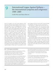
IN MEMORIAM: DR. PIERRE GLOOR 885FIG. 3. <strong>Gloor</strong> reading the electrocorticogram recorded from an epilepsy patient during surgery performed at the Neuro in the late 1980s.neocortical and thalamic networks to the generation ofspecific brain rhythms under normal and pathologic conditions.By all means, <strong>Gloor</strong>’s studies on patients were not limitedto technological advancements. Indeed, the outcomeof his clinical studies is absolutely remarkable. Just tomention a few, he established a number of prognosticfactors that would allow one to assess preoperatively thelikelihood of a good or unsatisfactory outcome of epilepsysurgery (11,12). In addition, through recording seizures bydepth electrodes and performing stimulation of temporallobe structures, he identified the anatomic substrates andthe nature of experiential phenomena in epilepsy patients,most often with temporal lobe epilepsy (13–15).<strong>Gloor</strong> began lecturing at McGill in 1954 to become fullProfessor in 1968 in the Department of Neurology andNeurosurgery. In 1998 he was designated McGill EmeritusProfessor. During his academic career, he was theDirector of the Laboratory of EEG and Clinical Neurophysiology.He was a key figure in the training of severalneurology trainees and epilepsy fellows, many of whomare now internationally renowned experts. He also tookan active part in teaching EEG technologists and in organizinga College program for them, resulting in Quebec,and Canada, having superbly trained EEG technologists.In 1988, the Quebec Association of EEG Technologistsorganized the “Prix d’Excellence <strong>Pierre</strong> <strong>Gloor</strong>.”His work in understanding and treating epileptic disordersearned him a worldwide reputation, as witnessed byseveral international recognitions that included the RobertBing Prize, the Michael Prize, the William G. LennoxAward, the Wilder Penfield Award, and the Milken FamilyFoundation Prize. <strong>Gloor</strong> was president of the Canadianas well as of the American Electroencephalographic Societyand of the American <strong>Epilepsy</strong> Society.<strong>Gloor</strong> was a scientist, a discoverer, a humanist, a trulydecent man, and from my personal experience, an invaluablementor. His life work has helped us to progress in understandingbrain function. No doubt his prodigious contributionswill be appreciated by the world neurosciencecommunity for years to come. Thus let me say once morewhat ended most of our conversations: “Thank you, Dr.<strong>Gloor</strong>.”Acknowledgment: I am indebted to Dan McIntyre for sharinga letter that was sent to him by <strong>Pierre</strong> <strong>Gloor</strong> and to Victor Eppand Toula Papadopoulos for their continuous editorial support.REFERENCES1. <strong>Gloor</strong> P. Amygdala: neurophysiology. In: Field J, Magoun HW,Hall EV, eds. Handbook of physiology: II. Washington: AmericanPhysiology Society, 1960:1395–420.2. <strong>Gloor</strong> P, Vera CL, Sperti L. Electrophysiological studies of hippocampalneurons, I: configuration and laminar analysis of the“resting” potential gradient, of the main transient response to perforantpath, fimbrial and mossy fiber volleys and of “spontaneous”activity. Electroencephalogr Clin Neurophysiol 1963;15:353–78.3. <strong>Gloor</strong> P, Sperti L, Vera C. Electrophysiological studies of hippocampalneurons, II: secondary postsynaptic events and single cell unitdischarges. Electroencephalogr Clin Neurophysiol 1963;15:379–402.4. <strong>Gloor</strong> P, Vera CL, Sperti L. Electrophysiological studies of hippocampalneurons, III: responses of hippocampal neurons to repetitiveperforant path volleys. Electroencephalogr Clin Neurophysiol1964;17:353–70.5. Murphy JT, Dreifuss JJ, <strong>Gloor</strong> P. Responses of hypothalamic neuronsto repetitive amygdaloid stimulation. Brain Res 1968;8:153–66.Epilepsia, Vol. 45, No. 7, 2004
886 M. AVOLI6. Dreifuss JJ, Murphy JT, <strong>Gloor</strong> P. Contrasting effects of two identifiedamygdaloid efferent pathways on single hypothalamic neurons.J Neurophysiol 1968;31:237–48.7. <strong>Gloor</strong> P. Evolution of the concept of the mechanism of generalizedepilepsy with bilateral spike and wave discharge. In: Wada JA, ed.Modern perspectives in epilepsy. Montreal: Eden Press, 1978:99–137.8. <strong>Gloor</strong> P. Generalized cortico-reticular epilepsies. Epilepsia1968;9:249–63.9. <strong>Gloor</strong> P. Neurophysiological bases of generalized seizures, termed“centrencephalic.” In: Gastaut H, Jasper H, Bancaud J, WaltregnyA., eds. The pathophysiogenesis of the epilepsies. Springfield, Ill:Charles C Thomas, 1969;18:209–36.10. <strong>Gloor</strong> P. The temporal lobe and limbic system. New York: OxfordUniversity Press, 1997.11. Bengzon A, Rasmussen T, <strong>Gloor</strong> P, et al. Prognostic factorsin the surgical treatment of temporal lobe epileptics. Neurology1968;18:717–31.12. So N, <strong>Gloor</strong> P, Quesney LF, et al. Depth electrode investigationsin patients with bitemporal epileptiform abnormalities. Ann Neurol1989;25:423–31.13. <strong>Gloor</strong> P. Experiential phenomena of temporal lobe epilepsy: factsand hypotheses. Brain 1990;113:1673–94.14. Palmini A, <strong>Gloor</strong> P, Jones-Gotman M. Pure amnestic seizures in temporallobe epilepsy: definition, clinical symptomatology and functionalanatomical considerations. Brain 1992;115:749–69.15. Fish DR, <strong>Gloor</strong> P, Quesney FL, et al. Clinical responses to electricalbrain stimulation of the temporal and frontal lobes in patients withepilepsy: pathophysiological implications. Brain 1993;116:397–414.Epilepsia, Vol. 45, No. 7, 2004