Atto 488 - Sigma-Aldrich
Atto 488 - Sigma-Aldrich
Atto 488 - Sigma-Aldrich

You also want an ePaper? Increase the reach of your titles
YUMPU automatically turns print PDFs into web optimized ePapers that Google loves.
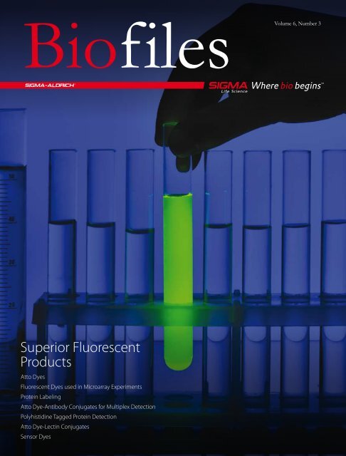
Biofiles<br />
Superior Fluorescent<br />
Products<br />
<strong>Atto</strong> Dyes<br />
Fluorescent Dyes used in Microarray Experiments<br />
Protein Labeling<br />
<strong>Atto</strong> Dye-Antibody Conjugates for Multiplex Detection<br />
Polyhistidine Tagged Protein Detection<br />
<strong>Atto</strong> Dye-Lectin Conjugates<br />
Sensor Dyes<br />
Volume 6, Number 3
Biofilesonline<br />
Your gateway to Biochemicals and Reagents<br />
for Life Science Research<br />
Biofiles Online allows you to:<br />
•<br />
•<br />
Easily navigate the content of<br />
the current Biofiles issue<br />
• Access any issue of Biofiles<br />
Subscribe for email notifications<br />
of future eBiofiles issues<br />
Register today for upcoming issues and<br />
eBiofiles announcements at<br />
sigma.com/biofiles<br />
Highlights from this issue:<br />
Superior Fluorescent Dyes<br />
New life science microscopy<br />
techniques require fluorescence labels<br />
that fulfill stringent application<br />
requirements. In this issue, we highlight<br />
the superior series of <strong>Atto</strong> dyes for the<br />
most sensitive applications.<br />
<strong>Atto</strong> dyes show extraordinary results in sensitive and target-specific<br />
applications such as microarray experiments, and a portfolio of<br />
<strong>Atto</strong> dye conjugates support analysis of proteins and carbohydrates.<br />
This issue also includes sensor probes for NO, oxygen, and ROS<br />
detection, which are of growing interest in cellular studies.<br />
Coming next issue:<br />
Insulin Resistance<br />
The next issue of Biofiles will focus on<br />
insulin resistance, a condition of reduced<br />
insulin responsiveness in key target<br />
tissues. The development of insulin<br />
resistance is crucial to the pathogenesis of<br />
metabolic syndrome and its constellation of associated diseases such as<br />
type II diabetes mellitus and atherosclerotic cardiovascular disease.<br />
Recent studies provide strong evidence that mitochondrial defects may<br />
underlie the development of most instances of insulin resistance.<br />
Understanding the mechanisms that lead to the loss of mitochondrial<br />
efficiency is essential for the development of effective<br />
treatment strategies.<br />
Biofilescontents<br />
Introduction 3<br />
<strong>Atto</strong> Dyes for Superior<br />
Fluorescent Imaging 5<br />
<strong>Atto</strong> Dyes 7<br />
<strong>Atto</strong> Dye Conjugates 7<br />
Analyzing Properties of<br />
Fluorescent Dyes Used<br />
for Labeling DNA in<br />
Microarray Experiments 9<br />
<strong>Atto</strong> Dyes and Tracy Dyes for<br />
Fluorescent Protein Labeling 14<br />
Fluorescent Multiplex<br />
Detection using Antibody <strong>Atto</strong><br />
Dye Conjugates 16<br />
Sensitive Detection of poly(His)tagged<br />
Proteins with Ni-NTA<br />
<strong>Atto</strong> Complex 18<br />
<strong>Atto</strong> Dye Lectin Conjugates<br />
for Fluorescent Carbohydrate<br />
Labeling 21<br />
Sensor Dyes 23<br />
NO Probes 23<br />
Oxygen Probes 25<br />
ROS Probes 25<br />
New Products for<br />
Detection Applications 26<br />
Cover: A fluorescent dye simplifies the<br />
recognition and location of a test tube,<br />
a cell, or a single molecule of interest.<br />
Technical content: Monika Bäumle, Ph. D.
Order sigma.com/order Technical service sigma.com/techinfo sigma.com/lifescience 3<br />
Introduction<br />
Monika Bäumle, Ph.D.<br />
Product Manager, Biochemistry<br />
monika.baeumle@sial.com<br />
Fluorescent techniques are widespread<br />
and fast-growing analytical methods<br />
used in life science. They allow sensitive<br />
and selective investigation of biological<br />
processes, diagnostic screening, kinetics,<br />
and conformational studies. Research is<br />
evolving from identification of a large<br />
number of target molecules to isolation and<br />
investigation at the level of a single molecule.<br />
Key innovations for microscopy and<br />
nanoscopy in the last decade have been:<br />
•<br />
Confocal laser scanning microscopy<br />
(CLSM)<br />
• Fluorescence correlation spectroscopy<br />
(FCS)<br />
• Total internal reflection of fluorescence<br />
(TIRF)<br />
• Stimulated emission depletion (STED)<br />
microscopy<br />
• Spectral precision distance microscopy<br />
(SPDM)<br />
• Scanning nearfield optical microscopy<br />
(SNOM)<br />
• Fluorescence photoactivation localization<br />
microscopy (FPALM)<br />
• microscopy (STORM). 1<br />
Stochastic optical reconstitution<br />
These super-resolution microscopic<br />
and spectroscopic techniques allow a<br />
tremendous increase in lateral resolution and<br />
enable scientists to have a new perspective<br />
of cellular studies. For example, STED<br />
microscopy, the revolutionary technique<br />
invented by Professor Stefan Hell, enables<br />
a microscopic resolution limit below the<br />
theoretical limit of resolution and permits<br />
more detailed studies in cellular processes. 2,3<br />
These techniques require suitable fluorescent<br />
dyes that fulfill the demands of the<br />
applications, including a high cross section<br />
for stimulated emission, an absence of<br />
excitation at the depletion wavelength, dye<br />
photostability at depletion and excitation<br />
wavelengths, low triplet-state formation, and<br />
low non-linear photobleaching. <strong>Atto</strong> dyes<br />
are a series of fluorophores that can be used<br />
in the most sensitive applications including<br />
STED microscopy, and are especially suitable<br />
for target-specific detection. In a study<br />
performed by Donnert, et al., <strong>Atto</strong> 532<br />
and <strong>Atto</strong> 647N were used for the study of<br />
synaptic vesicle proteins and demonstrated<br />
superior properties in two-color STED<br />
microscopy. 4 Due to its super resolution<br />
property, the two-color STED microscopy<br />
technique was able to reveal ring-shaped<br />
synaptophysin domains and differentiate<br />
colocalized proteins with greater resolution<br />
than confocal microscopy.<br />
With increasing instrument application<br />
limits, there is a growing demand for<br />
more sensitive, photostable and selective<br />
probes. Based on our extensive experience,<br />
expertise in organic synthesis and life<br />
sciences, and stringent quality assurance,<br />
<strong>Sigma</strong> offers a comprehensive selection<br />
of reagents for superior application results<br />
including the fluorescent <strong>Atto</strong> dyes series,<br />
MegaStokes dyes, and Chromeo Py-dyes.<br />
We are continuously increasing our portfolio<br />
of detection products and optimized<br />
protocols to meet the needs of fluorescence<br />
applications in life science research.
4<br />
In this issue, scientists from the Swiss<br />
Institute of technology ETH, Zurich,<br />
Switzerland, show extraordinary application<br />
results using the red-emitting dyes <strong>Atto</strong><br />
647N, <strong>Atto</strong> 633, and <strong>Atto</strong> 655 in microarray<br />
experiments. <strong>Atto</strong> dyes, kits for protein<br />
labeling with <strong>Atto</strong> dyes, and <strong>Atto</strong> dye<br />
conjugates used to detect recombinant<br />
polyhistidine-tagged proteins and<br />
carbohydrates are also reviewed.<br />
Core<br />
Additional sections in this issue review<br />
cellular processes and signaling by oxygencontaining<br />
molecules, and a new series<br />
of pI markers for isoelectric focusing (IEF)<br />
that are detectable by ultraviolet light for<br />
greater sensitivity.<br />
We hope you find our articles and products<br />
helpful and interesting. For more information<br />
on detection products available from <strong>Sigma</strong>,<br />
please visit sigma.com/fluorescence.<br />
Bioreagents.<br />
References<br />
(1) A guide to super-resolution fluorescence microscopy.<br />
Schermelleh, L., Heintzmann, R., Leonhardt, H., J. Cell.<br />
Biol., 190, 166–75 (2010).<br />
(2) Confocal Application Letter No. 32, Leica Microsystems<br />
(2009).<br />
(3) Breaking the diffraction resolution limit by stimulated<br />
emission: stimulated-emission-depletion fluorescence<br />
microscopy. Hell, S.W. and Wichmann, J., Opt. Lett., 19,<br />
780–2 (1994).<br />
(4) Two-color far-field fluorescence nanoscopy, Donnert,<br />
G., Keller, J., Wurm, C.A., Rizzoli, S.O., Westphal, V.,<br />
Schönle, A., Jah, R., Jakobs, S., Eggeling, C., Hell, S.W.,<br />
Biophysical Journal, 92, 67–9 (2007).<br />
<strong>Sigma</strong>® Life Science biochemicals bring<br />
you the most comprehensive range<br />
of high quality products for everyday<br />
research including agarose, antibiotics,<br />
buffers, and more.<br />
We offer biochemicals with:<br />
• Real-time product availability<br />
• Convenient packaging options<br />
• Competitive pricing<br />
• Product selection guides and other<br />
valuable research tools<br />
For more information visit,<br />
sigma.com/corebioreagents
Activated fluorescent dyes are routinely<br />
used to tag proteins, nucleic acids, and<br />
other biomolecules for use in life science<br />
applications including fluorescence<br />
microscopy, flow cytometry, fluorescence<br />
in situ hybridization (FISH), receptor binding<br />
assays, and enzyme assays. The <strong>Atto</strong> dyes<br />
are a series of fluorescent dyes that meet<br />
the critical needs of modern fluorescent<br />
technologies:<br />
• Stability - <strong>Atto</strong> 655 and <strong>Atto</strong> 647N are<br />
photostable and highly resistant to<br />
ozone degradation, making them ideal<br />
for microarray applications. See the<br />
related article "Analyzing Properties of<br />
Fluorescent Dyes used for Labeling DNA<br />
in Microarray Experiments" on page 9.<br />
• Long Signal Lifetimes - Signal decay<br />
times of 0.6–4.1 nanoseconds allow timegate<br />
studies to reduce autofluorescence<br />
background and scattering.<br />
• Reduced Background -Several <strong>Atto</strong> dyes<br />
employ excitation wavelengths greater<br />
than 600 nm, reducing background<br />
fluorescence from samples, Rayleigh and<br />
Raman scattering.<br />
• Selection - <strong>Atto</strong> dyes have strong<br />
fluorescent signals that cover visible and<br />
near-IR emission wavelengths.<br />
Long Signal Lifetimes<br />
<strong>Atto</strong> dyes exhibit longer fluorescence<br />
signal lifetimes (0.6–4.1 ns) in aqueous<br />
solution than either carbocyanine dyes<br />
or most of the autofluorescence inherent<br />
in cells and biomolecules. The signal<br />
from <strong>Atto</strong> dyes can be measured using<br />
pulsed laser excitation with a time-gated<br />
detection system to reduce interference<br />
Order sigma.com/order Technical service sigma.com/techinfo sigma.com/lifescience 5<br />
<strong>Atto</strong> Dyes for Superior Fluorescent Imaging<br />
<strong>Atto</strong> Dyes for Superior Fluorescent Imaging<br />
from fluorophores with shorter lifetimes,<br />
background autofluorescence, and Rayleigh<br />
and Ramen light scattering, improving<br />
overall sensitivity.<br />
Longer Excitation Wavelengths for<br />
Reduced Background<br />
Diode laser excitation at 635 nm and redabsorbing<br />
fluorescent dyes were shown<br />
to reduce autofluorescence of biological<br />
samples sufficiently so that individual<br />
antigen and antibody molecules could<br />
be detected in human serum samples. 1,2<br />
Excitation in the red spectral region also<br />
reduces cell damage when working with<br />
live cells. 3<br />
Many of <strong>Atto</strong> dyes (<strong>Atto</strong> 590 and above)<br />
can be excited using wavelengths greater<br />
than 600 nm. Using long-wavelength<br />
activated <strong>Atto</strong> dyes in conjunction with the<br />
appropriate excitation wavelength reduces<br />
autofluorescence due to sample, solvent,<br />
glass, or polymer support, and improves<br />
overall sensitivity in biological analysis<br />
and imaging techniques. The background<br />
fluorescence due to Rayleigh and Raman<br />
scattering are also dramatically reduced<br />
by use of longer wavelength excitation.<br />
λ Em Range from 479 to 764 nm for<br />
Fluorescent Multiplex Detection<br />
<strong>Atto</strong> dyes have strong fluorescent signals<br />
with most having molar absorptivity values<br />
>100,000 and low excitation/emission<br />
overlap, making <strong>Atto</strong> dyes ideal for multiplex<br />
techniques using visible and near-IR<br />
emission wavelengths.<br />
With excitation signal maxima ranging<br />
from 390 to 740 nm and good Stokes shift<br />
separation, there are <strong>Atto</strong> dyes suitable<br />
for use with any common excitation light<br />
source.<br />
Alternatives to Common<br />
Fluorophores<br />
With the extensive selection of <strong>Atto</strong> dyes<br />
available, any common excitation light<br />
source can be used, and <strong>Atto</strong> dyes can<br />
replace other fluorescent dyes commonly<br />
used in life science.<br />
Fluorophore Recommended <strong>Atto</strong> Dye<br />
Alexa Fluor® <strong>488</strong> <strong>Atto</strong> <strong>488</strong><br />
FITC <strong>Atto</strong> <strong>488</strong><br />
FAM <strong>Atto</strong> <strong>488</strong><br />
JOE <strong>Atto</strong> 520<br />
TET <strong>Atto</strong> 520<br />
Alexa Fluor 532 <strong>Atto</strong> 532<br />
HEX <strong>Atto</strong> 532, <strong>Atto</strong> Rho6G<br />
TAMRA <strong>Atto</strong> 550<br />
Cy®3 <strong>Atto</strong> 550<br />
Cy3.5 <strong>Atto</strong> 565<br />
ROX <strong>Atto</strong> 565, <strong>Atto</strong> Rho11<br />
Alexa Fluor 594 <strong>Atto</strong> 590 , <strong>Atto</strong> 594<br />
Texas Red® <strong>Atto</strong> 590<br />
Alexa Fluor 633 <strong>Atto</strong> 633, <strong>Atto</strong> Rho14<br />
Cy5 <strong>Atto</strong> 647, <strong>Atto</strong> 647N, <strong>Atto</strong> 655<br />
Alexa Fluor 647 <strong>Atto</strong> 647, <strong>Atto</strong> 647N, <strong>Atto</strong> 655<br />
Cy5.5 <strong>Atto</strong> 680, <strong>Atto</strong> 700
6<br />
Reactive <strong>Atto</strong> Dyes and<br />
Conjugates<br />
<strong>Atto</strong> dyes produce intense fluorescent<br />
signals due to strong absorbance and high<br />
quantum yields. Dyes are available as:<br />
• Free acid dyes for all routine staining<br />
applications<br />
• NHS-esters for use in common<br />
conjugation protocols<br />
• Maleimides for use in coupling to<br />
thiol-containing groups such as cysteine<br />
residues and thiol (-SH) tags added<br />
during automated synthesis<br />
• <strong>Atto</strong> Dyes conjugated to biotin,<br />
streptavidin, and antibodies are also<br />
available<br />
<strong>Atto</strong> 655, <strong>Atto</strong> 680, and <strong>Atto</strong> 700 are<br />
quenched by guanosine, tryptophan and<br />
related compounds through direct contact<br />
between the dye and the quenching<br />
agent and using an electron transfer<br />
process. Fluorescent quenching of dyes<br />
by tryptophan residues in proteins has<br />
been used to differentiate unbound (nonfluorescent)<br />
protein from protein-antibody<br />
(fluorescent) interactions. 1<br />
References<br />
(1) Neuweiler, H., et al., Detection of individual p53-<br />
autoantibodies by using quenched peptide-based<br />
molecular probes. Angew. Chemie, 41, 4769–73 (2002).<br />
(2) Sauer, M., et al., Detection and identification of individual<br />
antigen molecules in human serum with pulsed<br />
semiconductor lasers. Appl. Phys. B, 65, 427-31 (1997).<br />
(3) Terasaki, M., and Dailey, M. E. (1995) Confocal microscopy<br />
on living cells. In Handbook of Biological Confocal<br />
Microscopy. Pawley, J. B., Ed. 2 nd ed., pp 327–346, Plenum<br />
Press, NewYork.<br />
(4) Widengren, J. et al., Two new concepts to measure fluorescence<br />
resonance energy transfer via fluorescence<br />
correlation spectroscopy: Theory and experimental<br />
realizations. J. Phys. Chem. A, 105, 6851-66 (2001).<br />
(5) Buschmann, V., Weston, K.D., and Sauer, M., Spectroscopic<br />
study and evaluation of red-absorbing fluorescent<br />
dyes. Bioconjugate Chem., 14, 195–204 (2003).<br />
(6) Widengren, J., and Schwille, P., Characterization of<br />
photoinduced isomerization and back-isomerization<br />
of the cyanine dye Cy5 by fluorescence correlation<br />
spectroscopy. J. Phys. Chem. A, 104, 6416–28 (2000).<br />
<strong>Atto</strong> Dye<br />
λabs [nm]<br />
εmax [M cm ]<br />
-1 -1<br />
λem [nm]<br />
ηem [%]<br />
τem [ns]<br />
Free Acid NHS Ester Maleimide<br />
<strong>Atto</strong> 390 390 24000 479 90 3.8 89313 89204 89740<br />
<strong>Atto</strong> 425 436 45000 484 90 3.6 n/a 16805 49349<br />
<strong>Atto</strong> 465 453 75000 508 55 2.2 50712 53404 55607<br />
<strong>Atto</strong> <strong>488</strong> 501 90000 523 80 4.1 41051 41698 28562<br />
<strong>Atto</strong> 495 495 80000 527 45 2.4 16951 00379 41022<br />
<strong>Atto</strong> 520 516 110000 538 90 3.8 70706 77810 16590<br />
<strong>Atto</strong> 532 532 115000 553 90 3.8 06699 88793 68499<br />
<strong>Atto</strong> 550 554 120000 576 80 3.2 42424 92835 30730<br />
<strong>Atto</strong> 565 563 120000 592 90 4.0 75784 72464 18507<br />
<strong>Atto</strong> 590 594 120000 624 80 3.7 70425 79636 39887<br />
<strong>Atto</strong> 594 601 120000 627 85 3.5 08637 08741 08717<br />
<strong>Atto</strong> 610 615 150000 634 70 3.3 78493 93259 41061<br />
<strong>Atto</strong> 611X 611 100000 681 35 2.5 40049 18708 n/a<br />
<strong>Atto</strong> 620 619 120000 643 50 2.9 92716 67351 n/a<br />
<strong>Atto</strong> 633 629 130000 657 64 3.2 18620 01464 n/a<br />
<strong>Atto</strong> 647 645 120000 669 20 2.3 97875 07376 41784<br />
<strong>Atto</strong> 647N 644 150000 669 65 3.4 04507 18373 05316<br />
<strong>Atto</strong> 655 663 125000 684 30 1.9 93711 76245 80661<br />
<strong>Atto</strong> 665 663 160000 684 60 16851 04022 01407<br />
<strong>Atto</strong> 680 680 125000 700 30 1.8 94875 75999 04971<br />
<strong>Atto</strong> 700 700 120000 719 25 1.5 30674 16986 50611<br />
<strong>Atto</strong> 725 729 120000 752 10 0.5 47156 93725 n/a<br />
<strong>Atto</strong> 740 740 120000 764 10 0.6 91394 59808 n/a<br />
Fluorescent signal information for <strong>Atto</strong> dyes. λ abs - longest-wavelength absorption maximum; ε max - molar extinction coefficient at the longest-wavelength absorption maximum;<br />
λ em - fluorescence maximum; η em - fluorescence quantum yield; τ em - fluorescence decay time.
<strong>Atto</strong> Dyes<br />
Order sigma.com/order Technical service sigma.com/techinfo sigma.com/lifescience 7<br />
These <strong>Atto</strong> Dyes and Conjugates are our newest products, and we are continuously adding more products to the <strong>Atto</strong> dye portfolio.<br />
For further information and a comprehensive list of products visit sigma.com/atto.<br />
Name Recommended λ ex / λ em Cat. No. Qty<br />
<strong>Atto</strong> 580 Q - 03722-1MG-F 1 mg<br />
<strong>Atto</strong> 647N amine 644 / 669 nm 95349 Inquire<br />
<strong>Atto</strong> 647N azide 644 / 669 nm 91000 Inquire<br />
<strong>Atto</strong> 665 (free acid) 679 / 695 nm in 0.1 M phosphate pH 7.0 16851-1MG 1 mg<br />
<strong>Atto</strong> Oxa12 663 / 684 nm 92606 Inquire<br />
<strong>Atto</strong> Rho101 586 / 610 nm 77085 Inquire<br />
<strong>Atto</strong> Rho13 600 / 625 nm 16973 Inquire<br />
<strong>Atto</strong> Rho14 625 / 646 nm 59746 Inquire<br />
<strong>Atto</strong> Dye Conjugates<br />
Biotin and Streptavidin Conjugates<br />
Name Recommended λ ex / λ em Cat. No. Qty<br />
<strong>Atto</strong> 425-Biotin 437 / 483 nm in 0.1 M phosphate pH 7.0 28616-1MG-F 1 mg<br />
<strong>Atto</strong> 425-Streptavidin 437 / 477 nm in 0.1 M phosphate pH 7.0 09260-1MG-F 1 mg<br />
<strong>Atto</strong> <strong>488</strong>-Biotin 501 / 523 nm in 0.1 M phosphate pH 7.0 30574-1MG-F 1 mg<br />
<strong>Atto</strong> <strong>488</strong>-Streptavidin 501 / 523 nm in 0.1 M phosphate pH 7.0 49937-1MG 1 mg<br />
<strong>Atto</strong> 520-Biotin 520 / 540 nm in 0.1 M phosphate pH 7.0 01632-1MG-F 1 mg<br />
<strong>Atto</strong> 550-Biotin 554 / 576 nm in 0.1 M phosphate pH 7.0 28923-1MG-F 1 mg<br />
<strong>Atto</strong> 550-Streptavidin 554 / 576 nm in 0.1 M phosphate pH 7.0 96404-1MG 1 mg<br />
<strong>Atto</strong> 565-Biotin 565 / 590 nm in 0.1 M phosphate pH 7.0 92637-1MG-F 1 mg<br />
<strong>Atto</strong> 565-Streptavidin 565 / 590 nm in 0.1 M phosphate pH 7.0 56304-1MG-F 1 mg<br />
<strong>Atto</strong> 590-Biotin 590 / 625 nm in 0.1 M phosphate pH 7.0 43208-1MG-F 1 mg<br />
<strong>Atto</strong> 590-Streptavidin 590 / 619 nm in 0.1 M phosphate pH 7.0 40709-1MG-F 1 mg<br />
<strong>Atto</strong> 610-Biotin 610 / 635 nm in 0.1 M phosphate pH 7.0 43292-1MG-F 1 mg<br />
<strong>Atto</strong> 610-Streptavidin 610 / 635 nm in 0.1 M phosphate pH 7.0 56767-1MG-F 1 mg<br />
<strong>Atto</strong> 647N-Biotin 644 / 661 nm in 0.1 M phosphate pH 7.0 93606-1MG 1 mg<br />
<strong>Atto</strong> 647N-Streptavidin 644 / 669 nm in 0.1 M phosphate pH 7.0 94149-1MG 1 mg<br />
<strong>Atto</strong> 655-Streptavidin 655 / 680 nm in 0.1 M phosphate pH 7.0 02744-1MG-F 1 mg<br />
<strong>Atto</strong> 665 Biotin 679 / 695 nm in 0.1 M phosphate pH 7.0 01376-1MG 1 mg<br />
<strong>Atto</strong> 680-Biotin 680 / 695 nm in 0.1 M phosphate pH 7.0 55819-1MG-F 1 mg<br />
<strong>Atto</strong> 680-Streptavidin 680 / 695 nm in 0.1 M phosphate pH 7.0 16630-1MG-F 1 mg<br />
<strong>Atto</strong> Oxa12 Biotin 663 / 684 nm 61938 Inquire<br />
<strong>Atto</strong> Rho101 Biotin 586 / 610 nm 94379 Inquire<br />
<strong>Atto</strong> Rho12 Biotin 576 / 601 nm 91254 Inquire<br />
<strong>Atto</strong> Rho13 Biotin 600 / 625 nm 76141 Inquire<br />
<strong>Atto</strong> Rho14 Biotin 625 / 646 nm 76140 Inquire<br />
<strong>Atto</strong> Rho3B Biotin 565 / 592 nm 07876 Inquire<br />
<strong>Atto</strong> Rho6G-Biotin 535 / 560 nm 94169 Inquire<br />
<strong>Atto</strong> Thio12 Biotin 579 / 609 nm 87784 Inquire
8<br />
Reactive Dyes for Conjugation<br />
Name Recommended λ ex / λ em Cat. No. Qty<br />
<strong>Atto</strong> 655 NHS ester 655 / 680 nm in 0.1 M phosphate pH 7.0 76245-0.25MG-F<br />
76245-1MG-F<br />
<strong>Atto</strong> 665 maleimide 663 / 684 nm in 0.1 M phosphate pH 7.0 01407-1MG 1 mg<br />
<strong>Atto</strong> 665 NHS ester 663 / 684 nm in 0.1 M phosphate pH 7.0 04022-1MG 1 mg<br />
<strong>Atto</strong> Oxa12 NHS ester 663 / 684 nm 55785 Inquire<br />
<strong>Atto</strong> Rho101 maleimide 586 / 610 nm 73522 Inquire<br />
Other <strong>Atto</strong> Dye Conjugates<br />
Name Recommended λ ex / λ em Cat. No. Qty<br />
<strong>Atto</strong> <strong>488</strong> azide 501 / 523 nm 72709 Inquire<br />
<strong>Atto</strong> <strong>488</strong> iodoacetamide 501 / 523 nm 74402 Inquire<br />
<strong>Atto</strong> 647N iodoacetamide 644 / 669 nm 73353 Inquire<br />
Ovalbumin-<strong>Atto</strong> <strong>488</strong> conjugate 501 / 523 nm in PBS 41235-2MG 2 mg<br />
Ovalbumin- <strong>Atto</strong> 550 conjugate - 50693 Inquire<br />
Ovalbumin-<strong>Atto</strong> 594 conjugate 601 / 627 nm in PBS 76503-2MG 2 mg<br />
Phalloidin–<strong>Atto</strong> 390 390 / 472 nm in 0.1 M phosphate pH 7.0 50556-10NMOL 10 nmol<br />
Phalloidin–<strong>Atto</strong> 425 436 / 484 nm in 0.1 M phosphate pH 7.0 66939-10NMOL 10 nmol<br />
Phalloidin-<strong>Atto</strong> <strong>488</strong> 501 / 523 nm in 0.1 M phosphate pH 7.0 49409-10NMOL 10 nmol<br />
Phalloidin–<strong>Atto</strong> 532 532 / 553 nm in 0.1 M phosphate pH 7.0 49429-10NMOL 10 nmol<br />
Phalloidin–<strong>Atto</strong> 550 554 / 574 nm in 0.1 M phosphate pH 7.0 19083-10NMOL 10 nmol<br />
Phalloidin–<strong>Atto</strong> 565 563 / 592 nm in 0.1 M phosphate pH 7.0 94072-10NMOL 10 nmol<br />
Phalloidin–<strong>Atto</strong> 590 594 / 624 nm in 0.1 M phosphate pH 7.0 93042-10NMOL 10 nmol<br />
Phalloidin–<strong>Atto</strong> 633 629 / 651 nm in 0.1 M phosphate pH 7.0 68825-10NMOL 10 nmol<br />
Phalloidin–<strong>Atto</strong> 647N 644 / 669 nm in 0.1 M phosphate pH 7.0 65906 Inquire<br />
Phalloidin–<strong>Atto</strong> 655 663 / 684 nm in 0.1 M phosphate pH 7.0 18846-10NMOL 10 nmol<br />
Phalloidin–<strong>Atto</strong> 665 663 / 684 nm in 0.1 M phosphate pH 7.0 04497 Inquire<br />
Phalloidin–<strong>Atto</strong> Rho6G 535 / 560 nm in 0.1 M phosphate pH 7.0 55212-10NMOL 10 nmol<br />
Online Metabolomics Resource<br />
Use the IUBMB–<strong>Sigma</strong>-<strong>Aldrich</strong> ® Interactive<br />
Metabolic Pathway Chart to find the<br />
Metabolite Standards you need.<br />
The Metabolic Pathways Map contains over 500 hyperlinks to<br />
<strong>Sigma</strong> Life Science product listings. Just click on the metabolite<br />
name or the enzyme’s E.C. number to access product information.<br />
You can access the chart at<br />
sigma.com/metpath<br />
http://www.sigma-aldrich.com/ProductLookup.html?<br />
ProdNo=C7352&Brand=SIGMA<br />
0.25 mg<br />
1 mg
Dr. Jens Sobek, Catharine Aquino, Dr. Ralph<br />
Schlapbach<br />
Functional Genomics Center Zurich, Zurich,<br />
Switzerland<br />
Cy®3 and Cy5 are fluorescent dyes frequently<br />
used for microarray analysis. These two<br />
dyes have favorable properties, including<br />
relatively high fluorescence quantum yields<br />
and a minimal spectral overlap, making them<br />
suitable for multiplex detection applications.<br />
However, the Cy dyes and derivatives have<br />
low photostability compared to dyes that<br />
have been developed more recently.<br />
The object of our study is to replace Cy5 dye<br />
in the detection of arrayed oligonucleotides<br />
with dyes that (1) are more stable and<br />
(2) give a higher fluorescence signal in<br />
microarray experiments. For this study we<br />
have developed a model system that allows<br />
us to investigate the fluorescence and<br />
stability properties of oligonucleotide-dye<br />
conjugates hybridized to the slide surface<br />
without laborious, time-consuming sample<br />
preparation. We reduced the complexity<br />
of the experiment by spotting only a few<br />
oligonucleotides and by hybridizing a single<br />
dye-labeled complementary oligonucleotide<br />
to each slide. Here we present preliminary<br />
results that compare the established blue<br />
indocarbocyanine dyes Cy5, DY-647, and<br />
Alexa Fluor® 647, to promising candidates<br />
<strong>Atto</strong> 633 and <strong>Atto</strong> 647N. The results of the<br />
complete study will be published elsewhere. 1<br />
Order sigma.com/order Technical service sigma.com/techinfo sigma.com/lifescience 9<br />
Analyzing Properties of Fluorescent Dyes used for Labeling DNA in Microarray Experiments<br />
Analyzing Properties of Fluorescent Dyes Used for<br />
Labeling DNA in Microarray Experiments<br />
Experimental Details<br />
Materials<br />
Three 71-mer oligonucleotides (Operon,<br />
Cologne, Germany) that share a common<br />
13-mer binding region were used in this<br />
study. The common 13-mer region is<br />
located at the proximal 3′ end (71prox),<br />
near the center (71cent), or at the distal<br />
5′ end (71dist) of the oligonucleotide.<br />
13-mer oligonucleotide-dye conjugates<br />
(5′-dye-TGCCTGAAGCTAT; ON-Dye) that<br />
contain the sequence (ON) complementary<br />
to the binding region in the 71-mers were<br />
synthesized by Proligo (Hamburg, Germany)<br />
(<strong>Atto</strong> 633, <strong>Atto</strong> 647N), Microsynth (Balgach,<br />
Switzerland) (Cy5), Dyomics (Jena, Germany)<br />
(DY-647), and IBA (Göttingen, Germany)<br />
(Alexa Fluor 647). All dyes except Cy5<br />
were incorporated as the NHS ester via an<br />
aminohexyl linker in a separate reaction<br />
following oligonucleotide synthesis. For<br />
incorporation of Cy5, the phosphoramidite<br />
building block was used as received from<br />
Glen Research (Sterling, VA, USA). Water<br />
was deionized and filtered using a Milli-Q®<br />
Synthesis A10 purification system (Millipore,<br />
Billerica, MA, USA). All oligonucleotides<br />
were purified by HPLC. Sodium chloride<br />
(molecular biology grade), HEPES (N-[2hydroxyethyl]piperazine-N`-[2-ethanesulfonic<br />
acid], >99.5%), trisodium citrate (>99%),<br />
hydrochloric acid (p.a.), and sodium dodecyl<br />
sulfate (>98%) were obtained from <strong>Sigma</strong><br />
(Buchs, Switzerland).<br />
Array Production<br />
A sciFLEXARRAYER (Scienion, Berlin,<br />
Germany) and a Piezorray (Perkin Elmer,<br />
Downers Grove, IL, USA) non-contact<br />
dispensing instrument were used for<br />
microarray production. A dilution series<br />
(20, 10, 5, and 2.5 μM) of the three 71-mer<br />
oligonucleotides (71prox, 71cent, and<br />
71dist) in 3× saline-sodium citrate buffer<br />
(SSC) were spotted in 15 replicates<br />
(1.5 nL per spot) on epoxysilane coated slides<br />
(Scienion, Berlin, Germany). Oligonucleotides<br />
were immobilized to the slide surface by<br />
overnight incubation in a humidity chamber<br />
at room temperature. 2<br />
Hybridization<br />
Washing, blocking, hybridization, and drying<br />
steps were performed in an automated<br />
hybridization station (HS4800, Tecan,<br />
Salzburg, Austria), allowing 12 slides to be<br />
hybridized in parallel. 50 nM solutions of<br />
the 13-mer oligonucleotide-dye conjugates<br />
were hybridized to four replicate slides at<br />
30 °C in HEPES/SSC/SDS for 2 hours. After<br />
hybridization, the slides were washed<br />
under non-stringent conditions at 23 °C<br />
sequentially with 2× SSC / 0.2% SDS,<br />
0.2 × SSC / 0.2% SDS, and 0.2× SSC. The<br />
slides were then dried with nitrogen in the<br />
hybridization station to avoid air contact.<br />
After drying, the slides were quickly<br />
transferred to a plastic bag filled with<br />
nitrogen to reduce the time of air exposure<br />
to 10–15 seconds. Once in the bag, the<br />
slides were covered with blank glass slides<br />
to protect the dyes at the microarray surface<br />
from degradation by ozone and other<br />
oxidizing agents present in the air.
10<br />
Determination of Dye Stability<br />
To determine dye stability, the slides<br />
were scanned 12 times under a nitrogen<br />
atmosphere. Data provided by a nearby<br />
environmental measuring station<br />
(Stampfenbachplatz, Zurich) were used<br />
to estimate the atmospheric ozone<br />
concentration. For a correlation with these<br />
values, the concentration of atmospheric<br />
ozone in our laboratory was measured once<br />
using a spectrophotometer. 3 We found that<br />
the ozone concentration in our laboratory<br />
was ~75% of the concentration measured at<br />
Stampfenbachplatz. 4<br />
Scanning and Data Analysis<br />
All slides were scanned under identical<br />
conditions using a Tecan LS400 laser scanner.<br />
Arrays were protected by a cover glass and<br />
scanned with the hybridized side facing<br />
downwards. The scanner focus was set to<br />
the lower side of the slide and fluorescence<br />
was measured through the glass. Scans were<br />
evaluated using Array-Pro® Analyzer 4.5.1<br />
software (Media Cybernetics, Silver Spring,<br />
MD, USA). Reported mean fluorescence<br />
intensity values are calculated from four<br />
replicate slides.<br />
Absorption and Fluorescence Spectra<br />
Absorption and fluorescence spectra were<br />
taken in 1× SSC using a Tecan Safire2<br />
spectrometer. Fluorescence was excited at<br />
600 nm and detected at the surface of the<br />
solution (top reading mode).<br />
Results and Discussion<br />
Molecular structures of the dyes<br />
evaluated are shown in Figure 1. The core<br />
chromophore structure is identical for the<br />
classical indocarbocyanine dyes Cy5, DY-647,<br />
and Alexa Fluor 647. The compounds differ<br />
by the types and location of alkyl groups and<br />
the number of sulfonic acid groups present,<br />
which affects the overall charge of the core<br />
(+1, -1, and -3, respectively, for the three<br />
dyes). <strong>Atto</strong> 633 and <strong>Atto</strong> 647N belong to<br />
the class of carborhodamine dyes, and both<br />
carry a charge of +1. 5<br />
Indocarbocyanines Carborhodamines<br />
Figure 1. Chemical structures of indocarbocyanines and<br />
carborhodamines dyes.<br />
Dye R1 R2 R3 R4 Cy5 - (CH ) - 2 3<br />
OH<br />
(CH ) - 2 3<br />
COOH<br />
-<br />
DY-647 (CH 2 ) 3 -<br />
COOH<br />
Alexa<br />
Fluor<br />
647<br />
(CH 2 ) 3 -<br />
COOH<br />
-<br />
C H C H SO 2 5 2 5 3<br />
(CH ) - 2 3<br />
- SO3 (CH ) - 2 3<br />
- SO3 - SO3 Absorbance and fluorescence spectra for all<br />
five dyes are shown in Figure 2. An analysis<br />
of the spectra will be given in a forthcoming<br />
publication. 1<br />
A<br />
Molar Absorbance Coefficient/L/mol cm<br />
B<br />
Relative Fluorescence Intensity<br />
260000<br />
195000<br />
130000<br />
65000<br />
0<br />
50000<br />
40000<br />
30000<br />
20000<br />
10000<br />
0<br />
ON-Cy5<br />
ON-DY-647<br />
ON-Alexa Fluor 647<br />
ON-<strong>Atto</strong> 633<br />
ON-<strong>Atto</strong> 647N<br />
300 400 500 600 700<br />
Wavelength/nm<br />
Cy5<br />
DY-647<br />
Alexa Fluor 647<br />
<strong>Atto</strong> 633<br />
<strong>Atto</strong> 647N<br />
in 1xSSC<br />
l ex = 600 nm<br />
650 700 750 800 850<br />
Wavelength/nm<br />
Figure 2. Absorption (A) and fluorescence (B) spectra of<br />
oligonucleotide-dye conjugates (ON-Dye).<br />
Fluorescence Intensities<br />
A typical image of a slide hybridized with<br />
ON-<strong>Atto</strong> 647N is shown in Figure 3.<br />
The relative fluorescence intensity of<br />
each oligonucleotide-dye conjugate was<br />
measured after hybridization to the three<br />
71-mer oligonucleotide probes using<br />
15 replicate spots on four replicate slides,<br />
and the data for each conjugate was<br />
averaged (see Figure 4).<br />
Analysis of the fluorescence data<br />
shows a non-uniform pattern. The most<br />
intense signals were observed for the<br />
oligonucleotide-dye conjugates ON-Cy5<br />
and ON-<strong>Atto</strong> 647N hybridized to 71dist (see<br />
Figure 4A) and 71cent (see Figure 4B).<br />
The maximal intensity of ON-Cy5 and<br />
ON-<strong>Atto</strong> 647N was found to be ~50%<br />
higher than for the other three conjugates.<br />
Additionally, there were large differences in<br />
signal intensity between the five ON-dyes<br />
especially when hybridized to 71dist, and<br />
these differences correlate with dye charge.<br />
The positively charged dyes (Cy5, <strong>Atto</strong> 633,<br />
and <strong>Atto</strong> 647N) produced more intense<br />
fluorescent signals than DY-647 (charge<br />
-1) and Alexa Fluor 647 (charge -3). All<br />
five oligonucleotide-dye conjugates have<br />
comparable fluorescent intensities when<br />
hybridized to 71prox (see Figure 4C).<br />
To investigate the effect of dye charge on<br />
signal, a second set of hybridizations were<br />
performed using 5×SSC/0.1% SDS buffer at<br />
23 °C in the hybridization step. Under these<br />
less stringent conditions, no differences<br />
in fluorescence intensities for a given<br />
dye-labeled 13-mer were observed after<br />
hybridization to the three 71-mers (data not<br />
shown). The fluorescent intensities correlated<br />
with the dye charges, with Cy5 and <strong>Atto</strong><br />
647N conjugates again produced the highest<br />
intensities whereas those of the negatively<br />
charged dyes DY-647 and Alexa Fluor 647<br />
dyes were up to 60% lower.<br />
The data suggest that differences in<br />
fluorescence intensities at 30 °C result from a<br />
destabilization of the oligonucleotide hybrid<br />
caused by an interaction with the negative
dye charges. Such hybrid destabilization<br />
was observed by Waggoner and others in<br />
27-mer oligonucleotides multiply labeled<br />
with Cy3 at C-5 position of thymine, 6 in<br />
24-mers backbone-labeled with fluorescein<br />
and eosin, 7 or a rhodamine attached to<br />
the 2-position of a nonamer. 8 However, to<br />
the best of our knowledge, a destabilizing<br />
interaction caused by a labeling dye<br />
attached to one end of an oligonucleotide<br />
has not been reported previously.<br />
Our results suggest that, for applications<br />
using short oligonucleotides, positively<br />
charged labeling dyes (e.g., Cy5, <strong>Atto</strong> 647N,<br />
<strong>Atto</strong> 633) are recommended to avoid the<br />
observed destabilizing effects.<br />
71dist<br />
71cent<br />
71prox<br />
20 µM 10 µM 5 µM 2.5 µM<br />
Figure 3. Fluorescence image measured with a confocal<br />
microscope. The spot-to-spot distance is 500 μm.<br />
Order sigma.com/order Technical service sigma.com/techinfo sigma.com/lifescience 11<br />
A<br />
Relative Fluorescence Intensity<br />
B<br />
Relative Fluorescence Intensity<br />
C<br />
40000<br />
Relative Fluorescence Intensity<br />
60000<br />
45000<br />
30000<br />
15000<br />
0<br />
60000<br />
45000<br />
30000<br />
15000<br />
30000<br />
20000<br />
10000<br />
0<br />
3'-ACGGTCGGTTTAT<br />
5'-TGCCAGCCAAATAGGCTACC<br />
0<br />
Cy5<br />
Cy5<br />
Cy5<br />
71 mer<br />
71 mer<br />
DY-647<br />
DY-647<br />
Dye Investigation of Dye Stability<br />
3'-ACGGTCGGTTTAT<br />
CTATGCCAGCCAAATAGAA<br />
DY-647<br />
71dist 20 µM<br />
Alexa 647<br />
71prox 20 µM<br />
Alexa 647<br />
Alexa 647<br />
<strong>Atto</strong> 633<br />
71cent 20 µM<br />
3'-ACGGTCGGTTTAT<br />
AACATGCCAGCCAAATA<br />
<strong>Atto</strong> 633<br />
<strong>Atto</strong> 633<br />
<strong>Atto</strong> 647N<br />
<strong>Atto</strong> 647N<br />
Figure 4. Fluorescence intensities of oligonucleotide-dye<br />
conjugates (ON-Dye) after hybridization to complementary<br />
71-mer oligonucleotide (A, 71dist, B, 71cent, or C,<br />
71prox) immobilized on an epoxysilane coated surface.<br />
Dye<br />
71 mer<br />
<strong>Atto</strong> 647N<br />
Dye<br />
A limitation of many labeling dyes that<br />
absorb at long wavelengths, especially<br />
cyanine dyes including Cy5, DY-647,<br />
and Alexa Fluor 647, is an intrinsic low<br />
photostability and enhanced degradation by<br />
atmospheric ozone. 9 Rapid dye degradation<br />
due to high ozone concentration during<br />
slide scanning may result in invalid data. In<br />
Zurich, ozone concentrations of 10–30 ppb<br />
and 10-75+ ppb were measured during<br />
winter and summer, respectively. 4 A few<br />
minutes of exposure of unprotected slides<br />
at the highest ozone concentrations<br />
will result in a notable degradation of<br />
indocarbocyanine dyes.<br />
To compare the dye stability, we performed<br />
two sets of experiments. First, to determine<br />
photostability hybridized slides were<br />
scanned 12 times. Since photodegradation<br />
strongly increases with increasing ozone<br />
concentration we scanned the slides under<br />
nitrogen using a protective cover glass<br />
(see Experimental Details). Second, an<br />
alternative batch of hybridized slides was<br />
stored under ambient laboratory conditions<br />
in the dark and exposed to air containing<br />
ozone concentrations of ~15 ppb for<br />
30 minutes and 30 ppb for 150 minutes,<br />
respectively. Slides were scanned before and<br />
after this treatment under nitrogen. 10<br />
Scanning the slides 12 times resulted in<br />
signal degradation between 3% and 9%,<br />
with <strong>Atto</strong> 647N demonstrating the least<br />
degradation (see Figure 5). The most<br />
prominent signal intensity decrease was<br />
observed for Cy5 and <strong>Atto</strong> 633.
12<br />
Ratio Relative Fluorescence Intensity<br />
1.00<br />
0.95<br />
0.90<br />
0.85<br />
0.0<br />
Cy5<br />
DY-647<br />
Alexa Fluor 647<br />
<strong>Atto</strong> 633<br />
<strong>Atto</strong> 647N<br />
Figure 5. Dye degradation by irradiation. Hybridized slides<br />
were scanned 12 times. The ratio of fluorescence intensities<br />
of the first and last scan are plotted. To exclude the<br />
influence of ozone, slices were scanned under nitrogen.<br />
Exposure of cyanine dyes to ozone had<br />
a tremendous effect on fluorescence<br />
intensity of dyes at surfaces (see Figure<br />
6.). A 30 minute exposure of ON-Cy5 to a<br />
relatively low ozone concentration (15 ppb)<br />
resulted in a 30% loss in fluorescence<br />
intensity. Under these conditions, the<br />
sulfonated dyes (ON-DY-647 and ON-Alexa<br />
Fluor 647) are more stable. At a higher air<br />
ozone concentration (30 ppb) and with<br />
prolonged exposure (150 minutes), the<br />
signal intensity was reduced by 75% (Alexa<br />
Fluor 647), 79% (DY-647), and 89% (Cy5). The<br />
<strong>Atto</strong> dyes demonstrated only a minor loss<br />
of fluorescence intensity, with a reduction<br />
of 9% for <strong>Atto</strong> 647N, and of less than 1%<br />
for <strong>Atto</strong> 633. We even observed an initial<br />
increase in fluorescence intensity for the<br />
carborhodamine-based <strong>Atto</strong> dyes.<br />
<strong>Atto</strong> Dyes for Microarray Use<br />
A<br />
Ratio Relative Fluorescence Intensity<br />
B<br />
Ratio Relative Fluorescence Intensity<br />
1.4<br />
1.2<br />
1.0<br />
0.8<br />
0.6<br />
0.0<br />
1.0<br />
0.8<br />
0.6<br />
0.4<br />
0.2<br />
0.0<br />
Cy5<br />
Cy5<br />
DY-647<br />
DY-647<br />
Alexa Fluor 647<br />
Alexa Fluor 647<br />
<strong>Atto</strong> 633<br />
<strong>Atto</strong> 633<br />
<strong>Atto</strong> 647N<br />
<strong>Atto</strong> 647N<br />
Figure 6. Influence of atmospheric ozone on fluorescence<br />
intensity. Hybridized slides were exposed in the dark to<br />
ozone concentrations of A) 15 ppb (30 μg/m 3 ) for 30 minutes,<br />
and B) 25 ppb (50 μg/m 3 ) for 150 minutes. Intensities<br />
were measured before and after exposure.<br />
Conclusions<br />
We measured the relative fluorescence<br />
intensities and the stability of five<br />
oligonucleotide-dye conjugates after<br />
hybridization to three 71-mer oligonucleotides<br />
immobilized on a microarray glass surface.<br />
We observed fluorescence intensities that<br />
correlate to the overall charge of the dye, with<br />
more negatively charged dyes producing<br />
lower fluorescent intensity. We attribute<br />
this to a destabilization of the hybrid by<br />
the dye. This information may be valuable<br />
when selecting a dye for use in applications<br />
requiring oligonucleotides with low melting<br />
temperature.<br />
All dyes tested are sufficiently stable to<br />
repeated scanning at the appropriate<br />
excitation wavelength when protected from<br />
atmospheric ozone. <strong>Atto</strong> 633 and <strong>Atto</strong> 647N<br />
showed minor loss of fluorescence signal<br />
on exposure to ozone, making their use<br />
as labeling dyes in microarray applications<br />
optimal in this regard. We are aware that<br />
the results of this study do not indicate<br />
how successfully these dyes may be used<br />
in secondary DNA labeling reactions with<br />
aminoallyl substituted nucleotides and<br />
subsequent hybridization. Further work is in<br />
progress.<br />
References and Comments:<br />
1. Sobek, J., Aquino, C., Resch, U., Schlapbach, R., to be<br />
published.<br />
2. Sobek, J., Aquino, C., Schlapbach, R., Quality Considerations<br />
and Selection of Surface Chemistry for Glass-<br />
Based DNA, Peptide, Antibody, Carbohydrate, and Small<br />
Molecule Microarrays. Microarrays. Volume II, Applications<br />
and Data Analysis Series: Methods in Molecular Biology.<br />
Rampal, J.B. (Ed.), 2nd ed., Humana Press (2007).<br />
3. Many thanks to Dr. Jürg Brunner (Environment and<br />
Health Protection of the City of Zurich) for performing<br />
the measurements.<br />
4. www.stadtzuerich.ch/internet/ugz/home/fachbereiche/<br />
luftqualitaet/messwerte/tagesverlauf.html<br />
5. Thanks to Prof. Karl-Heinz Drexhage, ATTO-Tec (Siegen,<br />
Germany), for information on the compounds.<br />
6. Randolph, J. B., Waggoner, A. S., Stability, specificity and<br />
fluorescence brightness of multiply-labeled fluorescent<br />
DNA probes. Nucl. Acids Res., 25, 2923–2929 (1997).<br />
7. Ozaki, H., McLaughlin, W., The estimation of distances<br />
between specific backbone-labeled sites in DNA using<br />
fluorescence resonance energy transfer. Nucl. Acids Res.,<br />
20, 5205–5214 (1992).<br />
8. Aurup, H., Tuschl, T., Benseler, F., Ludwig, J. , Eckstein,<br />
F. Oligonucleotide duplexes containing 2′-amino-2′deoxycytidines:<br />
thermal stability and chemical reactivity.<br />
Nucl. Acids Res., 22, 20–24 (1994).<br />
9. Fare, T.L., et al., Effects of atmospheric ozone on microarray<br />
data quality. Anal. Chem., 75, 4672–4675 (2003).<br />
10. The dyes are stable for several weeks when slides are<br />
stored in the dark under a nitrogen atmosphere.<br />
Name Recommended λ ex / λ em Cat. No. Qty<br />
<strong>Atto</strong> 633 633 / 657 nm in 0.1 M phosphate pH 7.0 18620-1MG 1 mg<br />
<strong>Atto</strong> 633 NHS ester 630 / 650 nm in 0.1 M phosphate pH 7.0 01464-1MG-F 1 mg<br />
<strong>Atto</strong> 647N 644 / 667 nm in 0.1 M phosphate pH 7.0 04507-1MG-F 1 mg<br />
<strong>Atto</strong> 647N NHS ester 647 / 661 nm in 0.1 M phosphate pH 7.0 18373-1MG-F 1 mg
iomolecules<br />
Bioperfection.<br />
<strong>Sigma</strong> ® now offers over 10,000 Prestige Antibodies<br />
powered by Atlas Antibodies.<br />
1,700 new Prestige Antibodies are now available! Developed<br />
through the Human Protein Atlas Project, Prestige Antibodies are<br />
standardized in universal protocols to enhance the efficiency and<br />
effectiveness of your research. Each antibody is supported with<br />
over 700 immunohistochemistry, immunofluorescence and<br />
Western blot images that are all publicly available on the Human<br />
Protein Atlas website.<br />
sigma.com/prestige<br />
<strong>Sigma</strong>’s Prestige Antibodies ® could prove<br />
priceless to your research.<br />
Prestige Antibodies powered by Atlas Antibodies is a registered trademark of <strong>Sigma</strong>-<strong>Aldrich</strong> and <strong>Sigma</strong>-<strong>Aldrich</strong><br />
Biotechnolgy, LP. <strong>Sigma</strong> is a trademark of <strong>Sigma</strong>-<strong>Aldrich</strong> Biotechnology, L.P., an affiliate of <strong>Sigma</strong>-<strong>Aldrich</strong> Co.
14<br />
<strong>Sigma</strong> offers protein labeling kits based on<br />
two types of fluorescent dyes, the <strong>Atto</strong> dyes<br />
and the Tracy dyes (see Figure 1). Both series<br />
of kits provide an easy and reliable way to<br />
fluorescently label purified proteins, enzymes,<br />
and antibodies (see procedure in Figure 2).<br />
Figure 1. Protein Fluorescent Labeling Kit from <strong>Sigma</strong><br />
Buffer<br />
Step 2: Purification<br />
Step 1: Labeling<br />
Reaction mixture<br />
Equilibrate the<br />
column<br />
Dissolve Protein<br />
in buffer<br />
Protein NHS activated dye<br />
Add dye and<br />
stir for 2 h<br />
Add reaction<br />
mixture<br />
Start separation<br />
Collect purified<br />
labeled<br />
protein solution<br />
Figure 2. Illustration of fluorescent labeling of proteins<br />
using <strong>Atto</strong> dyes or Tracy dyes.<br />
<strong>Atto</strong> Dyes and Tracy Dyes for Fluorescent Protein Labeling<br />
<strong>Atto</strong> Dyes and Tracy Dyes for Fluorescent<br />
Protein Labeling<br />
<strong>Atto</strong> Dyes<br />
The <strong>Atto</strong> dye Protein Labeling Kits are based<br />
on high quality <strong>Atto</strong> dyes that produce<br />
extraordinary bright fluorescent signals with<br />
a narrow fluorescence emission spectrum.<br />
<strong>Atto</strong> dyes efficiently label proteins, and<br />
the resulting conjugates demonstrate<br />
high relative fluorescence intensity when<br />
compared to other commercially available<br />
fluorescence dyes. Six protein labeling kits<br />
are available, each containing a reactive <strong>Atto</strong><br />
dye specific for a commonly used excitation<br />
wavelength.<br />
• <strong>Atto</strong> <strong>488</strong> is a superior alternative to<br />
fluorescein, producing conjugates<br />
with more photostability and brighter<br />
fluorescence. It can be excited with the<br />
same light sources as fluorescein dyes or<br />
Alexa Fluor® <strong>488</strong>, and the same optical<br />
filter sets and instrument settings can be<br />
used to record the emission.<br />
• For longer wavelengths, <strong>Atto</strong> 550 is<br />
efficiently excited by HeNe lasers and can<br />
be used as an alternative to rhodamine<br />
dyes, Cy®3, or Alexa Fluor 550, offering<br />
more intense brightness and increased<br />
photostability.<br />
• <strong>Atto</strong> 594 is excited by HeNe lasers and<br />
can be used as alternative to Alexa Fluor<br />
594 or Texas Red®.<br />
• <strong>Atto</strong> 647N, an extraordinary highly<br />
fluorescent dye, <strong>Atto</strong> 655, and <strong>Atto</strong> 633<br />
are all well suited for excitation with HeNe<br />
or krypton lasers, similar to Cy5, Alexa<br />
Fluor 647, Alexa Fluor 633, and Alexa<br />
Fluor 655.<br />
The data in Tables 1 and 2 demonstrate<br />
the superior fluorescence of <strong>Atto</strong> dyelabeled<br />
proteins. Monoclonal anti-mouse<br />
IgG was labeled using <strong>Atto</strong> 550, Alexa Fluor<br />
555, or Cy3 dyes (see Table 1). The relative<br />
fluorescence intensity (RFI) signal of the <strong>Atto</strong><br />
dye-labeled anti-mouse IgG at the emission<br />
wavelength of 570-585 nm was 2 to 4-fold<br />
higher than the signals from the other dyeantibody<br />
conjugates tested.<br />
Dye Molar Ratio Dye/<br />
Antibody<br />
RFI*<br />
<strong>Atto</strong> 550 2.0 6000<br />
Alexa Fluor 555 3.0 1500<br />
Cy3 3.5 3000<br />
*RFI values are in terms of arbitrary units.<br />
Table 1. Relative Fluorescent Intensity (RFI) signals for<br />
solutions of dye-labeled anti-mouse IgG using dyes with<br />
λ ex of 544 nm. The RFI values were measured using a<br />
spectrofluorimeter and a HeNe laser with an excitation<br />
wavelength of 544 nm. Emission wavelength was<br />
optimized for each dye conjugate tested.<br />
In a second experiment, monoclonal antimouse<br />
IgG was labeled using <strong>Atto</strong> 647N,<br />
Alexa Fluor 647, or Cy5 (see Table 2). The RFI<br />
signals of the dye-labeled antibody solutions<br />
were detected using a spectro fluorimeter<br />
and a HeNe laser with an excitation<br />
wavelength of 633 nm; emission wavelength<br />
was optimized for each dye conjugate tested.<br />
The RFI signal of the <strong>Atto</strong> dye-labeled anti-<br />
IgG at the emission wavelength of 650–665<br />
nm was 6 to 13-fold higher than the signals<br />
from the other dye-antibody conjugates<br />
tested.
Dye<br />
Molar Ratio Dye/<br />
Antibody<br />
RFI *<br />
<strong>Atto</strong> 647N 4.4 130000<br />
Alexa Fluor 647 3.5 10000<br />
Cy5 3.5 20000<br />
*RFI values are in terms of arbitrary units.<br />
Table 2. RFI signals of solutions of dye-labeled antimouse<br />
IgG using dyes with λ ex of 633 nm. The signals<br />
were detected using a spectro fluorimeter and a HeNe<br />
laser with an excitation wavelength of 633 nm. Emission<br />
wavelength was optimized for each dye conjugate tested.<br />
Tracy Dyes<br />
Tracy 652, Tracy 645, and Tracy 690 are<br />
near infrared (NIR) fluorescent dyes that are:<br />
•<br />
•<br />
•<br />
•<br />
Able to retain chemical stability of the<br />
activated N-hydroxysuccinimide (NHS)<br />
ester under protein labeling condition<br />
(basic pH, which prevents decomposition<br />
or hydrolysis that could result in poor<br />
labeling efficiency).<br />
Photostable with bright fluorescence<br />
intensity (see Figure 3).<br />
Soluble in aqueous labeling media<br />
as the activated NHS ester, reducing<br />
precipitation that leads to inefficient<br />
protein labeling.<br />
Flexible for use in typical protein<br />
labeling protocols.<br />
<strong>Atto</strong> Dye Protein Labeling Kits<br />
Order sigma.com/order Technical service sigma.com/techinfo sigma.com/lifescience 15<br />
Tracy 652 and Tracy 645 surpass conventional<br />
fluorescent labels, such as Cy5, in all of the<br />
above criteria. Other, more modern labels of<br />
the corresponding type are also deficient in<br />
one or more of the above criteria. Tracy 690<br />
represents a high quality substitute for Cy5.5.<br />
fluorescence intensity [au]<br />
900<br />
800<br />
700<br />
600<br />
0 5 10 15<br />
time of irradiation [h]<br />
Tracy 652 (Cat. No. 38408)<br />
Cy5<br />
Figure 3. Comparison of Photostabilities of Tracy 652<br />
and Cy5. A 1 mM solution in methanol of each dye was<br />
irradiated in a white Pyrex flask by a 375 W white light<br />
lamp. The distance from lamp to probe was 20 cm and<br />
the probe maintained at room temperature. Fluorescence<br />
intensity (as au) was measured at λ ex,max and λ em,max for each<br />
dye.<br />
Protein Labeling Kits<br />
All <strong>Atto</strong> Dye and Tracy Dye Protein Labeling<br />
Kits include:<br />
• Five vials containing reactive fluorescent<br />
dye (optimized for 1 mg protein)<br />
• Sodium bicarbonate reaction buffer<br />
solution, pH 8.4<br />
• Phosphate buffer solution, pH 7.5<br />
• Protein purification set containing<br />
5 columns, 5 washing tubes and 5 sample<br />
collection tubes<br />
• Technical bulletin with labeling protocol<br />
Each kit contains sufficient amounts of<br />
reactive dye, buffers, and protein purification<br />
sets for performing five labeling reactions<br />
(1 mg protein each) and for the subsequent<br />
purification of the labeled proteins.<br />
Name Recommended λ ex / λ em Cat. No. Qty<br />
<strong>Atto</strong> <strong>488</strong> Protein Labeling Kit <strong>488</strong> / 520 nm in 0.1 M phosphate buffer, pH 7.0 (recommended) 38371-1KT 1 kit<br />
<strong>Atto</strong> 550 Protein Labeling Kit 544 / 576 nm in 0.1 M phosphate buffer, pH 7.0 (recommended) 51146-1KT 1 kit<br />
<strong>Atto</strong> 594 Protein Labeling Kit 594 / 625 nm in 0.1 M phosphate buffer, pH 7.0 (recommended) 68616-1KT 1 kit<br />
<strong>Atto</strong> 633 Protein Labeling Kit 633 / 661 nm in 0.1 M phosphate buffer, pH 7.0 (recommended) 51253-1KT 1 kit<br />
<strong>Atto</strong> 647N Protein Labeling Kit 647 / 661 nm in 0.1 M phosphate buffer, pH 7.0 (recommended) 76508-1KT 1 kit<br />
<strong>Atto</strong> 655 Protein Labeling Kit 647 / 680 nm in 0.1 M phosphate buffer, pH 7.0 (recommended) 73919-1KT 1 kit<br />
Tracy Dye Protein Labeling Kits<br />
Name Recommended λ ex / λ em Cat. No. Qty<br />
Tracy 645 Protein Labeling Kit 638 / 656 nm in PBS 91471-1KT 1 kit<br />
Tracy 652 Protein Labeling Kit 648 / 670 nm in PBS (0.1 M) 75824-1KT 1 kit<br />
Tracy 690 Protein Labeling Kit 688 / 715 nm in 0.1 M phosphate pH 7.0 39431 Inquire
16<br />
Immunoblotting (Western blot transfer) is a<br />
common technique in modern proteomics<br />
research. After electrophoresis, proteins<br />
are immobilized on a nitrocellulose or<br />
PVDF membrane and then probed with<br />
a primary antibody against the protein of<br />
interest. A secondary antibody conjugated<br />
to horseradish peroxidase or alkaline<br />
phosphatase is subsequently bound to<br />
the primary antibody. A chemiluminescent<br />
enzyme substrate is added, and the resulting<br />
light is detected using a CCD camera or<br />
X-ray film exposure. Typically, immunoblot<br />
detection targets a single protein. In<br />
certain applications, however, such as the<br />
comparison of a protein in its phosphorylated<br />
and nonphosphorylated states, or the<br />
determination of the relative abundance<br />
of a protein of interest to a control protein<br />
(e.g., actin), multiplex detection on a single<br />
membrane is desirable.<br />
<strong>Atto</strong> <strong>Atto</strong><br />
Secondary<br />
antibody<br />
Primary<br />
antibody<br />
Protein 1 Protein 2<br />
Figure 1. Schematic for multiplex application using two<br />
secondary antibodies with different <strong>Atto</strong> dyes.<br />
Fluorescent Multiplex Detection using Antibody <strong>Atto</strong> Dye Conjugates<br />
Fluorescent Multiplex Detection using Antibody<br />
<strong>Atto</strong> Dye Conjugates<br />
Two-Color Multiplex<br />
Immunoblotting<br />
In our first experiment, Protein 1 and Protein<br />
2 (500 ng each) were separated on a 4–20%<br />
SDS-PAGE (Tris-Glycine) gel. The proteins<br />
were transferred to a low-fluorescence PVDF<br />
membrane and blocked using 5% BSA in PBS.<br />
The membrane was incubated with antibodies<br />
to Protein 1 and Protein 2 (1:1000 each).<br />
Protein 1 was probed with a primary antibody<br />
developed in mouse, followed by goat<br />
anti-mouse-IgG-<strong>Atto</strong> 550 (Cat. No. 43394,<br />
diluted 1:1250) (λ = 532 nm, λ = 580 nm).<br />
ex em<br />
Protein 2 was probed with a primary antibody<br />
developed in rabbit, followed by goat antirabbit-IgG-<strong>Atto</strong><br />
633 (Cat. No. 41176, diluted<br />
1:1250) (λ = 633 nm, λ = 675 nm). Both<br />
ex em<br />
proteins are detected on a single membrane,<br />
when run in separate lanes and when<br />
combined (see Figure 2).<br />
100 -<br />
45 -<br />
30 -<br />
12 -<br />
Protein 1<br />
1 + 2<br />
Protein 2<br />
Protein 1: <strong>Atto</strong> 550<br />
Protein 2: <strong>Atto</strong> 633<br />
Figure 2. Immunoblot detection of Protein 1 and Protein<br />
2 using two primary antibodies and two anti-IgG-<strong>Atto</strong> dye<br />
conjugates. Both secondary antibodies were developed in<br />
the same species (goat). Imaging was done sequentially<br />
using a FLA-3000 Fuji laser scanner, first at an excitation<br />
wavelength of 532 nm with a 580 nm emission filter, then<br />
at an excitation wavelength of 633 nm with a 675 nm<br />
emission filter. The image overlay was done using a<br />
software tool. See text for further details.<br />
Routine Immunoblotting using<br />
<strong>Atto</strong> Dye Conjugated Antibodies<br />
<strong>Atto</strong> dye conjugated antibodies can also<br />
be used for routine immunoblotting. The<br />
limits of detection for amino-terminal FLAG-<br />
BAP fusion protein (Cat. No. P7582) using<br />
goat anti-mouse-IgG-<strong>Atto</strong> 647N (Cat. No.<br />
50185) and goat anti-mouse-IgG-<strong>Atto</strong> 550<br />
(Cat. No. 43394) conjugates are shown in<br />
separate experiments (see Figure 3). FLAG-<br />
BAP protein (5 - 500 ng) was separated<br />
on a 4-20% SDS-PAGE gel. The protein<br />
was transferred to low-fluorescence PVDF<br />
membranes and blocked overnight using 5%<br />
BSA in PBS. The membranes were incubated<br />
with monoclonal ANTI-FLAG® M2 antibody<br />
(Cat. No. F1804, diluted 1:1000). One<br />
membrane was detected using goat antimouse-IgG-<strong>Atto</strong><br />
647N (1:1250) (λ ex =<br />
633 nm, λ em = 675 nm). The minimum<br />
amount of FLAG-BAP protein detected was<br />
5 ng. A second membrane was detected<br />
using goat anti-mouse-IgG-<strong>Atto</strong> 550 (1:1250)<br />
(λ ex = 532 nm, λ em = 580 nm). The minimum<br />
amount of FLAG-BAP protein detected was<br />
20 ng. For the detection, we used a 532 nm<br />
laser combined with a 580 nm emission<br />
filter for <strong>Atto</strong> 550, and a 633 nm laser with<br />
a 675 nm emission filter for <strong>Atto</strong> 647N.<br />
Other imaging systems are possible with<br />
the corresponding excitation sources and<br />
emission filter settings.
Marker<br />
500 ng<br />
100 ng<br />
50 ng<br />
20 ng<br />
5 ng<br />
A B<br />
60--<br />
20--<br />
100 -<br />
45 -<br />
30 -<br />
Figure 3. Detection of FLAG-BAP protein (500 ng – 5 ng) by immunoblotting using <strong>Atto</strong><br />
dye labeled antibodies.<br />
Image A: Antibody used was goat anti-mouse-IgG-<strong>Atto</strong> 647N. Imaging was performed at<br />
an excitation wavelength of 633 nm with a 675 nm emission filter.<br />
Image B: Antibody used was goat anti-mouse-IgG-<strong>Atto</strong> 550. Imaging was performed<br />
at an excitation wavelength of 532 nm with a 580 nm emission filter. Imaging was performed<br />
on a FLA-3000 Fuji laser scanner. See text for further experimental details.<br />
<strong>Atto</strong> Dye Antibody Conjugates<br />
Marker<br />
Antibodies to IgG (Secondary Antibodies)<br />
Order sigma.com/order Technical service sigma.com/techinfo sigma.com/lifescience 17<br />
500 ng<br />
100 ng<br />
50 ng<br />
20 ng<br />
5 ng<br />
Additional Recommendations<br />
• Incubation with the primary antibodies can be done sequentially<br />
without pooling, so that the antibodies can be reused.<br />
• In order to avoid cross-reactivity, primary antibodies must be<br />
derived from different hosts, and secondary antibodies must be<br />
derived from the same host.<br />
• A low-fluorescence PVDF membrane such as Immobilon®-FL must<br />
be used to avoid high background.<br />
•<br />
Drying the membranes enhances the signal intensity.<br />
<strong>Atto</strong>-labeled antibodies offer new possibilities for immunoblot<br />
detection without enzymatic substrates as well as for single<br />
membrane multiplexing applications.<br />
Product Name Host Form Fluorescence (λ ex /λ em (nm) Cat. No.<br />
Anti-Human IgG (whole molecule)−<strong>Atto</strong> <strong>488</strong> goat affinity isolated antibody 500 / 522 in PBS 52526-1ML-F<br />
Anti-Mouse IgG - <strong>Atto</strong> 550 goat - 550 / 576 in PBS 43394-1ML-F<br />
Anti-Mouse IgG - <strong>Atto</strong> 594 goat - 603 / 625 in PBS 76085-1ML-F<br />
Anti-Mouse IgG - <strong>Atto</strong> 633 goat affinity isolated antibody 636 / 650 in PBS 78102-1ML-F<br />
Anti-Mouse-IgG - <strong>Atto</strong> 647N goat - 647 / 665 in PBS 50185-1ML-F<br />
Anti-Mouse IgG - <strong>Atto</strong> 655 goat - 655 / 677 in PBS 50283-1ML-F<br />
Anti-Mouse IgG-<strong>Atto</strong> 680 goat - 680 / 700 in PBS 03608-1ML<br />
Anti-Mouse IgG (whole molecule) - <strong>Atto</strong> <strong>488</strong> goat affinity isolated antibody 500 / 522 in PBS 62197-1ML-F<br />
Anti-Rabbit IgG F(ab′)2 fragment - <strong>Atto</strong> <strong>488</strong>, whole molecule goat - 500 / 522 in PBS 36098-1ML-F<br />
Anti-Rabbit IgG - <strong>Atto</strong> <strong>488</strong> goat - 485 / 552 in PBS 18772-1ML-F<br />
Anti-Rabbit-IgG - <strong>Atto</strong> 550 goat - 550 / 576 in PBS 43328-1ML-F<br />
Anti-Rabbit IgG - <strong>Atto</strong> 594 goat - 607 / 626 in PBS 77671-1ML-F<br />
Anti-Rabbit IgG - <strong>Atto</strong> 633 goat - 636 / 650 in PBS 41176-1ML-F<br />
Anti-Rabbit-IgG - <strong>Atto</strong> 647N goat - 647 / 665 in PBS 40839-1ML-F<br />
Anti-Rabbit IgG - <strong>Atto</strong> 655 goat - 655 / 679 in PBS 78519-1ML-F<br />
Anti-Rabbit IgG-<strong>Atto</strong> 680 goat - 680 / 700 in PBS 43269-1ML<br />
Anti-Rabbit IgG-<strong>Atto</strong> 740 goat - 740 / 764 in PBS 49559-1ML<br />
Additional Antibodies<br />
Product Name Host Form Fluorescence (λ ex /λ em (nm) Cat. No.<br />
<strong>Atto</strong> <strong>488</strong>-Anti-Peroxidase IgG goat - 504 / 524 in PBS 07812-0.5ML-F<br />
Anti-Nucleolin−<strong>Atto</strong> <strong>488</strong> rabbit affinity isolated antibody 500 / 522 in PBS N6288-25UL<br />
N6288-200UL
18<br />
Sensitive Detection of poly(His)-tagged Proteins<br />
with Ni-NTA <strong>Atto</strong> Complex<br />
State-of-the-art recombinant protein<br />
applications require reliable methods of<br />
detection. Polyhistidine is one of the most<br />
popular affinity tags incorporated into<br />
recombinant proteins. It can be inserted<br />
either at the N- or C-terminus, and expressed<br />
in a variety of hosts. Due to its small size, the<br />
polyhistidine tag serves as an elegant tool<br />
for both protein purification and detection.<br />
Immunodetection of His-tagged fusion<br />
proteins on a Western blot typically uses<br />
an antibody to polyhistidine, followed by<br />
a secondary antibody conjugated to an<br />
enzyme such as horseradish peroxidase or<br />
alkaline phosphatase. The protein is then<br />
detected with an appropriate enzyme<br />
substrate.<br />
Ni-NTA-<strong>Atto</strong> conjugates provide specific<br />
and highly sensitive detection of Histagged<br />
fusion proteins. The Ni-NTA-<strong>Atto</strong><br />
complex (Nα,Nα-bis(carboxymethyl)-l-lysine,<br />
Nickel(II) complex, conjugated to <strong>Atto</strong><br />
dye) is specific for polyhistidine tags with<br />
minimal crossreactivity. <strong>Atto</strong> dyes deliver<br />
strong fluorescent signals at commonly<br />
available wavelengths and with little<br />
quenching. Ni-NTA-<strong>Atto</strong> conjugates can be<br />
directly applied either to an SDS-PAGE gel<br />
or Western blot membrane for fluorescence<br />
imaging, and have been successfully used<br />
in living cells. 1,2 Detection with Ni-NTA-<strong>Atto</strong><br />
conjugates requires less incubation time<br />
than for protein-antibody binding.<br />
No secondary reaction is required, since the<br />
Sensitive Detection of poly(His)-tagged Proteins with Ni-NTA <strong>Atto</strong> Complex<br />
Ni-NTA complex is directly conjugated to<br />
the fluorophore. This results in a faster and<br />
more flexible procedure than the traditional<br />
immunodetection method (see Figure 1).<br />
CL<br />
His<br />
Protein<br />
Chemiluminescent<br />
detection<br />
Secondary<br />
Antibody<br />
Antibody to<br />
polyhistidine<br />
<strong>Atto</strong><br />
Ni-NTA<br />
His<br />
Protein<br />
Traditional Ni-NTA-<strong>Atto</strong> conjugate<br />
Figure 1. Comparison of protein detection by traditional<br />
antibody chemiluminescent immunodetection (left) to<br />
Ni-NTA-<strong>Atto</strong> conjugate detection (right). Immunodetection<br />
takes place on a transfer membrane after Western<br />
blotting, while the Ni-NTA-<strong>Atto</strong> conjugates may be used<br />
with either transfer membranes or directly on SDS-PAGE<br />
gels after fixing.<br />
Direct Detection of Histidine-<br />
Tagged Proteins on SDS-PAGE<br />
Gels<br />
Direct application of Ni-NTA-<strong>Atto</strong> conjugates<br />
to SDS-PAGE gels provides fast and easy<br />
detection for monitoring protein purification<br />
or protein expression at different points<br />
of time. A detection limit of 50 ng for Histagged<br />
p38 MAPK was observed using<br />
fluorescence imaging (see Figure 2).<br />
75-<br />
50-<br />
30-<br />
Marker<br />
500 ng<br />
250 ng<br />
100 ng<br />
50 ng<br />
25 ng<br />
p38 MAPK<br />
Figure 2. His-tagged p38 MAPK protein (500 ng –<br />
25 ng) was separated on a 4-20% Tris-glycine SDS-PAGE<br />
gel. The gel was fixed overnight in 40% ethanol/10%<br />
acetic acid, washed in water and incubated with Ni-NTA-<br />
<strong>Atto</strong> 647N (1:1000) in the dark. The gel was washed and<br />
then imaged using a FLA-3000 Fuji laser scanner with 633<br />
nm excitation and a 675 nm emission filter. Ni-NTA-<strong>Atto</strong><br />
647N (λ ex 647 nm, λ em 669 nm) is excited in the red region<br />
of the spectrum.<br />
Detection of Histidine-<br />
Tagged Proteins after Western<br />
Blot Transfer<br />
After electrophoresis, proteins may be<br />
transferred from SDS-PAGE gels to lowfluorescence<br />
PVDF-membranes, which are<br />
more robust to handling. Traditional Western<br />
blots work well for polyhistidine tags, but<br />
are time consuming. Ni-NTA-<strong>Atto</strong> conjugates<br />
combine the advantages of highly specific<br />
detection with a more rapid procedure.<br />
Application of Ni-NTA-<strong>Atto</strong> conjugates to the<br />
membrane yields sensitivity comparable to<br />
direct gel detection. Drying the membrane<br />
enhances the signal intensity (see Figure 3).<br />
In addition, the Ni-NTA-<strong>Atto</strong> conjugates may<br />
be stripped from the used membrane with a<br />
60–90 minute incubation in 20 mM EDTA.
Ni-NTA <strong>Atto</strong> Conjugates<br />
Comparison of procedures for the traditional<br />
Western immunoblot and Ni-NTA-<strong>Atto</strong><br />
conjugates is shown in Table 1. Direct SDS-<br />
PAGE gel detection is the most convenient<br />
as it omits the transfer step. Similar detection<br />
limits with Ni-NTA-<strong>Atto</strong> conjugates are<br />
observed for Western blot membranes<br />
compared to SDS-PAGE gels, but without risk<br />
of gel damage. In either case, the hands-on<br />
time is less than for the traditional antibody<br />
technique.<br />
Ni-NTA-<strong>Atto</strong> conjugates provide a<br />
valuable alternative to traditional antibody<br />
immunodetection for specific and sensitive<br />
detection of His-tagged fusion proteins. Use<br />
of Ni-NTA-<strong>Atto</strong> reduces experiment time<br />
Order sigma.com/order Technical service sigma.com/techinfo sigma.com/lifescience 19<br />
Name Recommended λ ex / λ em Cat. No. Qty<br />
NTA - <strong>Atto</strong> <strong>488</strong> 501 / 523 nm in PBS with 0.08% sodium azide 39625-250UG-F 250 µg<br />
NTA - <strong>Atto</strong> 550 554 / 576 nm in PBS with 0.08% sodium azide 94159-250UG-F 250 µg<br />
NTA - <strong>Atto</strong> 647 N 647 / 669 nm in PBS 02175-250UG-F 250 µg<br />
Traditional Immunoblotting<br />
Procedure<br />
SDS-PAGE<br />
Membrane transter<br />
(1-2 hr)<br />
Blocking with 5% BSA in PBS<br />
(1-14 hr [overnight])<br />
Western Blot using<br />
Ni-NTA-<strong>Atto</strong> conjugate<br />
SDS-PAGE<br />
Membrane transfer<br />
(1-2 hr)<br />
Rinse with PBST Blocking with 5% BSA in PBS<br />
(1-14 hr [overnight])<br />
Incubate with primary antibody<br />
(2-3 hr)<br />
Wash with PBST<br />
(335 min)<br />
Incubate with secondary<br />
antibody (1 hr)<br />
Wash with PBST<br />
(335 min)<br />
Chemiluminescent<br />
detection<br />
Rinse with PBST<br />
Incubate with Ni-NTA-<strong>Atto</strong><br />
1:1000-1:2500 in PBST<br />
(1hr, dark)<br />
Wash with PBST<br />
(0-1hr dark)<br />
Fluorescent imaging<br />
and eliminates time-consuming antibody<br />
validation experiments, saving both time<br />
and expense.<br />
References:<br />
(1) Single Molecule Detection with <strong>Atto</strong> 647N NTA, BioFiles,<br />
Issue 3, pp. 8–9, <strong>Sigma</strong>-<strong>Aldrich</strong> (2006).<br />
(2) Guignet, E.G., et al., Reversible site-selective labeling<br />
of membrane proteins in live cells. Nat. Biotechnol., 22,<br />
440–4 (2004).<br />
HIS-Select ®<br />
<strong>Sigma</strong>′s HIS-Select products have the ability<br />
to purify His-tagged proteins quickly and<br />
with high selectivity. This is made possible<br />
by the non-charged linkage chemistry<br />
that attaches the chelate to the agarose<br />
Direct Detection using<br />
Ni-NTA-<strong>Atto</strong> conjugate<br />
SDS-PAGE<br />
Gel fixation in 40% ethanol:<br />
10% acetic acid<br />
(1-14 hr [overnight])<br />
Wash with water<br />
(2330 min)<br />
Incubate with Ni-NTA-<strong>Atto</strong><br />
1:1000 - 1:2500 in PBST<br />
(1 hr, dark)<br />
Wash with water<br />
(1-2 hr, dark)<br />
Fluorescent<br />
imaging<br />
Total Time 5.5-20.5 hours Total Time 3-18 hours Total Time 4-18 hours<br />
Number of Steps 8 Number of Steps 6 Number of Steps 5<br />
Table 1. Comparison of the protocols for traditional Western immunoblot, Western blot using Ni-NTA-<strong>Atto</strong> conjugate, and<br />
direct detection on an SDA-PAGE gel using Ni-NTA-<strong>Atto</strong> conjugate. Ni-NTA-<strong>Atto</strong> stock solution is prepared by dissolving<br />
250 µg in 250 µL PBS.<br />
bead matrix. Most other immobilized metal<br />
affinity chromatography (IMAC) systems<br />
for His-tagged protein purification utilize a<br />
charged chelate linkage. Extraneous charges<br />
on the resin will attract any oppositely<br />
charged amino acid in a protein, thereby<br />
increasing non-specific binding. In addition,<br />
because the HIS-Select linker is hydrophilic,<br />
like the surface of most proteins, there is<br />
no interaction between the resin and nonspecific<br />
proteins due to polarity of the resin.<br />
The highly pure tetradentate chelate<br />
feature of the resin also aids in reducing<br />
non-specific binding and increasing binding<br />
capacity. Tetradentate chelates hold the<br />
metal ion at four coordination points<br />
opposed to three points like IDA type<br />
chelates. This makes the tetradentate chelate<br />
preferred over IDA type chelates because the<br />
metal ion is held tightly and this results in<br />
less metal leaching from the affinity gel.<br />
These technologies combined, the noncharged,<br />
hydrophilic linker and the highly<br />
75-<br />
50-<br />
30-<br />
Marker<br />
500 ng<br />
250 ng<br />
100 ng<br />
50 ng<br />
25 ng<br />
p38 MAPK<br />
Figure 3. His-tagged p38 MAPK protein (500 ng – 25 ng)<br />
was separated on a 4–20% SDS-PAGE gel. The protein was<br />
transferred to a low-fluorescence PVDF-membrane, blocked<br />
overnight with 5% BSA in PBS, rinsed with PBS-T, and<br />
incubated with Ni-NTA-<strong>Atto</strong> 647N (1:1,000) in the dark. The<br />
membrane was washed and then imaged using a FLA-3000<br />
Fuji laser scanner with 633 nm excitation and a 675 nm<br />
emission filter.
20<br />
pure tetradentate chelate, allow HIS-Select to<br />
provide superior selectivity and binding<br />
capacity for your His-tagged protein. Thus,<br />
HIS-Select allows you to eliminate timeconsuming<br />
secondary purification and<br />
allows for true one-step purification.<br />
Visit sigma.com/his_select to learn more<br />
about our products for histidine-tagged<br />
recombinant proteins.<br />
Columns<br />
HIS-Select® iLAP® 5 mL Column<br />
The HIS-Select iLAP (Integrated Lysis and<br />
Affinity Purification) Columns combine cell lysis<br />
and protein purification steps into one. These<br />
single-use, disposable columns are designed<br />
for the one-step purification of histidine-tagged<br />
proteins directly from a 5 mL bacterial culture.<br />
The single-step method uses <strong>Sigma</strong>′s patented<br />
iLAP technology, which allows quick and simple<br />
histidine-tagged protein purification from<br />
recombinant clones. Each column contains<br />
nickel chelate resin for the purification, as well<br />
as five lysis reagent tablets, which include all the<br />
necessary detergents and enzymes needed for<br />
efficient cell lysis and protein extraction while<br />
eliminating the need to harvest the cells from<br />
culture. Each column is capable of purifying at<br />
least 1 mg of histidine-tagged protein directly<br />
from 5 mL of bacterial culture in less than one<br />
hour. The procedure provided can be used to<br />
extract soluble proteins directly from growing<br />
cells and results in a nearly pure fusion protein<br />
preparation following elution from the column.<br />
This makes these columns ideal for confirming<br />
expression of a histidine-tagged target protein,<br />
for performing protein-protein interaction assays,<br />
or for rapidly screening colonies or multiple<br />
constructs (cultures). The resulting purified protein<br />
is compatible with protein assays, SDS-PAGE,<br />
Western blotting, and MALDI.<br />
Disposable gravity column<br />
Component<br />
Cellytic Express, 1 mL Tablets (<strong>Sigma</strong> C5491)<br />
store at: 2–8 °C<br />
H9913-25EA 25 ea<br />
HIS-Select® Spin Columns<br />
Pre-packed and ready-to-use HIS-Select Spin<br />
Columns allow for fast and consistent purification<br />
of small-scale crude cell extracts containing<br />
histidine-tagged proteins. The spin columns<br />
contain a matrix of silica particles and uses the<br />
HIS-Select patented non-charged, hydrophilic<br />
nickel chelating linkage chemistry. HIS-Select Spin<br />
Columns are highly selective for histidine-tagged<br />
proteins and exhibit very low non-specific binding<br />
of other proteins. These convenient Spin Columns<br />
allow for fast, one-step purification for the isolation<br />
of at least 150 μg of histidine-tagged protein in<br />
less than 15 minutes.<br />
Kit contains 3 collection tubes for every spin<br />
column.<br />
• High Selectivity for higher purity<br />
• Convenient and Ready-to-Use<br />
• Consistent protein binding each use<br />
Compatible with CelLytic cell lysis reagents<br />
•<br />
store at: 2–8 °C<br />
H7787-10EA 10 ea<br />
H7787-50EA 50 ea<br />
Plates<br />
HIS-Select® High Capacity (HC) Nickel<br />
coated plates<br />
HIS-Select High Capacity (HC) Coated Plates<br />
are coated with a proprietary high-density<br />
nickel chelate matrix. This patent pending high<br />
capacity coating provides a greater per-well<br />
binding capacity than any other commercial<br />
histidine binding plate and can be used in a<br />
variety of applications including in-well protein<br />
quantification, high throughput purification,<br />
and post-capture analysis such as MALDI. These<br />
plates are also pre-treated to reduce non-specific<br />
binding. Manufactured under ISO 9002 in <strong>Sigma</strong>'s<br />
GMP facility.<br />
flat bottom<br />
micrograms of<br />
protein per well<br />
3<br />
2<br />
1<br />
0<br />
HIS-Select ®<br />
HC<br />
Competitor Competitor<br />
Q<br />
P<br />
Plate Type<br />
Competitor<br />
P HB<br />
HIS-Select High Capacity Plates have higher protein<br />
binding capacity than the competition<br />
HIS-Select HC plates, along with plates from Competitors<br />
Q and P, were tested for protein binding; amount of<br />
captured protein determined by BCA assay.<br />
store at: 2–8 °C<br />
� 96-well, clear, polystyrene<br />
S5563-1EA 1 ea<br />
HIS-Select® High Sensitivity (HS) Nickel<br />
Coated Plates<br />
HIS-Select Nickel coated High Sensitivity multiwell<br />
plates are manufactured by utilizing a unique noncharged<br />
hydrophilic linkage for the nickel-chelate.<br />
This proprietary coating technology reduces nonspecific<br />
binding of recombinant fusion proteins<br />
as detected with conventional nickel chelate<br />
resins. HIS-Select Nickel coated high sensitivity<br />
coated plates are designed for low-level detection<br />
of recombinant fusion proteins with histidinecontaining<br />
tags. Captured HIS-tagged proteins<br />
can be detected by standard ELISA methods.<br />
Manufactured under ISO 9002 in <strong>Sigma</strong>′s GMP<br />
facility, HIS-Select coated High Sensitivity plates<br />
can detect ≤1 ng/well of tagged fusion protein.<br />
flat bottom<br />
store at: 2–8 °C<br />
� 96-well, clear strip-well plate<br />
S5688-1EA 1 ea
Lectins are ubiquitous proteins or<br />
glycoproteins that can be isolated from<br />
plant and animal sources and can bind<br />
to specific carbohydrate moieties. Due to<br />
their high affinity to sugar residues, lectins<br />
have become important tools for sensitive<br />
detection of cellular carbohydrates, revealing<br />
subtle alteration in glycosylation between<br />
otherwise indistinguishable cells. This allows<br />
identification of cellular surface structures,<br />
e.g. cell surface, cytoplasm, and nuclear<br />
structures. Furthermore, lectin affinity binding<br />
allows for the detection of degeneration of<br />
tissue as well as pathogenic infestations such<br />
as fungi.<br />
Histochemical studies are of importance<br />
in the histological and pathological<br />
investigation of tissue in clinical research.<br />
Lectin histochemistry can be performed on<br />
living cells in suspension, on cell smears,<br />
tissue imprints, fixed tissue sections,<br />
or fresh cryostat sections. <strong>Atto</strong> dyelabeled<br />
lectins have many applications,<br />
including carbohydrate, mitogenicity,<br />
and histochemical studies. <strong>Atto</strong> dyes<br />
have very bright fluorescent signals and<br />
high photostability, which enable a direct<br />
one-step tissue-binding protocol. Timeconsuming<br />
multistage amplification<br />
procedures are not required for <strong>Atto</strong> dyelectin<br />
conjugates. Here, we demonstrate a<br />
highly specific identification of pathogenic<br />
fungi on human tissue via direct fluorescence<br />
detection using a fluorescently labeled lectin<br />
(see Figure 1).<br />
Order sigma.com/order Technical service sigma.com/techinfo sigma.com/lifescience 21<br />
<strong>Atto</strong> Dye Lectin Conjugates for Fluorescent Carbohydrate Labeling<br />
<strong>Atto</strong> Dye Lectin Conjugates for Fluorescent<br />
Carbohydrate Labeling<br />
<strong>Atto</strong> dye<br />
Lectin<br />
Carbohydrate residue<br />
Figure 1. Schematic of direct binding of fluorescent <strong>Atto</strong><br />
dye conjugated lectins.<br />
Histological Detection of<br />
Pathogenic Fungi in Human<br />
Tissue<br />
Lectin histology was performed on both<br />
polymer and paraffin embedded human<br />
skin tissue. The lectin conjugate used was<br />
Phytolacca americana-<strong>Atto</strong> <strong>488</strong> (Cat. No.<br />
39905). The conjugate was diluted 100<br />
times in PBS buffer, pH 7.4, before incubating<br />
with each specimen for 30 minutes. After<br />
washing to remove any unbound lectin and<br />
counterstaining the nuclei with DAPI (Cat. No.<br />
32670), the samples were examined using a<br />
microscope equipped for epifluorescence with<br />
a 450–490 nm excitation bandpass filter and a<br />
520–560 nm barrier (emission) filter.<br />
The images obtained show a very specific<br />
labeling of pathogenic fungi infecting human<br />
tissue (see Figure 2). The image demonstrates<br />
the fine filaments of the fungi containing<br />
typical mycelium, and individual fungi cells are<br />
clearly visible. A slightly higher fluorescence<br />
is observed in the separating cross-walls<br />
between two cells (septa), which is due to a<br />
higher concentration of target carbohydrates.<br />
Very low background is observed.<br />
Figure 2. Fluorescent microscopy of human skin tissue<br />
section (paraffin fixation) with fungal infection. The target<br />
carbohydrate chitotriose of the pathogenic fungi are specifically<br />
bound to lectin from Phytolacca americana <strong>Atto</strong> <strong>488</strong><br />
conjugate (green). The nuclei are counterstained with DAPI<br />
(blue). Image by J. Zbären, Inselspital, Bern.<br />
Fungal cell walls contain chitin, a polymer<br />
of β-(1→4) linked N-acetyl-d-glucosamine,<br />
while animal and plant cells do not synthesize<br />
chitin. The lectin Phytolacca americana targets<br />
the fungal carbohydrate fragment chitotriose<br />
[(β-N-Acetyl-d-glucosamine) ; (GlcNAc) ]<br />
3 3<br />
shown in green (λ 485 nm). Due to the lack<br />
ex<br />
of the target carbohydrate chitotriose in the<br />
skin tissue, no specific interaction between the<br />
lectin Phytolacca americana and the tissue<br />
is observed.<br />
The bright and stable fluorescence properties<br />
of the <strong>Atto</strong> <strong>488</strong> dye provide a strong<br />
fluorescent signal without requiring additional<br />
amplification steps. Further experiments<br />
with staining different fungal infected tissues<br />
were carried out. Similar results confirm this<br />
approach to be a successful and reliable<br />
way to detect fungi. This application may<br />
encourage scientists to investigate further<br />
histological phenomena by using lectin<br />
interactions.<br />
Reference<br />
Lectin Methods and Protocols; J. M. Rhodes; J. D. Milton;<br />
Humana Press, Totowa, New Jersey, 1997.
22<br />
<strong>Atto</strong>-Dye Lectin Conjugates<br />
Description λ ex /λ em (nm) Carbohydrate Specificity Cat. No. Qty<br />
Concanavalin A-<strong>Atto</strong> 565 conjugate 563 / 592 nm in PBS α-d-Mannose; α−d-Galactose 69535-1MG 1 mg<br />
Lectin from Artocarpus integrifolia-<strong>Atto</strong> 594<br />
conjugate<br />
601 / 627 nm in PBS α-Galactose-O-Me 76158-1MG 1 mg<br />
Lectin from Phaseolus vulgaris-<strong>Atto</strong> <strong>488</strong><br />
conjugate<br />
501 / 523 nm in PBS Oligosaccharide 75319-1MG 1 mg<br />
Lectin from Phaseolus vulgaris-<strong>Atto</strong> 550<br />
conjugate<br />
554 / 576 nm in PBS Oligosaccharide 90852-1MG 1 mg<br />
Lectin from Phaseolus vulgaris-<strong>Atto</strong> 647N<br />
conjugate<br />
644 / 669 nm in PBS Oligosaccharide 77363-1MG 1 mg<br />
Lectin from Phytolacca americana-<strong>Atto</strong> <strong>488</strong><br />
conjugate<br />
501 / 523 nm in PBS (GlcNAc) 3 39905-1MG 1 mg<br />
Lectin from Phytolacca americana-<strong>Atto</strong> 550<br />
conjugate<br />
554 / 576 nm in PBS (GlcNAc) 3 94816-1MG 1 mg<br />
Lectin from Phytolacca americana-<strong>Atto</strong> 647N<br />
conjugate<br />
644 / 669 nm in PBS (GlcNAc) 3 03065-1MG 1 mg<br />
Lectin from Triticus vulgaris-<strong>Atto</strong> <strong>488</strong><br />
conjugate<br />
501 / 523 nm in PBS (GlcNAc) , NeuNAc 2 16441-1MG 1 mg<br />
Lectin from Triticus vulgaris-<strong>Atto</strong> 532<br />
conjugate<br />
532 / 558 nm in PBS (GlcNAc) , NeuNAc 2 68917-1MG 1 mg<br />
Lectin from Ulex europaeus-<strong>Atto</strong> <strong>488</strong><br />
conjugate<br />
501 / 523 nm in PBS α-l-Fucose 19337-0.5MG 500 µg<br />
8 Lectin from Ulex europaeus-<strong>Atto</strong> 550<br />
conjugate<br />
554 / 576 nm in PBS α-l-Fucose 94165-0.5MG 500 µg<br />
Lectin from Ulex europaeus-<strong>Atto</strong> 594<br />
conjugate<br />
601 / 627 nm in PBS α-l-Fucose 73873-0.5MG 500 µg<br />
biohighlight<br />
Nancy-520<br />
A non-toxic and ultrasensitive stain for DNA detection.<br />
• For the detection of dsDNA on agarose<br />
gel electrophoresis and for the<br />
determination of DNA in solutions<br />
• Higher sensitivity compared to<br />
ethidium bromide and SYBR® Green I<br />
• Fast and reliable staining (1 h)<br />
sigma.com/nancy520<br />
SYBR® Green is a registered trademark of Molecular Probes, Inc.<br />
• Lower mutagenicity than ethidium<br />
bromide and SYBR Green I according<br />
to Ames test<br />
• Product Number 01494
Sensor Dyes<br />
NO Probes<br />
Sensor Dyes<br />
In 1998 R.F. Furchgott, L.J. Ignarro, and<br />
F. Murad received the Nobel Prize in<br />
Physiology and Medicine for their discoveries<br />
concerning nitric oxide as a signaling<br />
molecule in the cardiovascular system.<br />
This indicates the importance the scientific<br />
community has placed on the role of nitric<br />
oxide (NO) as a signal transporter in neurons,<br />
endothelial cells and in the immune system.<br />
NO has been implicated in vasodilation, 1<br />
neurotransmission, 2 cytotoxicity, immune<br />
response, 3 and inflammation. 4–6<br />
Within cells, nitric oxide synthase (NOS)<br />
catalyzes the conversion of arginine to<br />
citrulline plus NO in the presence of<br />
molecular oxygen, tetrahydrobiopterin,<br />
NADPH, and flavin cofactors. Nε-hydroxy-l arginine is an intermediate in this reaction.<br />
NO has an in vivo half-life of only a few<br />
seconds, 7 however, since it is soluble in both<br />
aqueous and lipophilic media, it readily<br />
diffuses through the cytoplasm and across<br />
cell membranes. NO has effects on neuronal<br />
transmission as well as synaptic plasticity<br />
in the central nervous system. NO activates<br />
guanylate cyclase in the vasculature, thus<br />
increasing the synthesis of cGMP which<br />
in turn induces smooth smooth muscle<br />
relaxation and vasodilation. NO toxicity<br />
is related to its ability to combine with<br />
superoxide anions to form peroxynitrite, an<br />
oxidizing free radical that can damage DNA<br />
and other cellular constituents.<br />
The NO molecule is unstable and occurs<br />
in very low concentrations in biological<br />
systems. In order to detect it in real time,<br />
methods with high sensitivities and<br />
specificities are required. Fluorescence<br />
microscopy seems to be the method<br />
Order sigma.com/order Technical service sigma.com/techinfo sigma.com/lifescience 23<br />
of choice, provided suitable NO probes<br />
are available. This high spatial-temporal<br />
resolution technique allows for reliable<br />
imaging of in situ NO concentrations.<br />
Research into the design of sensitive NO<br />
probes revealed a reaction involving vicinal<br />
aromatic diamino compounds that allow for<br />
selective trapping and detection of NO. 8,9<br />
Figure 1 illustrates this reaction using the<br />
novel NO probe 5,6-diaminofluorescein<br />
diacetate (DAF-2-DA) as an example.<br />
H 2N<br />
NH2<br />
AcO O<br />
OAc<br />
O<br />
H 2N<br />
intracellular<br />
esterases NO / O2 O<br />
O<br />
NH2<br />
HO O<br />
OH<br />
O<br />
N<br />
N<br />
NH<br />
HO O<br />
OH<br />
DAF-2-DA DAF-2 DAF-2 T<br />
Figure 1. Principle of intracellular NO measurement,<br />
shown with the probe DAF-2 DA [10] . Lipophilic, nonfluorescent<br />
DAF-2-DA permeates the cell membrane and<br />
is then hydrolyzed by cytosolic esterases to the weakly<br />
fluorescent probe DAF-2. The presence of NO radicals<br />
transforms it to the strongly fluorescent triazole DAF-2 T.<br />
Naphthalenes<br />
2,3-Diaminonaphthalene (DAN) was<br />
established as a sensitive probe to measure<br />
NO-concentration in vitro and under<br />
physiological conditions. 11 The respective<br />
2,3-naphthotriazole (NAT) was detected<br />
in the low nM concentration range. DAN,<br />
however, proved to be ill-suited to NO<br />
detection in cells due to rapid leakage<br />
through cell membranes after loading.<br />
High background fluorescence in biological<br />
matrices was also experienced due to<br />
its rather short excitation and emission<br />
wavelengths.<br />
DAN-1 and its cell membrane permeable<br />
precursor DAN-1-EE were developed. 12<br />
DAN-1 is formed by the hydrolysis of DAN-<br />
1-EE by cytosolic esterases. DAN-1-T, the<br />
O<br />
O<br />
fluorescent reaction product of DAN-1 with<br />
NO, showed considerably improved leakage<br />
behavior (see Figure 2).<br />
fluorescence intensity [a.u.]<br />
700<br />
600<br />
500<br />
400<br />
300<br />
200<br />
100<br />
0<br />
0 10 20 30 40 50 60 70<br />
[min]<br />
Figure 2. Time course of formation of fluorescent DAN-1-T<br />
from DAN-1 upon addition of final concentration of 5 mM<br />
(green line) or 50 mM (red line) respectively of a NO forming<br />
agent (NONOate) [12] .<br />
Xanthenes<br />
Fluorescein and rhodamine-based probes<br />
have been developed in order to eliminate<br />
some drawbacks of naphthalene-based<br />
NO probes, such as cytotoxicity, strong<br />
autofluorescence, a small extinction<br />
coefficient, poor solubility in neutral<br />
buffer and short excitation/emisson<br />
wavelengths. 10,13–16<br />
The limit of detection (LOD) of NO by DAF-2<br />
is 5 nM in vitro, in the absence of absorbing<br />
side products. Oxidized forms of NO such<br />
as NO2- and NO3- , as well as reactive oxygen<br />
- species such as O , H2O , and ONOO 2 2 - do<br />
not react with DAF-2 to give a fluorescent<br />
product. Therefore, under physiological<br />
conditions fluorescent DAF-2 T is only<br />
formed in the presence of NO.<br />
A fluorescent response was detected when<br />
cultured, smooth aortic rat muscle cells<br />
loaded with DAF-2-DA were viewed using a<br />
confocal laser scanning microscope in the<br />
presence of endotoxins and cytokines. The<br />
addition of l-arginine resulted in a sharp<br />
increase of the fluorescence signal, whereas
24<br />
probe<br />
Table 1. Spectral properties of DAF and DAR probes a)b)<br />
a) Data from reference. 11<br />
b) Data at 20 °C in 0.1 M phosphate buffer, pH 7.4; quantum yields derived by comparison with known<br />
quantum yield of rhodamine B in ethanol.<br />
Table 2. General Properties of DAF and DAR probes a)b)<br />
a) Data from references. 10-12<br />
b) Based on fluorescence intensity measurements after exposure to sunlight for 3 h.<br />
fluorescence intensity (a.u.)<br />
1000<br />
900<br />
800<br />
700<br />
600<br />
500<br />
400<br />
300<br />
200<br />
100<br />
0<br />
2 3 4 5 6 7 8 9 10 11<br />
pH<br />
Figure 3. pH dependence of fluorescence intensity (a.u.) of DAF-2 (green), DAR-1 (orange) and DAR-2 (pink) [11] .<br />
Fluorogenic Probes<br />
λ abs, max [nm] ε[x10 4 M -1 cm -1 ] λ abs, max [nm] rel. quantum yields<br />
diamine triazole diamine triazole triazole diamine triazole<br />
DAF-2 486 491 7.9 7.3 513 0.005 0.92<br />
DAR-1 550 556 12 8.7 575 0.004 0.25<br />
DAR-2 549 552 10 7.1 571 0.006 0.34<br />
DAF’s DAF's DAR’s DAR's<br />
λ ex [nm] ~490 ~550<br />
λ em [nm] ~515 (green) ~575 (red)<br />
suitable pH range > pH 6 > pH 4<br />
detection limit for NO ~5 nM (DAF-2, in vitro) ~10 nM (DAR-M, in vitro)<br />
photostability b) photobleached more than 70% not photobleached<br />
distribution nucleus/cytosol equal cytosol > nucleus<br />
supplementary addition of N-monomethyll-arginine<br />
(l-NMMA) resulted in no signal<br />
increase, clearly demonstating the inhibition<br />
of NO-synthase.<br />
The fluorescein type probe (DAF-2) shows<br />
a bright fluorescence signal, while the<br />
rhodamine type probes (DAR-1 and<br />
DAR-2) excel with high photostability and<br />
their applicability over a broad pH range (see<br />
Table 1, Table 2 and Figure 3).<br />
References<br />
1. Palmer, R.M., et al., Nature, 327, 524 (1987).<br />
2. J. Garthwaite, J., et al., Nature, 336, 385 (1988).<br />
3. Marletta, M.A., et al., Biochemistry, 27, 8706 (1988).<br />
4. Stuehr, D.J., Ann. Rev. Pharmacol. Toxicol., 37, 339 (1997).<br />
5. Marletta, M.A., et al., Curr. Opin. Chem. Biol., 2, 656 (1998).<br />
6. Geller, D.A. and Billiar, T.R., Cancer Metastasis Rev., 17, 7<br />
(1998).<br />
7. Mordvintcev, P., et al., Anal. Biochem., 199, 142 (1991).<br />
8. Misko, T.P., et al., Anal. Biochem., 214, 11 (1993).<br />
9. Miles, A.M., et al., Methods Enzymol., 268, 105 (1996).<br />
10. Kojima, H., et al., Anal. Chem., 70, 2446 (1998).<br />
11. Andrew. P.J., et al., FEBS Lett., 408, 319 (1997).<br />
12. Kojima, H., et al., Biol. Pharm. Bull., 20, 1229 (1997).<br />
13. Kojima, H., et al., Tetrahedron Lett,. 41, 69 (2000).<br />
14. Kojima, H., et al., Anal. Chem., 73, 1967 (2001).<br />
15. Kojima, H., et al., Chem. Pharm. Bull., 46, 373 (1998).<br />
16. Nakatsubo, N., et al., FEBS Lett., 427, 263 (1998).<br />
Name Description Cat. No. Qty<br />
DAF-FM ≥98% (HPLC) D1821-1MG 1 mg<br />
DAF-FM solution 5 mM in DMSO, ≥98% (HPLC) D2196-1MG 1 mg<br />
DAF-FM DA ≥98% (HPLC) D1946-1MG 1 mg<br />
DAF-FM DA solution 5 mM in DMSO, ≥98% (HPLC) D2321-1MG 1 mg<br />
4,5-Diaminofluorescein diacetate solution 5 mM in DMSO D225-1MG 1 mg<br />
4,5-Diaminofluorescein-Isopropanol adduct (1:2) powder D2938-1MG 1 mg<br />
5,6-Diaminofluorescein diacetate BioReagent, suitable for fluorescence, ≥98.0% (HPLC) 50277-1MG-F 1 mg<br />
Diaminorhodamine-4M AM solution 5 mM in DMSO D9194-1MG 1 mg
Oxygen Probes<br />
Order sigma.com/order Technical service sigma.com/techinfo sigma.com/lifescience 25<br />
The detection of molecular oxygen plays an important role in many applications and biological processes. The probes are oxygen-sensitive<br />
indicators that change their fluorescense intensity or emission maximum after binding to molecular oxygen. The sensors are reversible and are<br />
stable against photobleaching.<br />
Name Description Cat. No. Qty<br />
N,N′-Dimethyl-9,9′-biacridinium dinitrate - M8010-500MG<br />
M8010-1G<br />
M8010-5G<br />
meso-Tetraphenyl-tetrabenzoporphine Palladium Complex ≥97.0% 76724-5MG 5 mg<br />
Tris(4,7-diphenyl-1,10-phenanthroline)ruthenium(II)<br />
bis(hexafluorophosphate) complex<br />
for fluorescence, ≥95.0% (HPCE) 85793-1MG-F 1 mg<br />
Tris(4,7-diphenyl-1,10-phenanthroline)ruthenium(II)<br />
dichloride complex<br />
for fluorescence 76886-1MG-F 1 mg<br />
ROS Probes<br />
Reactive oxygen probes (ROS) are associated with research in many diseases like inflammatory and cancer, where increased levels of ROS are<br />
produced in cells. The study of ROS requires photostable sensors with minimal autooxidation and low production rates of by-products. 1<br />
Name Description Cat. No. Qty<br />
2′,7′-Dichlorofluorescin diacetate ≥97% D6883-50MG<br />
D6883-250MG<br />
Dihydroethidium ≥95% D7008-10MG 10 mg<br />
Dihydroethidium suitable for fluorescence, BioReagent, ≥95% (HPCE) 37291-25MG<br />
37291-100MG<br />
Dihydrorhodamine 123 ≥95% D1054-2MG<br />
D1054-10MG<br />
Reference<br />
1. Xu, H., et al. A real-time ratiometric method for the determination of molecular oxygen inside living cells using sol-gel-based spherical optical nanosensors with applications to rat C6<br />
glioma. Anal. Chem., 73, 4124–33 (2001).<br />
500 mg<br />
1 g<br />
5 g<br />
50 mg<br />
250 mg<br />
25 mg<br />
100 mg<br />
2 mg<br />
10 mg
26<br />
New Products for Detection Applications<br />
UV-detectable IEF Markers<br />
Isoelectric focusing (IEF) is a powerful<br />
analytical tool for the separation of proteins.<br />
In order to ensure the high performance<br />
of analysis, isoelectric point (pI) standards<br />
are needed. In addition to classical protein<br />
based standards, low molecular weight<br />
compounds have been developed and<br />
succesfully examined in capillary IEF and<br />
IEF-gel electrophoresis. Gel electrophoresis,<br />
the common technology for IEF, minimizes<br />
convection and introduces an additional<br />
gel-sieving effect to separate proteins by<br />
size. However, it has several disadvantages<br />
such as lengthy analysis time, limited<br />
IEF Markers<br />
New Products for Detection Applications<br />
resolution, and difficulty in detection. These<br />
challenges have been largely overcome by<br />
the development of IPG-strips (immobilized<br />
pH gradients) for use in high-resolution<br />
pI-separation.<br />
Capillary electrophoresis is a high-resolution<br />
approach to separate molecules based<br />
on the pI point that can be automated.<br />
Separation is carried out in fused-silica<br />
capillaries with internal diameters of<br />
25 - 75 μm. The electrophoresis takes<br />
place in free solution and the capillary<br />
controls convection currents. After focusing<br />
is complete, the solutes are pumped out<br />
of the capillary. UV absorption is the most<br />
Name Cat. No.<br />
UV-VIS detectable IEF Marker solution, pI 3.7, 1 mg/mL in 10 mM NaOH, 0.45 μm filtered 91589-200UL<br />
UV-VIS detectable IEF Marker solution, pI 5.3, 1 mg/mL in 10 mM NaOH, 0.45 μm filtered 15877-200UL<br />
UV-VIS detectable IEF Marker solution, pI 6.2, 1 mg/mL in 10 mM NaOH, 0.45 μm filtered 67673-200UL<br />
UV-VIS detectable IEF Marker solution, pI 6.6, 1 mg/mL in 10 mM NaOH, 0.45 μm filtered 90423-200UL<br />
UV-VIS detectable IEF Marker solution, pI 7.0, 1 mg/mL in 10 mM NaOH, 0.45 μm filtered 73743-200UL<br />
UV-VIS detectable IEF Marker solution, pI 7.2, 1 mg/mL in 10 mM NaOH, 0.45 μm filtered 68599-200UL<br />
UV-VIS detectable IEF Marker solution, pI 7.5, 1 mg/mL in 10 mM NaOH, 0.45 μm filtered 93001-200UL<br />
UV-VIS detectable IEF Marker solution, pI 8.4, 1 mg/mL in 10 mM NaOH, 0.45 μm filtered 19107-200UL<br />
UV-VIS detectable IEF Marker solution, pI 9.0, 1 mg/mL in 10 mM NaOH, 0.45 μm filtered 75718-200UL<br />
UV-VIS detectable IEF Marker solution, pI 9.6, 1 mg/mL in 10 mM NaOH, 0.45 μm filtered 52033-200UL<br />
UV-VIS detectable IEF Marker solution, pI 10.1, 1 mg/mL in 10 mM NaOH, 0.45 μm filtered 39881-200UL<br />
UV-VIS detectable IEF Marker solution, pI 10.4, 1 mg/mL in 10 mM NaOH, 0.45 μm filtered 36035-200UL<br />
popular detection method for capillary<br />
IEF. These UV-detectable pI markers are<br />
recommended for use in HPCE and IEF<br />
gels and cover a pI range of 3.7-10.4. The<br />
markers have an extraordinarily strong<br />
absorbance rate and high stability that<br />
enables sensitive detection.
MegaStokes Dyes for<br />
Click Labeling<br />
MegaStokes dyes are characterized by a<br />
broad shift between absorption and emission<br />
maximum. For most dyes, Stokes shifts are<br />
approximately 20 nm – 40 nm. This new<br />
series of MegaStokes dyes covers a broad<br />
wavelength region and has Stokes shift values<br />
up to 120 nm. Dyes with larger Stokes shifts<br />
are recommended for multiplex applications<br />
and resonance electron transfer (FRET). The<br />
MegaStokes dyes are modified with reactive<br />
azide or alkyne moieties to support copperfree<br />
or copper-catalyzed click reactions.<br />
Experimental studies on Chinese hamster<br />
ovary (CHO) has shown no toxicity of these<br />
dyes up to concentration of 50 μM. 3<br />
References<br />
1. Monofunctional carbocyanine dyes for bio- and<br />
bioorthogonal conjugation. Shao, F., et al., Bioconjugate<br />
Chem., 19, 2487 (2008).<br />
2. I n vivo imaging of membrane-associated glycans in<br />
developing zebrafish. Laughlin, S.T., et al., Science, 320,<br />
664 (2008).<br />
3. Clickable fluorophores for biological labeling-with or<br />
without copper. Kele, P., et al., Org. Biomol. Chem., 7,<br />
3486.B (2009)<br />
Alkyne cyanine dye 718 8<br />
O<br />
O S<br />
O<br />
H3C HN<br />
CH3<br />
N<br />
CH<br />
H3C<br />
N<br />
CH 3<br />
O<br />
S OH<br />
O<br />
2-{2-[3-{2-[1,3-Dihydro-3,3-dimethyl-1-(4-sulfo butyl)-<br />
2H-indo l-2-yl idene]ethyl idene]}-2-(2-pro pyn-1-ylamino)-1-cyclopenten-1-yl]ethenyl}-3,3-dimethyl-1-(4sulfo<br />
butyl)-3H-indolium inner salt<br />
[1188292-54-7] C 40 H 49 N 3 O 6 S 2 FW 731.96<br />
� for copper catalyzed click labeling<br />
fluorescence..............................λ ex 672 nm, λ em 718 nm<br />
in ethanol<br />
store at: −20 °C<br />
30154-10MG 10 mg<br />
Alkyne MegaStokes dye 608 8<br />
H3C<br />
N<br />
CH3 N N H<br />
PF6 -<br />
1-{3-{[4-(2-Cyclo octyn-1-yl methyl)benzoyl]amino}<br />
pro pyl}-4-{2-[4-(dimethyl amino)phenyl]ethenyl}pyridinium<br />
hexa fluoro phos phate<br />
[1188292-56-9] C 34 H 40 F 6 N 3 OP FW 651.67<br />
O<br />
Order sigma.com/order Technical service sigma.com/techinfo sigma.com/lifescience 27<br />
� for copper-free click labeling<br />
fluorescence..............................λ ex 486 nm, λ em 608 nm<br />
in ethanol<br />
store at: −20 °C<br />
79249-10MG 10 mg<br />
Alkyne MegaStokes dye 673 8<br />
H 3C<br />
H3C<br />
N<br />
O<br />
O<br />
N<br />
O S O<br />
O<br />
4-{2-[7-(Diethyl amino)-2-oxo-2H-1-benzo pyran-3-yl]<br />
ethenyl}-1-(4-pentyn-1-yl)-3-sulfo-pyri dinium inner salt<br />
C 25 H 26 N 2 O 5 S FW 466.55<br />
� for copper catalyzed click labeling<br />
fluorescence..............................λ ex 542 nm, λ em 673 nm<br />
in ethanol<br />
store at: −20 °C<br />
52225-10MG 10 mg<br />
Alkyne MegaStokes dye 735 8<br />
H3C<br />
H3C<br />
N<br />
O<br />
N<br />
O S O<br />
O<br />
4-{2-[6-(Diethyl amino)-2-benzo furan yl]ethenyl}-1-<br />
(4-pentyn-1-yl)-3-sulfo-pyri dinium inner salt<br />
C 24 H 26 N 2 O 4 S FW 438.54<br />
� for copper-free click labeling<br />
fluorescence..............................λ ex 586 nm, λ em 735 nm<br />
in ethanol<br />
store at: −20 °C<br />
79252-10MG 10 mg<br />
Azide cyanine dye 728 8<br />
O<br />
O S<br />
O<br />
H3C<br />
CH3 N<br />
NH<br />
N 3<br />
H 3C<br />
N<br />
CH3<br />
CH<br />
CH<br />
O<br />
S OH<br />
O<br />
2-{2-{2-[(3-Azido pro pyl)amino]-3-{2-[1,3-dihydro-3,3dimethyl-1-(4-sulfo<br />
butyl)-2H-indo l-2-yl idene]ethylidene}-1-cyclopenten-1-yl}ethenyl}-3,3-dimethyl-1-(4sulfo<br />
butyl)-3H-indolium inner salt<br />
[1188332-22-0] C 40 H 52 N 6 O 6 S 2 FW 777.01<br />
� for copper catalyzed click labeling<br />
fluorescence..............................λ ex 670 nm, λ em 728 nm<br />
in ethanol<br />
store at: −20 °C<br />
89524-10MG 10 mg<br />
Azide MegaStokes dye 673 8<br />
H3C<br />
H3C<br />
N<br />
O<br />
O<br />
N +<br />
O S O<br />
O- 1-(3-Azido pro pyl)-4-[2-[7-(diethyl amino)-2-oxo-2H-1benzo<br />
pyran-3-yl]ethenyl]-3-sulfo-pyri dinium inner salt<br />
C 23 H 25 N 5 O 5 S FW 483.54<br />
� for copper catalyzed click labeling<br />
fluorescence..............................λ ex 542 nm, λ em 673 nm<br />
in ethanol<br />
store at: −20 °C<br />
90561-10MG 10 mg<br />
Azide MegaStokes dye 735 8<br />
H3C<br />
H3C<br />
N<br />
O<br />
N<br />
O S O<br />
O<br />
1-(3-Azido pro pyl)-4-{2-[6-(diethyl amino)-2-benzofuran<br />
yl]ethenyl}-3-sulfo-pyri dinium inner salt<br />
C 22 H 25 N 5 O 4 S FW 455.53<br />
� for copper catalyzed click labeling<br />
fluorescence..............................λ ex 586 nm, λ em 735 nm<br />
in ethanol<br />
store at: −20 °C<br />
87564-10MG 10 mg<br />
N-Hydroxy succini mide (NHS) 8<br />
activated MegaStokes dye 605<br />
H3C<br />
N<br />
CH3 N +<br />
PF6 -<br />
O<br />
O<br />
N<br />
O<br />
4-{2-[4-(Dimethyl amino)phenyl]ethenyl]-1-{5-[(2,5dioxo-1-pyrrolidin<br />
yl)oxy]-5-oxo pentyl}pyri dinium<br />
hexa fluoro phos phate<br />
C 24 H 28 F 6 N 3 O 4 P FW 567.46<br />
fluorescence..............................λ ex 490 nm, λ em 605 nm<br />
in ethanol<br />
store at: −20 °C<br />
93543-10MG 10 mg<br />
N3<br />
N 3<br />
O
28<br />
Chromeo Py-Dyes for Protein Labeling<br />
Chromeo Py-dyes are a new class of amino-reactive fluorescent protein stains that have a spectral shift and dramatic increase of quantum yield<br />
upon binding to proteins. Unbound Py-dye is hydrolyzed during labeling, eliminating the need for post-labeling purification. Chromeo Py-dyes<br />
are stable in aqueous solutions and use a simple one-step reaction under ambient temperature. Chromeo Py-dyes are licensed from Active<br />
Motif, Inc.<br />
Name Recommended λ ex / λ em Cat. No. Qty<br />
Chromeo P540 576 / 614 nm in 0.1 M phosphate pH 7.0, ethanolamine 72701-1MG 1 mg<br />
Chromeo P465 460 / 707 nm in 0.1 M phosphate pH 7.0, ethanolamine 08747-1MG 1 mg<br />
Chromeo P503 540 / 638 nm in 0.1 M phosphate pH 7.0, ethanolamine 30693-1MG 1 mg<br />
MTS-derivatives<br />
Using MTS (methanethiosulfonate)<br />
derivatives is a powerful strategy to<br />
covalently label biomolecules. The MTS<br />
group rapidly reacts with thiol groups<br />
of proteins and requires less material<br />
for complete reaction than comparable<br />
maleimide, iodoacetate or NHS-ester<br />
conjugation reactions.<br />
N-[2-Meth ane thio sul fonyl ethyl]-7- 8<br />
methoxy cou marin-4-acet amide<br />
MTS-EMCA<br />
H3CO<br />
O<br />
N<br />
H<br />
O O<br />
C 15 H 17 NO 6 S 2 FW 371.43<br />
corresponds for coupling to thiols<br />
O<br />
S S CH3<br />
O<br />
fluorescence..............................λ ex 326 nm, λ em 389 nm in 0.1 M<br />
phosphate pH 7.0<br />
store at: −20 °C<br />
89446-1MG 1 mg<br />
1-Pyrenyl methyl meth ane thio- 8<br />
sulfo nate<br />
O<br />
S S CH3 O<br />
Meth ane sul fono thio ic acid S-(1-pyrenyl methyl) ester<br />
[384342-65-8] C 18 H 14 O 2 S 2 FW 326.43<br />
corresponds for coupling to thiols<br />
fluorescence..............................λ ex 374 nm, λ em 398 nm in 0.1 M<br />
phosphate pH 7.0<br />
store at: −20 °C<br />
89858-1MG 1 mg<br />
2-(1-Pyrenyl amino carbo nyl)ethyl 8<br />
meth ane thio sulfo nate<br />
HN<br />
O<br />
O<br />
S S CH3<br />
O<br />
Meth ane sul fono thio ic acid S-[3-oxo-3-(1-pyrenylamino)pro<br />
pyl] ester<br />
C 20 H 17 NO 3 S 2 FW 383.48<br />
� ≥90% (TLC)<br />
corresponds for coupling to thiols<br />
fluorescence..............................λ ex 348 nm, λ em 384 nm in 0.1 M<br />
phosphate pH 7.0<br />
store at: −20 °C<br />
89447-1MG 1 mg<br />
2-[3-(1-Pyrenyl)pro pyl carbox amido]- 8<br />
ethyl meth ane thio sulfo nate<br />
Py rene-7-MTS<br />
C 23 H 23 NO 3 S 2 FW 425.56<br />
O<br />
H<br />
N<br />
� ≥95% (TLC)<br />
corresponds for coupling to thiols<br />
O<br />
S S CH3 O<br />
fluorescence..............................λ ex 348 nm, λ em 378 nm in 0.1 M<br />
phosphate pH 7.0<br />
store at: −20 °C<br />
90701-1MG 1 mg<br />
N-[4-(Amino sul fonyl)-2,1,3-benzoxa- 8<br />
diazol-7-yl]-2-amino ethyl meth ane thio<br />
sulfo nate<br />
HN<br />
O S O<br />
NH2<br />
O<br />
S S CH3 O<br />
N<br />
O<br />
N<br />
ABD-MTS<br />
C H N O S FW 352.41<br />
9 12 4 5 3<br />
� ≥90% (TLC)<br />
corresponds for coupling to thiols<br />
fluorescence..............................λ 424 nm, λ 593 nm in 0.1 M<br />
ex em<br />
phosphate pH 7.0<br />
store at: −20 °C<br />
92014-1MG 1 mg<br />
Hydroperoxide Detection<br />
Europium-tetra cycline com plex 8<br />
reagent<br />
EuTc; Eu 3 Tc<br />
The basic principle of the assay is based upon the<br />
following equilibrium:<br />
EuTc (weakly fluorescent) + H O ↔ EuTc-H O 2 2 2 2<br />
(strongly fluorescent)<br />
The dye europium-tetracycline (EuTc) is ideal<br />
for use in assays for H O and peroxide-related<br />
2 2<br />
enzymes. EuTc alone is weakly fluorescent but<br />
combines rapidly with free H O to form a EuTc-<br />
2 2<br />
H O complex that is 15-fold brighter than EuTc<br />
2 2<br />
alone. Measuring fluorescent intensity makes it<br />
possible to quantify the amount of H O present<br />
2 2<br />
in a sample.<br />
fluorescence..............................λ 386 nm, λ 617 nm±5 nm<br />
ex em<br />
(H O -bound)<br />
2 2<br />
store at: −20 °C<br />
02816-1EA 1 ea
Conjugates<br />
NTA - Tracy 690 8<br />
Tracy 690 - Nitrilo tri acetic acid, com plex ed to<br />
Ni 2+ - ion; Nα, Nα-Bis(carboxy methyl)-l-lysine Nickel(II)<br />
com plex - Tracy 690 conjugate<br />
� ≥90% (HPCE)<br />
fluorescence..............................λ ex 688 nm, λ em 715 nm±5 nm in<br />
0.1 M phosphate pH 7.0<br />
store at: 2–8 °C<br />
04691-250UG 250 μg<br />
Ovalbumin-Tracy 652 conjugate 8<br />
free of unconjugated dye<br />
fluorescence..............................λ ex 652 nm, λ em 672 nm in PBS<br />
store at: −20 °C<br />
09209-2MG 2 mg<br />
Streptavidin−Gold from Streptomyces 8<br />
avidinii<br />
For detection of biotinylated compounds.<br />
� particle size 40 nm<br />
in 50 mM phosphate, 75 mM NaCl buffer, pH 7.4,<br />
with 20% glycerol<br />
store at: 2–8 °C<br />
07211-1ML 1 mL<br />
� particle size 20 nm<br />
in 50 mM potassium phosphate, 75 mM NaCl<br />
buffer, pH 7.4 with 20% glycerol<br />
absorption /520 nm..............................≤3.0<br />
store at: 2–8 °C<br />
53134-1ML 1 mL<br />
Lectin from Ulex europaeus-<strong>Atto</strong> 550 8<br />
conjugate<br />
Ulex europaeus agglutinin-<strong>Atto</strong> 550 conjugate<br />
fluorescence..............................λ ex 554 nm, λ em 576 nm in PBS<br />
carbohydrate specificity.............................., α-l-Fucose<br />
94165-0.5MG 500 μg<br />
Order sigma.com/order Technical service sigma.com/techinfo sigma.com/lifescience 29<br />
Additional Products<br />
Iris 7-WS carboxy lic acid 8<br />
O<br />
O S<br />
O<br />
HC<br />
CH3<br />
CH3<br />
N<br />
HN<br />
O<br />
O<br />
O<br />
CH3<br />
H3C<br />
N<br />
OH<br />
O<br />
S OK<br />
O<br />
2-{2-{2-[4-(4-Carboxy butyryl amino)phen oxy]-3-{2-<br />
[1-(hex-5-ynyl)-1,3-dihydro-3,3-dimethyl-5-sulfoindo<br />
l-2-yl idene]ethyl idene}cyclo hex-1-enyl}vinyl}-<br />
1-(hex-5-ynyl)-3,3-dimethyl-3H-indolium-5-sulfonate<br />
monopotassium salt<br />
C 53 H 58 KN 3 O 10 S 2 FW 1000.27<br />
fluorescence..............................λ ex 777 nm, λ em 796 nm in 0.1 M<br />
phosphate pH 7.0<br />
store: at −20 °C<br />
00409-1MG 1 mg<br />
Iris 7-WS NHS ester 8<br />
O<br />
O S<br />
O<br />
HC<br />
CH3<br />
CH3<br />
N<br />
HN<br />
O<br />
O<br />
H 3C<br />
O<br />
CH3<br />
N<br />
O<br />
O N<br />
O<br />
CH<br />
O<br />
S OK<br />
O<br />
2-{2-{2-{4-[4-(2,5-Dioxo pyrrolidin yloxy carbo nyl)butyrylamino]phenoxy}-3-{2-[1-(hex-5-ynyl)-1,3-dihydro-3,3dimethyl-5-sulfo<br />
indo l-2-yl idene]ethyl idene}cyclo hex-<br />
1-enyl}vinyl}-1-(hex-5-ynyl)-3,3-dimethyl-3H-indolium-<br />
5-sulfo nate monopotassium salt<br />
C 57 H 61 KN 4 O 12 S 2 FW 1097.34<br />
The NHS group conjugates to amino groups under<br />
mild condition and enables convenient labeling of<br />
proteins, antibodies, and other amine-containing<br />
biomolecules.<br />
fluorescence..............................λ 777 nm, λ 796 nm in 0.1 M<br />
ex em<br />
phosphate pH 7.0<br />
store: at −20 °C<br />
68226-1MG 1 mg<br />
CH<br />
Iris 7G-WS carboxy lic acid 8<br />
O<br />
O S<br />
O<br />
HC<br />
CH 3<br />
N<br />
CH3<br />
HN<br />
S<br />
O<br />
H 3C<br />
O<br />
CH3<br />
N<br />
OH<br />
O<br />
S OK<br />
O<br />
2-{2-{2-[4-(4-Carboxy butyryl amino)phenyl thio]-<br />
3-{2-[1-(hex-5-ynyl)-1,3-dihydro-3,3-dimethyl-5sulfo<br />
indo l-2-yl idene]ethyl idene}cyclo hex-1-enyl}<br />
vinyl}-1-(hex-5-ynyl)-3,3-dimethyl-3H-indolium-5-sulfonate<br />
monopotassium salt<br />
C H KN O S FW 1016.34<br />
53 58 3 9 3<br />
fluorescence..............................λ 804 nm, λ 823 nm in 0.1 M<br />
ex em<br />
phosphate pH 7.0<br />
store at: −20 °C<br />
14955-1MG 1 mg<br />
Iris 7G-WS NHS ester 8<br />
O<br />
O S<br />
O<br />
HC<br />
CH3 CH3 N<br />
HN<br />
S<br />
O<br />
O<br />
CH3<br />
H3C<br />
N<br />
O<br />
O N<br />
O<br />
CH<br />
O<br />
S OK<br />
O<br />
2-{2-{2-[4-(4-(2,5-Dioxo pyrrolidin yloxy carbo nyl)butyrylamino)phenyl<br />
thio]-3-{2-[1-(hex-5-ynyl)-1,3-dihydro-<br />
3,3-dimethyl-5-sulfo indo l-2-yl idene]ethyl idene}<br />
cyclo hex-1-enyl}vinyl}-1-(hex-5-ynyl)-3,3-dimethyl-3Hindolium-5-sulfo<br />
nate monopotassium salt<br />
C 57 H 61 KN 4 O 11 S 3 FW 1113.41<br />
The NHS group conjugates to amino groups under<br />
mild condition and enables convenient labeling of<br />
proteins, antibodies, and other amine-containing<br />
biomolecules.<br />
fluorescence..............................λ 804 nm, λ 823 nm in 0.1 M<br />
ex em<br />
phosphate pH 7.0<br />
store at: −20 °C<br />
69242-1MG 1 mg<br />
d-Luciferin potassium salt 8<br />
HO<br />
N<br />
S<br />
Firefly luciferin potassium salt; (S)-2-(6-Hydroxy-<br />
2-benzo thia zolyl)-2-thia zol ine-4-carboxy lic<br />
acid potassium salt; 4,5-Dihydro-2-(6-hydroxy-2benzo<br />
thia zolyl)-4-thia zole carboxy lic acid potassium<br />
salt<br />
[115144-35-9] C H KN O S FW 318.41<br />
11 7 2 3 2<br />
� ≥98.0% (HPLC)<br />
Suitable as substrate for luciferase (EC 1.13.12.7)<br />
store at: −20 °C<br />
50227-1MG 1 mg<br />
N<br />
S<br />
O<br />
OK<br />
CH
30<br />
(2-Pyri dyl)dithio bimane 8<br />
N<br />
H3C<br />
S S<br />
O<br />
N<br />
N<br />
O<br />
CH3<br />
CH3<br />
2,3,6-Tri methyl-5-[(2-pyridinyl dithio)methyl]-1H,7Hpyrazolo[1,2-a]pyra<br />
zole-1,7-dione<br />
C 15 H 15 N 3 O 2 S 2 FW 333.43<br />
corresponds for coupling to thiols<br />
fluorescence..............................λ ex 406 nm, λ em 481 nm in 0.1 M<br />
phosphate pH 7.0<br />
store at: −20 °C<br />
89856-1MG 1 mg<br />
Hexa relin 8<br />
HN<br />
HN<br />
O NH2 O<br />
N<br />
H<br />
O<br />
O<br />
N<br />
H<br />
H<br />
N<br />
HN<br />
CH3 O<br />
H3C NH2<br />
O<br />
N<br />
H<br />
H<br />
N<br />
NH2 N<br />
l-His tidyl-2-methyl-d-tryptophyl-l-alanyl-l-tryptophyld-phenyl<br />
alanyl-l-lysin amide; Examorelin<br />
[140703-51-1] C 47 H 58 N 12 O 6 FW 887.04<br />
� ≥90% (HPLC)<br />
solubility<br />
H 2 O..............................1 mg/mL, clear, colorless<br />
store at: −20 °C<br />
80666-50MG 50 mg<br />
Free Biological Detergents Poster<br />
Complete Overview of Over 180 Detergents<br />
Organized by nature of the hydrophilic head group<br />
Includes information on:<br />
• Recommended applications<br />
• Quality<br />
• Molecular weight<br />
• CMC (critical micellular concentration)<br />
• Aggregation number<br />
• HLB (hydrophile-lipophile balance)<br />
Cloud point<br />
•<br />
To order the Biological Detergents poster go to<br />
sigma.com/detergents<br />
bioreagents<br />
Biological Detergents<br />
Detergent<br />
Lipid<br />
Protein<br />
CMC (mM)<br />
Aggregation Number<br />
HLB<br />
Cloud Point (°C)<br />
Average Micellar<br />
Weight<br />
Diagnostic Applications<br />
Molecular Biology<br />
Cell Culture<br />
Electrophoresis/Chromatography<br />
Membrane Protein Solubilization<br />
Enzymology<br />
Antigen/Vaccine Preparation<br />
Drug Delivery/Liposomes<br />
Mass Spectroscopy<br />
Microbiology<br />
Name Description Brand Cat. No. CAS MW<br />
Anionic Detergents<br />
Chenodeoxycholic acid � 95% <strong>Sigma</strong> C9377 474-25-9 392.57 3 * * * * x x x<br />
Choleate sodium salt crude ox bile extract <strong>Sigma</strong> S9875 * * * * *<br />
Cholic acid � 99.0% T Fluka 27010 81-25-4 408.57 9-15 2-3 18 * 900-1300 x x x x x x x x<br />
ox or sheep bile, � 98% <strong>Sigma</strong> C1129<br />
Cholic acid sodium salt Bioreagent, suitable for cell culture <strong>Sigma</strong> C9282 206986-87-0 430.55 9-15 2-3 18 * 900-1300 x x x x x x x x<br />
� 97.0% NT dried material <strong>Sigma</strong> 27029<br />
BioXtra, � 99% <strong>Sigma</strong> C6445<br />
ox or sheep bile, � 99% <strong>Sigma</strong> C1254<br />
Deoxycholic acid BioXtra, � 99% TLC and titration <strong>Sigma</strong> D4297 83-44-3 392.57 2-6 3-12 16 * 1200-5000 x x x x x x x x<br />
� 99% TLC and titration <strong>Sigma</strong> D2510<br />
Deoxycholic acid sodium salt � 98.0% NT dry matter <strong>Sigma</strong> 30970 302-95-4 414.55 2-6 3-12 16 * 1200-5000 x x x x x x x x<br />
� 97% titration <strong>Sigma</strong>-<strong>Aldrich</strong> D6750<br />
Glycocholic acid hydrate � 97% TLC <strong>Sigma</strong> G2878 475-31-0 465.62 7.1 2.1 * * 1000 x x<br />
Glycocholic acid sodium salt hydrate � 97% TLC <strong>Sigma</strong> G7132 863-57-0 487.6 7.1 2.1 * * 1000 x x<br />
Lauroylsarcosine � 95% <strong>Sigma</strong> L5000 97-78-9 271.4 14.6 2 * * 600 x x x x<br />
� 98.0% GC <strong>Sigma</strong>-<strong>Aldrich</strong> 61739<br />
Lauroylsarcosine sodium salt 30% aqueous solution, � 97.0% <strong>Sigma</strong> 61747 137-16-6 293.38 14.6 2 * * 600 x x x x<br />
BioUltra, for molecular biology, � 99.0% <strong>Sigma</strong> 61743<br />
BioXtra, � 97% TLC <strong>Sigma</strong> L5777<br />
� 94% <strong>Sigma</strong> L5125<br />
BioReagent, for molecular biology, � 94% <strong>Sigma</strong> L9150<br />
Lithium dodecyl sulfate BioReagent, for molecular biology, ~ 99% GC <strong>Sigma</strong> L9781 2044-56-6 272.33 7-10 * * * * x x x x x<br />
� 98.5% GC <strong>Sigma</strong>-<strong>Aldrich</strong> L4632<br />
BioXtra, � 98.5% GC <strong>Sigma</strong>-<strong>Aldrich</strong> L5901<br />
SDS, Sodium dodecyl sulfate � 98.0% GC, dust-free pellets <strong>Sigma</strong> 75746 151-21-3 288.38 7-10 62 40 >100 18000 x x x x x x x x x<br />
BioUltra, for molecular biology, 10% water <strong>Sigma</strong> 71736<br />
� 98.0% GC <strong>Sigma</strong> 71729<br />
puriss. p.a., for ion pair chromatography, � 99.0 Fluka 71726<br />
BioUltra, for molecular biology, � 99.0% <strong>Sigma</strong> 71725<br />
BioUltra, for molecular biology, 20% water Fluka 05030<br />
BioReagent, for molecular biology, � 98.5% GC <strong>Sigma</strong> L4390<br />
BioReagent, suitable for electrophoresis, � 98.5% GC <strong>Sigma</strong> L3771<br />
� 90% <strong>Sigma</strong>-<strong>Aldrich</strong> 62862<br />
ACS reagent, � 99.0% <strong>Sigma</strong>-<strong>Aldrich</strong> 436143<br />
92.5-100.5% based on total alkyl sulfate content <strong>Sigma</strong>-<strong>Aldrich</strong> L5750<br />
ReagentPlus ® , � 98.5% GC <strong>Sigma</strong>-<strong>Aldrich</strong> L4509<br />
BioXtra, � 99.0% GC <strong>Sigma</strong>-<strong>Aldrich</strong> L6026<br />
Sodium deoxycholate monohydrate BioUltra, � 99.0% NT <strong>Sigma</strong> 30968 145224-92-6 432.57 2-6 3-12 16 * 1200-5000 x x x x x x x x<br />
BioXtra, � 99% titration <strong>Sigma</strong> D5670<br />
Taurochenodeoxycholic acid sodium salt �95% <strong>Sigma</strong> T6260 6009-98-9 521.69 4 * 20.1 * * x x<br />
Taurocholic acid sodium salt hydrate BioXtra, � 95% TLC <strong>Sigma</strong> T9034 345909-26-4 537.68 3-11 4 * * 2100 x x x<br />
� 97.0% TLC <strong>Sigma</strong> 86339<br />
� 95% TLC <strong>Sigma</strong> T4009<br />
Taurodeoxycholic acid sodium salt monohydrate BioXtra, � 97% TLC <strong>Sigma</strong> T0557 207737-97-1 521.69 1-4 6 * * 3100 x x x x<br />
� 95% TLC <strong>Sigma</strong> T0875<br />
Tauroursodeoxycholate sodium salt ~ 90% <strong>Sigma</strong> T0266 14605-22-2 521.69 4 * 20.1 * * x<br />
Ursodeoxycholic acid � 99% <strong>Sigma</strong> U5127 128-13-2 392.57 7 * * * * x<br />
Cationic Detergents<br />
CTAB, Hexadecyltrimethyl ammonium bromide puriss. p.a., for ion pair chromatography, � 99.0 Fluka 52367 57-09-0 364.45 092 170 10 * 62000 x x x x x<br />
BioUltra, for molecular biology, � 99.0% <strong>Sigma</strong> 52365<br />
BioReagent, for molecular biology, � 99% <strong>Sigma</strong> H6269<br />
BioXtra, � 99% <strong>Sigma</strong> H9151<br />
� 98%, powder <strong>Sigma</strong> H5882<br />
TTAB ,Trimethyl-tetradecylammonium bromide puriss. p.a., for ion pair chromatography, � 99.0 Fluka 87208 1119-97-7 336.39 4-5 80 * * 27000 x<br />
� 99% <strong>Sigma</strong> T4762<br />
Non-ionic Detergents<br />
APO-10, Dimethyldecylphosphine oxide purum, � 98.0% GC Fluka 40108 2190-95-6 218.32 4.6 131 * * 28597 x x<br />
Big CHAP Deoxy, deoxy-BigCHAP � 90% HPLC <strong>Sigma</strong> 14840 86303-23-3 862.06 1.1-1.4 8-16 * * 10500 x<br />
> 65% TLC <strong>Sigma</strong> D9414<br />
Brij ® 35 30% (w/v) solution <strong>Sigma</strong>-<strong>Aldrich</strong> B4184 9002-92-0 1200 0.09 40 16.9 >100 48000 x x x x x x<br />
Stein-Moore chromatography suitable <strong>Sigma</strong>-<strong>Aldrich</strong> P1254<br />
Brij 35 P ~ 10% water solution <strong>Sigma</strong> 16012 9002-92-0 1200 0.09 40 16.9 >100 48000 x x x x x x<br />
main component: tricosaethylene <strong>Sigma</strong> 16005<br />
Brij 58 average M n ~ 1124 <strong>Sigma</strong> P5884 9004-95-9 ~1124 (avg.) 0.08 70 15.7 >100 79000 x x x x x x<br />
CYMAL-1 ® , Cyclohexylmethyl-β-D-maltoside � 99.0% TLC Fluka 29467 260804-64-6 438.47 340 * * * * x x<br />
CYMAL-2 ® , Cyclohexylmethyl-β-D-maltoside � 99.0% TLC <strong>Sigma</strong> 29395 260804-65-7 452.49 120 * * * * x x<br />
CYMAL-5 ® , Cyclohexylmethyl-β-D-maltoside � 98.0% TLC <strong>Sigma</strong> 96193 250692-65-0 494.57 2.4-5 66 * * 32600 x x<br />
CYMAL-6 ® , Cyclohexylmethyl-β-D-maltoside � 99.0% TLC <strong>Sigma</strong> 29396 228579-27-9 508.6 0.56 63 * * 32000 x x<br />
Decanoylsucrose, Sucrose monodecanoate <strong>Sigma</strong> S1266 31835-06-0 496.55 2.5 * * * * x x<br />
Decyl β-D-glucopyranoside ~ 98% GC <strong>Sigma</strong> D5394 58846-77-8 320.42 2.2 * * * * x<br />
Decyl β-D-maltopyranoside 10 mM solution Fluka 07509 82494-09-5 482.56 1.6-1.8 * * * * x<br />
� 98% GC <strong>Sigma</strong> D7658<br />
Decyl β-D-1-thiomaltopyranoside � 99.0% TLC <strong>Sigma</strong> 30727 148565-56-4 498.63 0.9 * * * * x x<br />
Digitonin <strong>Sigma</strong> D141 11024-24-1 1229.31 0.2-0.73 60 * * 74000 x x x x x x<br />
~ 50% TLC <strong>Sigma</strong> D5628<br />
Dodecanoylsucrose, Sucrose monolaurate � 97.0% TLC <strong>Sigma</strong> 84110 25339-99-5 524.6 0.3 * * * * x x<br />
Dodecyl β-D-glucopyranoside � 98% GC <strong>Sigma</strong> D8035 59122-55-3 348.47 0.13 * * * 70000 x<br />
Dodecyl β-D-maltoside BioXtra, � 98% GC <strong>Sigma</strong> D5172 69227-93-6 510.62 0.15-0.19 98 * * 50000 x x x x x x x<br />
� 98% GC <strong>Sigma</strong> D4641<br />
Genapol ® C-100 <strong>Sigma</strong> 61028 9002-92-0 627 0.075 * * * *<br />
Genapol X-080 <strong>Sigma</strong> 48750 9043-30-5 553 0.06-0.15 * * * *<br />
Genapol X-100 <strong>Sigma</strong> G6923 9043-30-5 641 0.15 88 * * 56000<br />
HECAMEG, Methyl 6-O-(N-heptylcarbamoyl)-β-D-glucopyranoside � 97.0% TLC <strong>Sigma</strong> 67328 115457-83-5 335.39 19.5 * * * *<br />
Heptyl β-D-glucopyranoside � 98.0% TLC <strong>Sigma</strong> 51980 78617-12-6 278.34 79 * * * *<br />
Cell Lipid Membrane<br />
with Surface Proteins<br />
� �<br />
Membrane with Detergents<br />
Low Detergents Concentration (Below CMC)<br />
+ + +<br />
Detergents Micelles Protein-detergents<br />
High Detergents Concentration (At or greater than CMC)<br />
CMC (mM)<br />
Aggregation Number<br />
HLB<br />
Cloud Point (°C)<br />
Average Micellar<br />
Weight<br />
Diagnostic Applications<br />
Molecular Biology<br />
Cell Culture<br />
Electrophoresis/Chromatography<br />
Membrane Protein Solubilization<br />
Enzymology<br />
Antigen/Vaccine Preparation<br />
Drug Delivery/Liposomes<br />
Mass Spectroscopy<br />
Microbiology<br />
Name Description Brand Cat. No. CAS MW<br />
Non-ionic Detergents<br />
Heptyl �-D-thioglucopyranoside � 99% GC <strong>Sigma</strong> H3264 85618-20-8 294.41 30 * * * * x<br />
Hexadecyl �-D-maltoside � 99.0% TLC <strong>Sigma</strong> 52318 98064-96-1 566.72 0.0006 * * * * x x<br />
Hexyl �-D-glucopyranoside � 98.0% TLC <strong>Sigma</strong> 53180 59080-45-4 264.32 250 * * * *<br />
Igepal ® CA-630 BioReagent, suitable for electrophoresis, 2-D electrophoresis <strong>Sigma</strong> I7771 9036-19-5 603 (avg.) 0.08 * 13 63-69 * x x x x x x x x<br />
CMC 0.083 mM Fluka 56741<br />
BioReagent, for molecular biology <strong>Sigma</strong> I8896<br />
viscous liquid <strong>Sigma</strong>-<strong>Aldrich</strong> I3021<br />
MEGA-10, Decanoyl-methylglucamine � 98% GC <strong>Sigma</strong> D6277 85261-20-7 349.46 6-7 * * * * x x x x x x<br />
MEGA-8, Octanoyl-methylglucamine 500 mM solution <strong>Sigma</strong> 39490 85316-98-9 321.41 58 * * * * x x x x<br />
~ 98% <strong>Sigma</strong> O3129<br />
MEGA-9, Nonanoyl-methylglucamine ~ 98% <strong>Sigma</strong> N1138 85261-19-4 335.44 19-25 * * * * x x x x x<br />
Nonaethylene glycol monododecyl ether <strong>Sigma</strong> P9641 3055-99-0 582.81 0.08 110 * * 64000 x x<br />
Nonidet P 40 Substitute ampule, ~ 10% water solution <strong>Sigma</strong> 74388 9016-45-9 680 0.059 ~132 * 45-50 ~90000 x x x x x x x x<br />
mixture of 15 homologues <strong>Sigma</strong> 74385<br />
Nonyl �-D-glucopyranoside ~ 98% GC <strong>Sigma</strong> N7507 69984-73-2 306.4 6.5 * * * *<br />
Nonyl �-D-1-thiomaltoside � 98.0% TLC <strong>Sigma</strong> 74436 148565-55-3 484.6 3.2 * * * *<br />
Octaethylene glycol monododecyl ether � 98% GC <strong>Sigma</strong> P8925 3055-98-9 538.75 0.11 123 * * 66000 x x x<br />
� 98.0% GC <strong>Sigma</strong> 74680<br />
Octyl �-D-1-thioglucopyranoside � 98% GC <strong>Sigma</strong> O6004 85618-21-9 308.43 9 * * * * x x x<br />
Octyl �-D-maltoside � 99.0% HPLC <strong>Sigma</strong> 19181 82494-08-4 454.51 23.4 84 * * 38000<br />
Octyl-�-D-glucopyranoside BioReagent, suitable for electrophoresis, 50% (w/v) aq. solution <strong>Sigma</strong> O3757 29836-26-8 292.37 20-25 84 * >100 24500-25001 x x x x x x x<br />
� 98% GC <strong>Sigma</strong>-<strong>Aldrich</strong> O8001<br />
BioXtra, > 98% GC <strong>Sigma</strong>-<strong>Aldrich</strong> O9882<br />
Pluronic ® F-127 BioReagent, suitable for cell culture <strong>Sigma</strong> P2443 9003-11-6 12500 (avg.) 4-11 * * * *<br />
Pluronic F-68 10%, solution, sterile-filtered, BioReagent, for insect <strong>Sigma</strong> P5556 9003-11-6 8350 (avg.) 0.04 * 29 >100 * x x x x<br />
cell culture solid, BioReagent, suitable for cell culture <strong>Sigma</strong> P1300<br />
tested, insect cell culture tested solid, BioReagent, <strong>Sigma</strong> P7061<br />
suitable for cell culture<br />
Polysorbate ® 20 (see also Tween ® 20) Ph Eur Fluka 44112 9005-64-5 1228 (avg.) 0.06 * 16.7 76 * x x x x x x x<br />
Polysorbate 80 (see also Tween ® 80) Ph Eur Fluka 59924 9005-65-6 1310 (avg.) 0.012 60 15 65 79000 x x x x x x<br />
Saponin BioReagent, for molecular biology <strong>Sigma</strong> 47036 8047-15-2 1000-2000 0.001-0.01% * * * * x x x x x<br />
Sapogenin content 20-35% <strong>Sigma</strong> S4521<br />
Sapogenin content � 10% <strong>Sigma</strong> S7900<br />
<strong>Sigma</strong> 84510<br />
Thesit ® BioXtra, for membrane research, 10% water soution <strong>Sigma</strong> 88317 9002-92-0 0.1 * * * * x x x x x<br />
for membrane research <strong>Sigma</strong> 88315<br />
Triton ® X-100 ampule, ~ 10% water solution <strong>Sigma</strong> 93427 9002-93-1 ~628 (avg.) 0.2-0.9 100-155 13.5 65 80000 x x x x x x x<br />
BioReagent, suitable for electrophoresis <strong>Sigma</strong> T8532<br />
BioReagent, for molecular biology <strong>Sigma</strong> 93426<br />
BioUltra, for molecular biology, ~ 10% in water <strong>Sigma</strong> 93443<br />
BioReagent, for molecular biology <strong>Sigma</strong> T8787<br />
laboratory grade <strong>Sigma</strong>-<strong>Aldrich</strong> X100<br />
peroxide- and carbonyl-free <strong>Sigma</strong>-<strong>Aldrich</strong> X100PC<br />
BioXtra <strong>Sigma</strong>-<strong>Aldrich</strong> T9284<br />
Triton X-100 reduced <strong>Sigma</strong> X100RS 92046-34-9 ~628 (avg.) 0.2-0.9 100-155 13.5 65 80000 x x x x x x x<br />
Triton X-114 ampule, ~ 10% water solution <strong>Sigma</strong> 93428 9036-19-5 ~537 (avg.) 0.2 * 12.4 23 * x x<br />
<strong>Sigma</strong> 93422<br />
laboratory grade <strong>Sigma</strong>-<strong>Aldrich</strong> X114<br />
Tween ® 20 Fluka 65698 9005-64-5 1228 (avg.) 0.06 * 16.7 76 * x x x x x x x<br />
<strong>Sigma</strong> T2700<br />
viscous liquid, cell culture tested <strong>Sigma</strong> P2287<br />
BioReagent, suitable for electrophoresis, <strong>Sigma</strong> P5927<br />
BioReagent, for molecular biology, viscous liquid <strong>Sigma</strong> P9416<br />
250-450mPa.s 25°C <strong>Sigma</strong> 93773<br />
average M n ~ 1,228 <strong>Sigma</strong>-<strong>Aldrich</strong> 274348<br />
Low-peroxide; Low-carbonyls, BHT as antioxidant <strong>Sigma</strong>-<strong>Aldrich</strong> P6585<br />
Low-peroxide; Low-carbonyls <strong>Sigma</strong>-<strong>Aldrich</strong> P8341<br />
BioXtra <strong>Sigma</strong>-<strong>Aldrich</strong> P7949<br />
viscous liquid <strong>Sigma</strong>-<strong>Aldrich</strong> P1379<br />
Tween 40 viscous liquid <strong>Sigma</strong> P1504 9005-66-7 1283 (avg.) 0.027 * * * * x x x x x x<br />
Tween 80 BioReagent, suitable for insect cell culture, viscous liquid <strong>Sigma</strong> P4675 9005-65-6 1310 (avg.) 0.012 60 15 65 79000 x x x x x x<br />
BioReagent, for molecular biology, syrup <strong>Sigma</strong> P5188<br />
BioReagent, suitable forcell culture, viscous liquid <strong>Sigma</strong> P4780<br />
375-480mPa.s 25°C <strong>Sigma</strong> 93781<br />
BioXtra <strong>Sigma</strong>-<strong>Aldrich</strong> P8074<br />
10%, Low peroxide <strong>Sigma</strong>-<strong>Aldrich</strong> P8192<br />
viscous liquid <strong>Sigma</strong>-<strong>Aldrich</strong> P1754<br />
viscous liquid, Low Peroxide <strong>Sigma</strong>-<strong>Aldrich</strong> P6349<br />
viscous liquid, Preservative Free, Low-peroxide;<br />
Low carbonyls <strong>Sigma</strong>-<strong>Aldrich</strong> P6474<br />
Undecyl �-D-maltoside � 99.0% TLC <strong>Sigma</strong> 94206 170552-39-3 496.59 0.59 * * * * x x<br />
������������ ����������<br />
ASB 14-4 <strong>Sigma</strong> 77644 448.7 * * * * * x<br />
ASB C7BzO, 3-(4-Heptyl) phenyl 3-hydroxy propyl)<br />
dimethylammonio propane sulfonate <strong>Sigma</strong> C0856 399.6 * 10000 x x x<br />
ASB-C8Ø, 3-{N,N-Dimethyl-N-[3-(4-octylbenzoylamino)<br />
propyl]ammonio}propanesulfonate <strong>Sigma</strong> 49845 216667-49-1 4406 * * * * * x x<br />
CHAPS � 98% TLC <strong>Sigma</strong>-<strong>Aldrich</strong> C3023 75621-03-3 614.88 6 10 * >100 6150 x x x x x x x<br />
100 mM solution <strong>Sigma</strong> 19899<br />
BioReagent, suitable for electrophoresis, � 98% TLC <strong>Sigma</strong> C9426<br />
BioXtra, � 98% TLC <strong>Sigma</strong>-<strong>Aldrich</strong> C5070<br />
CHAPSO BioXtra <strong>Sigma</strong> C4695 82473-24-3 630.88 8 11 * 90 7000 x x x x<br />
� 98% <strong>Sigma</strong> C3649<br />
DDMAB � 97.0% <strong>Sigma</strong> 00413 15163-30-1 299.49 4.3 * * * * x<br />
EMPIGEN ® BB detergent active substance ~ 35% water <strong>Sigma</strong> 45165 66455-29-6 272 1.6-2.1 * * * * x x x<br />
SB3-8, Dimethyloctylammonio propanesulfonate �98% <strong>Sigma</strong> O6626 15178-76-4 279.44 330 * * * * x x<br />
SB3-10, Decyldimethylammonio propanesulfonate �98% <strong>Sigma</strong> D4266 15163-36-7 307.49 25-40 41 * * 12600 x x<br />
SB3-12, Dodecyl-N,dimethyl-3-ammonio-1-propanesulfonate BioXtra <strong>Sigma</strong> D0431 14933-08-5 335.55 2-4 55 * * 18500 x x x x x x<br />
� 97.0% CHN dried material <strong>Sigma</strong> 40232<br />
SB3-14, Dimethylmyristylammonio propanesulfonate BioXtra <strong>Sigma</strong> T0807 14933-09-6 363.6 0.1-0.4 83 * * 30200 x x x x x<br />
� 99% TLC <strong>Sigma</strong> T7763<br />
purum, � 98.0% T Fluka 40772<br />
SB3-16, Dimethylpalmitylammonio propanesulfonate <strong>Sigma</strong> H6883 2281-11-0 391.65 0.01-0.06 155 * * 60700 x x x x<br />
SB3-18, Dimethyloctadecylammonio propanesulfonate purum, � 99.0% TLC <strong>Aldrich</strong> 41570 13177-41-8 419.71 * * * * * x x<br />
�������������� �������������� ��������������<br />
NDSB 195, Dimethylethylammoniumpropane sulfonate <strong>Sigma</strong> D0195 160255-06-1 195.3 n/a * * * * x x<br />
NDSB 211, Dimethyl-(2-hydroxyethyl)-(3-sulfopropyl)ammonium <strong>Sigma</strong> N0665 38880-58-9 211.3 n/a * * * * x x<br />
NDSB 221, 3-(1-Methylpyridinium)-1-propane sulfonate <strong>Sigma</strong> N0790 1670788-56-7 221.3 n/a * * * * x x<br />
NDSB 256-4T, 3-(4-tert-Butyl-1-pyridinio)-1-propanesulfonate <strong>Sigma</strong> 53487 257.4 n/a * * * * x x<br />
Detergents are water-soluble, surface-active agents composed of a hydrophilic head group and a hydrophobic or lipophilic tail group.<br />
Due to their amphiphilic character, detergent molecules aggregate in solution to form micelles. They can also align at aqueous/non-aqueous<br />
interfaces, reducing surface tension, increasing miscibility, and stabilizing emulsions.<br />
Categories<br />
Physical Properties<br />
In order to help you choose a detergent for a particular application, detergents are arranged into four categories, based on the nature of the hydrophilic head group:<br />
� ��������� � Gentle detergents used for solubilizing proteins while maintaining native subunit structure, enzymatic activity, or other functions.<br />
� ��� � Critical Micellular Concentration is the concentration at which micelles begin to form (i.e. the maximum monomer concentration). It should be noted that micelles cannot<br />
� ������� � Strong detergents that often completely disrupt cell membranes and fully denature proteins. They are sensitive to pH, ionic strength, and the nature of the<br />
form, even above this concentration, if the temperature is too low. The minimum temperature for self-aggregation is called the critical micellar temperature (CMT). CMC values<br />
counter-ion and can interfere with charge-based analytical methods.<br />
are a guide to detergent hydrophobic binding strength and stability. The lower the CMC, the more stable the micelle is, and the more slowly molecules are incorporated into or<br />
� �������� � Strong detergents with properties similar to those for anionic detergents. These are used in DNA purification, as surfactants in drug/vaccine delivery systems,<br />
removed from the micelle. The higher the CMC, the weaker the binding and the easier the removal of the detergent, such as by dialysis.<br />
and in cleaning and disinfecting applications.<br />
� ����������� ������ is the average number of monomers in a micelle. A low aggregation number and high CMC favor removal by dialysis.<br />
� ������������ � Electrically neutral detergents that not only protect the native state of proteins but also prevent non-specific aggregation. They are often useful alternatives � ��� is the hydrophile-lipophile balance. It defines the hydrophilic character of the detergent. A low HLB favors removal of the detergent by reverse-phase chromatography.<br />
to nonionic detergents in ion-exchange chromatography, electrophoresis, and isoelectric focusing.<br />
� ����� ����� temperature is the temperature at which a detergent solution begins to look cloudy due to aggregation into larger structures that scatter light. The “cloud point”<br />
� ������������� ������������� � Although not detergents, these reagents possess hydrophilic groups similar to those of zwitterionic detergents but with much shorter<br />
phenomenon interferes with applications that require optical clarity, but can be used to advantage in removing a detergent from aqueous solution.<br />
hydrophobic chains. They may improve the yield of membrane proteins when used with detergents and reportedly prevent aggregation of denatured proteins.<br />
Sulfobetaines do not form micells.<br />
©2010 <strong>Sigma</strong>-<strong>Aldrich</strong> Co. All rights reserved. SIGMA, SAFC, SIGMA-ALDRICH, ALDRICH, FLUKA, and SUPELCO are trademarks belonging to <strong>Sigma</strong>-<strong>Aldrich</strong> Co. and its affiliate <strong>Sigma</strong>-<strong>Aldrich</strong> Biotechnology, L.P. EMPIGEN is a registered trademark of Albright & Wilson UK Ltd. Tween is a registered trademark of Croda International PLC. Thesit is a registered trademark of Desitin Arzneimittel GmbH. Pluronic is a registered trademark of BASF. SE Igepal is a registered trademark of Rhodia Operations. Brij is a registered trademark of Uniqema Americas LLC. Genapol is a registered trademark of Clariant Produkte GmbH. CYMAL is a registered trademark of Affymetrix Inc.<br />
MWT<br />
74561-510322<br />
1080<br />
Monascin 8<br />
Don’t see the Detergent you are looking for? Go to our<br />
online product catalog for additional information on<br />
over 500 Detergents.<br />
H3C<br />
O<br />
H<br />
O<br />
O CH3<br />
O<br />
O<br />
CH3<br />
Monascoflavin; (3S,3aR,9aR)-3a,4,8,9a-Tetra hydro-<br />
9a-methyl-3-(1-oxo hexyl)-6-(1E)-1-prop enyl-2Hfuro[3,2-g][2]benzo<br />
pyran-2,9(3H)-dione; (3S,3aR,9aR)-<br />
3-Hexa noyl-9a-methyl-6-((E)-prop enyl)-3a,4,8,9atetra<br />
hydro-3H-furo[3,2-g]iso chrome ne-2,9-dione<br />
[21516-68-7] C 21 H 26 O 5 FW 358.43<br />
� ≥97.0% (HPLC)<br />
store at: 2–8 °C<br />
52442-1MG 1 mg
<strong>Sigma</strong>-<strong>Aldrich</strong> and Grace Bio-Labs<br />
<strong>Sigma</strong>-<strong>Aldrich</strong> customers can now count on<br />
the Proteomics and Cell Biology expertise<br />
of <strong>Sigma</strong>-<strong>Aldrich</strong> with the planar thin-film<br />
expertise of Grace Bio-Labs and SuperNova<br />
technology. We now offer the entire product<br />
portfolio from Grace Bio-Labs, including<br />
the innovative, next generation array,<br />
SuperNova!<br />
Microarray technology is a multi-plex, highcontent<br />
tool critical to research in areas<br />
that range from protein-protein interactions<br />
to medicinal chemistry to personalized<br />
medicine. Grace Bio-Labs’ new SuperNova<br />
technology provides the superior sensitivity<br />
and ultra-low background that is paramount<br />
in delivering super-clean actionable data.<br />
Grace Bio-Labs offers a broad product<br />
platform in the following areas:<br />
Order sigma.com/order Technical service sigma.com/techinfo sigma.com/lifescience 31<br />
<strong>Sigma</strong>-<strong>Aldrich</strong> now offers the Grace Bio-Labs<br />
Product Portfolio<br />
SuperNova Technology InfoSite<br />
Microarrays<br />
ProPlate® Systems<br />
FlexWell Systems<br />
ONCYTE® Substrates<br />
EliSpot Slides<br />
Microfluid Flow Cells<br />
Chambers<br />
Chips<br />
Schematics<br />
Hybridization/Incubation Systems<br />
HybriWell<br />
FastWells<br />
HybriSlips<br />
Imaging and Microscopy<br />
Perfusion chambers<br />
Spacers<br />
Imaging chambers<br />
Visit sigma-aldrich.com/gbl for more information<br />
Cell Culture<br />
CultureWell<br />
SecureSlip<br />
MultiSlip<br />
Protein Crystalization<br />
ProCrystal<br />
ProPlate and ONCYTE are registered trademarks of Grace Bio-Labs, Inc. FlexWell, EliSpot, HybriWell, FastWells, HybriSlip, CultureWell, SecureSlip, MultiSlip and ProCrystal are trademarks of Grace Bio-Labs, Inc.
<strong>Sigma</strong>-<strong>Aldrich</strong> ® Worldwide Offices<br />
Argentina<br />
Free Tel: 0810 888 7446<br />
Tel: (+54) 11 4556 1472<br />
Fax: (+54) 11 4552 1698<br />
Australia<br />
Free Tel: 1800 800 097<br />
Free Fax: 1800 800 096<br />
Tel: (+61) 2 9841 0555<br />
Fax: (+61) 2 9841 0500<br />
Austria<br />
Tel: (+43) 1 605 81 10<br />
Fax: (+43) 1 605 81 20<br />
Belgium<br />
Free Tel: 0800 14747<br />
Free Fax: 0800 14745<br />
Tel: (+32) 3 899 13 01<br />
Fax: (+32) 3 899 13 11<br />
Brazil<br />
Free Tel: 0800 701 7425<br />
Tel: (+55) 11 3732 3100<br />
Fax: (+55) 11 5522 9895<br />
Canada<br />
Free Tel: 1800 565 1400<br />
Free Fax: 1800 265 3858<br />
Tel: (+1) 905 829 9500<br />
Fax: (+1) 905 829 9292<br />
Chile<br />
Tel: (+56) 2 495 7395<br />
Fax: (+56) 2 495 7396<br />
People’s Republic of China<br />
Free Tel: 800 819 3336<br />
Tel: (+86) 21 6141 5566<br />
Fax: (+86) 21 6141 5567<br />
Czech Republic<br />
Tel: (+420) 246 003 200<br />
Fax: (+420) 246 003 291<br />
Denmark<br />
Tel: (+45) 43 56 59 00<br />
Fax: (+45) 43 56 59 05<br />
Finland<br />
Tel: (+358) 9 350 9250<br />
Fax: (+358) 9 350 92555<br />
France<br />
Free Tel: 0800 211 408<br />
Free Fax: 0800 031 052<br />
Tel: (+33) 474 82 28 88<br />
Fax: (+33) 474 95 68 08<br />
Enabling Science to<br />
Improve the Quality of Life<br />
Germany<br />
Free Tel: 0800 51 55 000<br />
Free Fax: 0800 64 90 000<br />
Tel: (+49) 89 6513 0<br />
Fax: (+49) 89 6513 1169<br />
Hungary<br />
Ingyenes telefonszám: 06 80 355 355<br />
Ingyenes fax szám: 06 80 344 344<br />
Tel: (+36) 1 235 9055<br />
Fax: (+36) 1 269 6470<br />
India<br />
Telephone<br />
Bangalore: (+91) 80 6621 9400<br />
New Delhi: (+91) 11 4358 8000<br />
Mumbai: (+91) 22 4087 2364<br />
Hyderabad: (+91) 40 4015 5<strong>488</strong><br />
Kolkata: (+91) 33 4013 8000<br />
Fax<br />
Bangalore: (+91) 80 6621 9550<br />
New Delhi: (+91) 11 4358 8001<br />
Mumbai: (+91) 22 2579 7589<br />
Hyderabad: (+91) 40 4015 5466<br />
Kolkata: (+91) 33 4013 8016<br />
Ireland<br />
Free Tel: 1800 200 888<br />
Free Fax: 1800 600 222<br />
Tel: (+353) 402 20370<br />
Fax: (+ 353) 402 20375<br />
Israel<br />
Free Tel: 1 800 70 2222<br />
Tel: (+972) 8 948 4222<br />
Fax: (+972) 8 948 4200<br />
Italy<br />
Free Tel: 800 827 018<br />
Tel: (+39) 02 3341 7310<br />
Fax: (+39) 02 3801 0737<br />
Japan<br />
Tel: (+81) 3 5796 7300<br />
Fax: (+81) 3 5796 7315<br />
Korea<br />
Free Tel: (+82) 80 023 7111<br />
Free Fax: (+82) 80 023 8111<br />
Tel: (+82) 31 329 9000<br />
Fax: (+82) 31 329 9090<br />
Luxembourg<br />
Tel: (+32) 3 899 1301<br />
Fax: (+32) 3 899 1311<br />
Malaysia<br />
Tel: (+60) 3 5635 3321<br />
Fax: (+60) 3 5635 4116<br />
Mexico<br />
Free Tel: 01 800 007 5300<br />
Free Fax: 01 800 712 9920<br />
Tel: (+52) 722 276 1600<br />
Fax: (+52) 722 276 1601<br />
The Netherlands<br />
Free Tel: 0800 022 9088<br />
Free Fax: 0800 022 9089<br />
Tel: (+31) 78 620 5411<br />
Fax: (+31) 78 620 5421<br />
New Zealand<br />
Free Tel: 0800 936 666<br />
Free Fax: 0800 937 777<br />
Tel: (+61) 2 9841 0555<br />
Fax: (+61) 2 9841 0500<br />
Norway<br />
Tel: (+47) 23 17 60 00<br />
Fax: (+47) 23 17 60 10<br />
Poland<br />
Tel: (+48) 61 829 01 00<br />
Fax: (+48) 61 829 01 20<br />
Portugal<br />
Free Tel: 800 202 180<br />
Free Fax: 800 202 178<br />
Tel: (+351) 21 924 2555<br />
Fax: (+351) 21 924 2610<br />
Russia<br />
Tel: (+7) 495 621 5828<br />
Fax: (+7) 495 621 6037<br />
Singapore<br />
Tel: (+65) 6779 1200<br />
Fax: (+65) 6779 1822<br />
Slovakia<br />
Tel: (+421) 255 571 562<br />
Fax: (+421) 255 571 564<br />
South Africa<br />
Free Tel: 0800 1100 75<br />
Free Fax: 0800 1100 79<br />
Tel: (+27) 11 979 1188<br />
Fax: (+27) 11 979 1119<br />
Order/Customer Service (800) 325-3010 • Fax (800) 325-5052<br />
Technical Service (800) 325-5832 • sigma-aldrich.com/techservice<br />
Development/Custom Manufacturing Inquiries (800) 244-1173<br />
Safety-related Information sigma-aldrich.com/safetycenter<br />
Spain<br />
Free Tel: 900 101 376<br />
Free Fax: 900 102 028<br />
Tel: (+34) 91 661 99 77<br />
Fax: (+34) 91 661 96 42<br />
Sweden<br />
Tel: (+46) 8 742 4200<br />
Fax: (+46) 8 742 4243<br />
Switzerland<br />
Free Tel: 0800 80 00 80<br />
Free Fax: 0800 80 00 81<br />
Tel: (+41) 81 755 2511<br />
Fax: (+41) 81 756 5449<br />
Taiwan<br />
SAFC Hitech<br />
Tel: (+886) 7 695 5066<br />
Fax: (+886) 7 695 5088<br />
Thailand<br />
Tel: (+66) 2 126 8141<br />
Fax: (+66) 2 126 8080<br />
United Kingdom<br />
Free Tel: 0800 717 181<br />
Free Fax: 0800 378 785<br />
Tel: (+44) 1747 833 000<br />
Fax: (+44) 1747 833 313<br />
United States<br />
Toll-Free: 800 325 3010<br />
Toll-Free Fax: 800 325 5052<br />
Tel: (+1) 314 771 5765<br />
Fax: (+1) 314 771 5757<br />
Vietnam<br />
Tel: (+84) 3516 2810<br />
Fax: (+84) 6258 4238<br />
Internet<br />
sigma-aldrich.com<br />
©2011 <strong>Sigma</strong>-<strong>Aldrich</strong> Co. All rights reserved. SIGMA, SAFC, SIGMA-ALDRICH, ALDRICH, SUPELCO, ANTI-FLAG, PRESTIGE ANTIBODIES, HIS-Select and iLAP, are trademarks of <strong>Sigma</strong>-<strong>Aldrich</strong> Biotechnology, L.P., an affiliate of <strong>Sigma</strong>-<br />
<strong>Aldrich</strong> Co. Where bio begins and CelLytic are trademarks of <strong>Sigma</strong>-<strong>Aldrich</strong> Biotechnology L.P., an affiliate of <strong>Sigma</strong>-<strong>Aldrich</strong> Co. FLUKA is a registered trademark of Fluka GmbH. ALEXA FLUOR, SYBR and TEXAS RED are registered<br />
trademarks of Molecular Probes, Inc. or its affiliates. FAM, JOE, TET, HEX, TAMRA and ROX are trademarks of Life Technologies Corporation or its affiliates. CY is a registered trademark of GE Healthcare UK Limited. MILLI-Q and<br />
IMMOBILON are registered trademarks of Millipore Corporation. ARRAY-PRO is a registered trademark of Media Cybernetics, Inc. Piezorray is a trademark of PerkinElmer Inc. Safire2 is a trademark of Tecan Group AG. Chromeo<br />
is a trademark of Active Motif, Inc. <strong>Sigma</strong> brand products are sold through <strong>Sigma</strong>-<strong>Aldrich</strong>, Inc. <strong>Sigma</strong>-<strong>Aldrich</strong>, Inc. warrants that its products conform to the information contained in this and other <strong>Sigma</strong>-<strong>Aldrich</strong> publications.<br />
Purchaser must determine the suitability of the product(s) for their particular use. Additional terms and conditions may apply. Please see reverse side of the invoice or packing slip.<br />
World Headquarters<br />
3050 Spruce St.<br />
St. Louis, MO 63103<br />
(314) 771-5765<br />
sigma-aldrich.com<br />
NJG<br />
75435-511543<br />
1041