Volume 2 - Issue 1 (Nov-Jan) - IJMD
Volume 2 - Issue 1 (Nov-Jan) - IJMD
Volume 2 - Issue 1 (Nov-Jan) - IJMD

Create successful ePaper yourself
Turn your PDF publications into a flip-book with our unique Google optimized e-Paper software.
<strong>Volume</strong> 2, <strong>Issue</strong> 1<strong>Nov</strong> 2011 - <strong>Jan</strong> 2012The Effect of CPP-ACP on Remineralization of Artificial Caries likelesions: An Invitro study (Page 366)Comparative Efficacy Evaluation of Articaine as Buccal Infiltration and Lignocaine asIANB in the Mandibular first Molar with Irreversible Pulpitis (Page 370)Pharmacovigilance: A tool for health safety (Page 374)Sialolith: A Case Report with Review of Literature (Page 377)Myth of Endodontics in Oral Focal Infection (Page 380)The Scope and Limitations of Adult Orthodontics (Page 383)Esthetic Enhancement of Discolored Teeth by Macroabrasion Microabrasion andits psychological impact on patients - A case series (Page 388)Roll Flap Technique for Anterior Implant Esthetics (Page 393)Management of Frontal Sinus Outer Table Injury with Involvement of the NasofrontalDuct with Review (Page 396)Capillary Heamangioma as a rare benign tumour of Gingival Origin : A Case Report (Page 399)Prosthodontic Management of a Completely Edentulous Patient with Bell’s Palsy (Page 404)Finger Prostheses - Overcoming a Social Stigma: Clinical Case Reports (Page 407)Bordetella avium and Bacillus megaterium in Endodontic Infection (Page 411)Peripheral Ossifying Fibroma - Report of a case (Page 415)http://www.ijmdent.com/archives.phphttp://ebook.ijcpgroup.com/ijmd_6_11.aspx
Indian Journal ofMultidisciplinary DentistryExecutive EditorS BhuminathanProsthodonticsMahesh VermaSrinisha JRaghavendra Jayesh SSanjna NayarConservative Dentistry/EndodonticsSukumaran VGSubbiya ASwaminathan S (Singapore)ImplantologyJohn W Thurmond (USA)GeneticsAravind RamanathanOncologyAbraham Kuriakose MDr Sanjiv ChopraProf. of Medicine & Faculty DeanHarvard Medical SchoolGroup Consultant EditorDr Deepak ChopraChief Editorial Advisor<strong>IJMD</strong>’s Editorial PanelEditor-in-ChiefKMK Masthan<strong>IJMD</strong> Advisory BoardOral and MaxillofacialSurgeryRamakrishna ShenoiVijay EbenezerRaj Kutta (USA)Oral Pathology andMicrobiologyVinay K HazareyIpe Vargese VPuneet AhujaSangeeta P WanjariOrthodonticsKrishna Nayak USDhandapani GMurali RVDeepak CPharmacologyMuthiah NSElumalai MIJCP’s Editorial Panel<strong>Volume</strong> 2, <strong>Issue</strong> 1<strong>Nov</strong>ember 2011 to <strong>Jan</strong>uary 2012Associate EditorN Aravindha BabuGeneral MedicineRajendran SMPeriodonticsChandrasekaran SCAsh Vasanthan (USA)Oral Medicine andRadiologyNalini AswathPanjab V WanjariPraveen BNMubeenPedodonticsKrishan GaubaAshima GaubaBiochemistryJulius AMicrobiologyMahalakshmi KDr KK AggarwalCMD, Publisher and GroupEditor-in-ChiefDr Veena AggarwalJoint MD & Group Executive EditorAnand Gopal BhatnagarEditorial Anchor<strong>IJMD</strong> is included in the databases of Genamics JournalSeek along with UlrichInternational periodical directory and Index Copernicus International, Ltd. HINARI and EBSCO PublishingAdvisory BodiesHeart Care Foundation of India, Non-Resident Indians Chamber of Commerce & Industry,World Fellowship of Religions
From the Editor-in-chiefxxxxxxxxxThis issue denotes our successful completionof six issues in one year i.e. First <strong>Volume</strong>and the smooth sail of our journal into itsSecond <strong>Volume</strong>. The topic I have chosen to discussin this editorial is the shift of trend of the dental andmedical academics towards research. When I attendedthe Research Council Meet of Bharath University, Irealized that the universities, Governing Councils andMinistries are intent upon implementing research aspart of the teacher’s life and that this mindset is notgoing to be blown away by the passage of time. Theslogans raised by the members,’’Publish or Perish’’ and‘’If and only if you are a researcher you may continueas the HOD’’ were as explicit as a stick behindthe donkey. My discussions with the staff of otherUniversities revealed that they too had received severewarnings and intended punishments if research workswere not taken up immediately by the staff members.It is quite right to expect us to produce somethingwhen they pay for it but not fair to expect us to getit done yesterday.I am not against research and, in fact, support itwhole-heartedly, since I myself have agreed to take upthe responsibility of running Oral Cancer ResearchCentre at my college. But we entered into the teachinginstitutions with the intention of teaching because itgives us immense pleasure to be associated with thetask of shaping the minds of a dental student andhis/her academics. Most of the teachers I know areextremely proud of teaching and I have seen severalof them foregoing golden opportunities of monetarilygainful ventures just for the sake of continuing to teach.Hence, in a way, no one should try to turn teachersaway from teaching with the intention of convertingthem into researchers. G.K.Chesterton’s saying’’ Donot free a camel of the burden of his hump; you may befreeing him from being a camel’’ is to be rememberedwhile attempting such radical endeavors.In dentistry and medicine we teach establishedprotocols, time-tested procedures and conventionalpractice modalities. Research is, at its core values,contradictory to what we cherish and convey inDr KMK MasthanProfessor and Head,Department of Oral Pathology and MicrobiologySree Balaji Dental College and HospitalChennaiteaching. It is aimed to prove that a so called constantis a variable. So how are we going to solve this tug withteaching and research at two ends of the rope?One way is to segment the faculty members into twoclasses namely teachers or researchers. Another is totrain the existing teachers into willing researchers in astep by step manner. Let me indulge in a parable toelucidate what I try to convey. In a nearby state, myfriend in his early fifties works as a village medical officer.He is quite busy managing routine cases, vaccinationsand minor trauma etc. Then the Health Ministry ofthat state had a brainstorm and wanted to sensitizeall the state medical personnel in the identificationand diagnosis of HIV and AIDS. The concernedofficials tried several measures to make the medicaland paramedical personnel to attend workshops andsymposia with severe warnings for those who did notattend. There was only marginal response. After duedeliberations and a frank dialogue with non-attendees,they struck pay dirt. A scheme was announced whereinthe attending medical personnel can come with his/herentire family, stay in government guest houses, attendthe update workshops with an added remunerationof eight hundred to three thousands per day. Thisworkshop was held during weekends in the statecapital, every week for the next three months or so.The response was tremendous. My friend shamelesslyadmitted it was the best holiday his family had had foryears and he looked forward to attending more suchworkshops. Hence I sincerely believe this attitude, if itIndian Journal of Multidisciplinary Dentistry, Vol. 2, <strong>Issue</strong> 1, <strong>Nov</strong>ember 2011 to <strong>Jan</strong>uary 2012363
From the Editor-in-chiefwere to be adapted by Universities and Ministries willproduce much faster results regarding research. Theycould offer to pay substantially, say about 50 thousands,for every research paper submitted. When I proposedthis suggestion at a meeting held for improving researchactivities, the responses were• We will be flooded with too many papers.• We can’t control the quality.• Do we have to stoop down to pay to get researchdone?• We can’t pay that much, probably around one ortwo thousands.So the mindset of those who insist on research is notto get research publications but to get them at littleor no cost and the teachers must feel it as a prestigeand not to consider it as a way to make money. Myopinion is you will get a stampede of researchers if theauthorities are ready to pay unconditionally for it. Yourephrase the terms of research into a paying job and, inno time, you can see existing teachers infusing fervorinto it enthusiastically. If the incentives were adequate,within one or two years a stage may be reached whichis better illustrated by the story of a wagon driver andhis passengers. As the wagon driver said when theycame to a long, hard hill,’ Them that is going with us,get out and push. Them that ain’t,get out of the way’’.Those of us who may opt not to enter research mightget pushed to the side in the stampede.Some may feel differently to my Machiavellian views.I also realize that it is not wise to attempt to throwstones from within a glasshouse. But somebody has todare to touch the core issue and my suggestions reflectthe broad sentiments of dental and medical teachersand are likely to definitely pave the way to get moreresearch works done. I leave the readers with thisthought and expect to hear their opinions at ijmdent@gmail.com.Best Wishes.364Indian Journal of Multidisciplinary Dentistry, Vol. 2, <strong>Issue</strong> 1, <strong>Nov</strong>ember 2011 to <strong>Jan</strong>uary 2012
From the Desk of IJCP Group Editor-in-ChiefxxxxxxxxxSystemic conditions may affect oral healthSystemic conditions may affect oral health and/or the deliveryof dental care if the systemic condition increases the risk ofodontogenic infectionDr KK AggarwalPadma Shri and Dr BC Roy National AwardeeSr. Physician and Cardiologist, Moolchand MedcityPresident, Heart Care Foundation of IndiaGroup Editor-in-Chief, IJCP GroupEditor-in-Chief, eMedinewSChairman Ethical Committee, Delhi Medical CouncilDirector, IMA AKN Sinha Institute (08-09)Hony. Finance Secretary, IMA (07-08)Chairman, IMA AMS (06-07)President, Delhi Medical Association (05-06)emedinews@gmail.comhttp://twitter.com/DrKKAggarwalKrishan Kumar Aggarwal (Facebook)Conditions that are associated with increased risk of infectionConditionOral manifestationor complicationDental managementChildhood cancersChemotherapeuticimmunosuppressionGraft-versus-hostdiseaseHIV infectionDiabetes mellitusCongenital heartdiseaseIn-dwelling catheters/central linesSickle cell anemiaDecreased immunefunction, leading togingival inflammationand alveolar bone lossOral ulceration;infection byopportunisticorganismsMucositis, xerostomia,oral painsSevere acute andchronic gingivaland periodontalinflammation,exceeding thatexpected for localirritants presentIncreased gingivaland periodontalinflammation andincreased risk ofodontogenic infectionsCyanotic friable oraltissues; ingress ofmicroorganismsimplicated in subacutebacterial endocarditisRisk of infection ofdevice from oralbacteremia introducedby dental treatmentNonspecific oralfindings, but atincreased risk forinfection secondaryto dental treatment insome casesCasamassimo, PS. Pediatr Clin North Am 2000; 47:1149Reduction of plaque andmicroorganisms throughoral hygiene and use ofchemotherapeutic agents,such as chlorhexidineReduction of plaque andmicroorganisms throughoral hygiene and use ofchemotherapeutic agents,such as chlorhexidineArtificial saliva,chemotherapeutic agents toreduce plaque, pain controlmeasuresReduction of plaque andmicroorganisms throughoral hygiene and use ofchemotherapeutic agents,such as chlorhexidineReduction of plaque andmicroorganisms throughoral hygiene and use ofchemotherapeutic agents,such as chlorhoxidineAdherence to AmericanHeart Association guidelinesfor prevention of infectiveendocarditis [1]Adherence to AmericanHeart Association guidelinesfor prevention of infectiveendocarditis [1]Prophylaxis with appropriateantibiotic when needed fordental treatmentIndian Journal of Multidisciplinary Dentistry, Vol. 2, <strong>Issue</strong> 1, <strong>Nov</strong>ember 2011 to <strong>Jan</strong>uary 2012365
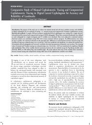
ORIGINAL RESEARCHThe Effect of CPP-ACP on Remineralization ofArtificial Caries like lesions: An Invitro studyYoshaskam Agnihotri*, Namratha Lakshmi Pragada**, Gaurav Patri*, PK Thajuraj †AbstractThe aim of the study was to investigate the efficacy of casein phosphopeptide-amorphous calcium phosphate (CPP-ACP)containing tooth mousse on the remineralization of enamel lesions and to compare its efficacy to fluoride containing toothpaste. Thirty premolar teeth were placed in demineralizing solution for 96 hours to produce artificial caries-like lesions.They were sectioned into half and ground sections were prepared. Samples were randomly assigned into three groups:Group A: Nonfluoridated toothpaste (negative control), Group B: Fluoridated toothpaste and Group C: Tooth Moussecontaining CPP-ACP. Group C showed a significant decrease in lesion depth after the specified treatment followed byGroup B whereas, Group A demonstrated an increase in lesion depth. CPP-ACP containing tooth mousse remineralized initialenamel lesions and showed a higher remineralizing potential than fluoridated toothpaste.Key words: Remineralization, CPP-ACP, demineralizationDental caries a common tooth malady hassignificantly declined over the past fewdecades, largely due to the use of fluorides intoothpastes. Fluoride has been proven to reduce cariesin both the primary and permanent dentitions whenused in a variety of ways. 1In recent years, casein phosphopeptide-amorphouscalcium phosphate (CPP-ACP) nanocomplexeshas demonstrated anticariogenic properties in bothlaboratory animal and human in situ experiments. 2CPP can stabilize calcium phosphate in amorphouscalcium phosphate solution and has been shownin vitro to localize on the tooth surface, preventingdemineralization and helping in remineralization. 3CPP-ACP stabilized calcium phosphate solutions havealso shown remineralization of subsurface lesions andstabilization of free calcium and phosphate ions. CPP-ACP (tooth mousse) has even shown a greater capacityto neutralize acids than fluoridated toothpastes. 4 Theacid resistance of enamel exposed to CPP-ACP wasincreased by the addition of fluoride. 4,5*Senior Lecturer, Dept. of Conservative Dentistry and Endodontics**Senior Lecturer, Dept. of Prosthodontics†Professor and Head, Dept. of Conservative Dentistry and EndodonticsHi-Tech Dental College and Hospital, BhubaneswarAddress for correspondenceDr Yoshaskam AgnihotriE-mail: drlee2@gmail.comThe aim of this study was to investigate the efficacyof CPP-ACP containing tooth mousse on theremineralization of enamel lesions and to compare itsremineralization ability with that of fluoride containingtooth paste.Material and MethodsThirty sound extracted premolars were cleansed of softtissue debris and inspected for cracks, hypoplasia andwhite spot lesions. The teeth were then coated with anail varnish, leaving a narrow window, approximately1 mm wide, on the sound, intact surface of the buccalenamel. 6 Each tooth was subsequently immersed inthe demineralizing solution 7 (2.2 mM CaCl 2, 2.2mM KH 2PO 4, 0.05M acetic acid having pH adjustedto 4.4 and 1 M KOH) for four days to producelesions 120-200 µm deep. 6 The teeth were sectionedlongitudinally through the lesions in two halves andground sections were made which was visualizedunder polarized light microscopy and the depth ofthe lesions was measured using a microtome andimage ‘J’ software.Thirty sections were randomly assigned to threetreatment groups as follows: (1) Group A: Negativecontrol nonfluoridated toothpaste (Dabur promise,India); (2) Group B: Fluoridated toothpaste (Pepsodenttooth paste, Hindustan lever, India); (3) Group C:CPP-ACP as toothpaste (tooth mousse, GC Corp,Tokyo, Japan).366Indian Journal of Multidisciplinary Dentistry, Vol. 2, <strong>Issue</strong> 1, <strong>Nov</strong>ember 2011 to <strong>Jan</strong>uary 2012
original researchToothpaste and tooth mousse supernatants inGroups A, B, C were prepared by suspending 15 gof the respective toothpaste⁄tooth mousse in 45 ml ofdeionized water in order to achieve 1:3 (toothpaste:Deionized water) ratio, these suspensions were thenthoroughly stirred and mechanically agitated. Thesections were placed in the pH cycling system on anorbital shaker for 10 days. 8,9 Each cycle involved threehours of demineralization twice-daily with two hoursof remineralization in between. The remineralizingsolution 7 contained 1.5 mM CaCl 2, 0.9 mM NaH 2PO 4,0.15 M KCL and had a pH of 7.0. Specimens inGroups A, B, and C were treated for 60 seconds withtoothpaste supernatant (5 ml/section) before the firstdemineralizing cycling, and both before and after thesecond demineralizing cycles. After the 10-day pHcycle the nail polish was carefully removed from thespecimens using acetone. Ground sections were revisualizedunder polarized light microscopy. 10After imbibition of the sections in water, polarizedlight microscopy (PLM) was employed to qualitativelyevaluate the body of the lesions in each of the enamelsections. Depth of lesions was measured by usingmicrotome and image ‘J’ software and values werecompared with the previous ones.ResultsThe mean and standard deviation (SD) of pretreatmentlesion depth from each group rangedfrom 0.234 ± 0.043 mm to 0.244 ± 0.066 mm.No statistically significant difference was notedamong these pre-treatment lesion depths (p = 0.9996,ANOVA). The paired ‘t’ test showed that Groups Band C had a significant decrease in lesion depth afterthe specified treatment, whereas Group A demonstrateda significant increase in lesion depths (Table 1).Table 1.GroupsAfterdemineralizationAfterremineralizationChange insize usingpaired ‘t’testGroup A 0.234 ± 0.043 0.262 ± 0.035 +0.028(95% conf.)Group B 0.244 ± 0.039 0.230 ± 0.044 -0.014(95% conf.)Group C 0.242 ± 0.066 0.222 ± 0.050 -0.020(95% conf.)Group AGroup BGroup CFigure 3. Group C, Images of demineralization andRemineralization under polarized light microscope.DiscussionThe recent approach in caries management is thenoninvasive method. 11 Non-cavitated and cavitatedlesions extending up to dentinoenamel junction canbe arrested if the cariogenic challenges of certainmicroenvironment are sufficiently controlled and iftherapeutic agents are applied for tissue healing. 12Professional fluoride-delivery methods, such as gels,varnishes, fluoride releasing materials, are commonlyapplied to remineralize high-risk tooth areas. Bioactiveagents based on milk products have now been developedto release elements that enhance remineralization ofthe enamel and dentine, under cariogenic conditions.This agent (commercially available as Tooth Mousse,GC International, Itabashi-ku, Tokyo, Japan)is based on a nanocomplex of the milk proteinCPP-ACP and has shown to promote remineralizationof the carious lesions in ‘Invitro’ and ‘in vivo’ studiesby maintaining a supersaturated state of enamelmineral. 13 It has been proposed that the anticariogenicmechanism of CPP-ACP is due to localization ofACP at the tooth surface which then buffers the freeIndian Journal of Multidisciplinary Dentistry, Vol. 2, <strong>Issue</strong> 1, <strong>Nov</strong>ember 2011 to <strong>Jan</strong>uary 2012367
original researchcalcium and phosphate ion activities, thereby helpingto maintain a state of supersaturation with respectto the enamel, 14 thus depressing demineralizationand promoting remineralization. Studies have shownthat higher concentration of CPP-ACP elicit higherremineralization. 15,16CPP-ACP can be incorporated into the pelliclein exchange for albumin to inhibit the adherenceof S. mutans and S. sobrinus thus producing bothneutralization and enhancement of remineralization(Schupbach et al). Therefore, CPP-ACP can beexpected to be effective in high-risk children who havenot developed good oral hygiene habits. 17Tooth mousse can be used to prevent root caries asits application prevented demineralization of dentindue to buffering capacity of the agent. Casein buffersplaque acid directly or indirectly through bacterialcatabolism. This agent also releases basic amino acidswhich accept proton ions thus when applied on rootdentin acts as an inert barrier preventing diffusionof protons. This agent also has the ability to releasecalcium thus depressing demineralization. 18CPP-ACP when used in combination with fluoridesshowed better results and lower caries score than whenused individually. Our study also substantiates thatwhen CPP-ACP was used after fluoridated paste thebenefits of both the agents are enhanced.The findings of our study shows that when CPP-ACP was applied, the increase in remineralization anddecrease in lesion depth was greater as compared tofluoridated paste and nonfluoridated paste showed anincrease in lesion depth and demineralization.ConclusionBased on the data obtained it can be concluded thatCPP-ACP effectively decreases the lesion depth betterthan fluoridated toothpaste and nonfluoridated toothpaste which showed no improvement in the lesionsize. Efficiency of remineralization can be increasedwhen CPP-ACP and fluoridated tooth pastes are usedtogether.References1.Wefel JS, Jensen ME, Triolo PT, Faller RV, HoganMM, Bowman WD. De/remineralization from sodiumfluoride dentifrices. Am J Dent 1995;8(4):217-20.2.3.4.5.6.7.8.9.10.11.12.13.14.15.Reynolds EC. Remineralization of enamel subsurfacelesions by casein phosphopeptide-stabilized calciumphosphate solutions. J Dent Res 1997;76(9):1587-95.Iijima Y, Cai F, Shen P, Walker G, Reynolds C, ReynoldsEC. Acid resistance of enamel subsurface lesionsremineralized by a sugar-free chewing gum containingcasein phosphopeptide-amorphous calcium phosphate.Caries Res 2004;38(6):551-6.Kariya S, Sato T, Sakaguchi Y, Yoshii E. Fluoride effecton acid resistance capacity of CPP-ACP containingmaterial. Abstract 2045 - 82nd General Session of theIADR 2004, Honolulu, Hawaii.Yamaguchi K, Miyazaki M, Takamizawa T, Inage H,Moore BK. Effect of CPP-ACP paste on mechanicalproperties of bovine enamel as determined by anultrasonic device. J Dent 2006;34(3):230-6.Kumar VL, Itthagarun A, King NM. The effect ofcasein phosphopeptide-amorphous calcium phosphateon remineralization of artificial caries-like lesions: an invitro study. Aust Dent J 2008;53(1):34-40.ten Cate JM, Duijsters PP. Alternating demineralizationand remineralization of artificial enamel lesions. CariesRes 1982;16(3):201-10.Itthagarun A, Wei SH, Wefel JS. Morphology of initiallesions of enamel treated with different commercialdentifrices using a pH cycling model: scanning electronmicroscopy observations. Int Dent J 1999;49(6):352-60.Itthagarun A, Wei SH, Wefel JS. The effect of differentcommercial dentifrices on enamel lesion progression: anin vitro pH-cycling study. Int Dent J 2000;50(1):21-8.Arends J, ten Bosch JJ. Demineralization andremineralization evaluation techniques. J Dent Res1992;71 Spec No:924-8.Rahiotis C, Vougiouklakis G. Effect of a CPP-ACP agenton the demineralization and remineralization of dentinein vitro. J Dent 2007;35(8):695-8.Burke FJ. From extension for prevention to preventionof extension: (minimal intervention dentistry). DentUpdate 2003;30(9):492-8, 500, 502.Reynolds EC, Cai F, Shen P, Walker GD. Retention inplaque and remineralization of enamel lesions by variousforms of calcium in a mouthrinse or sugar-free chewinggum. J Dent Res 2003;82(3):206-11.Reynolds EC, Cain CJ, Webber FL, Black CL, Riley PF,Johnson IH, et al. Anticariogenicity of calcium phosphatecomplexes of tryptic casein phosphopeptides in the rat. JDent Res 1995;74(6):1272-9.Reynolds EC. The prevention of sub-surfacedemineralization of bovine enamel and change in plaque368Indian Journal of Multidisciplinary Dentistry, Vol. 2, <strong>Issue</strong> 1, <strong>Nov</strong>ember 2011 to <strong>Jan</strong>uary 2012
original research16.composition by casein in an intra-oral model. J Dent Res1987;66(6):1120-7.Schüpb ach P, Neeser JR, Golliard M,Rouvet M, Guggenheim B. Incorporation ofcaseinoglycomacropeptide and caseinophosphopeptideinto the salivary pellicle inhibits adherence of mutansstreptococci. J Dent Res 1996;75(10):1779-88.17. Gagnaire V, Pierre A, Molle D, Leonil J. Phosphopeptidesinteracting with colloidal calcium phosphate isolated bytryptic hydrolysis of bovine casein micelles. J Dairy Res1996;63(3):405-22.Indian Journal of Multidisciplinary Dentistry, Vol. 2, <strong>Issue</strong> 1, <strong>Nov</strong>ember 2011 to <strong>Jan</strong>uary 2012369
ORIGINAL RESEARCHComparative Efficacy Evaluation of Articaine as Buccal Infiltration andLignocaine as IANB in the Mandibular first Molar with Irreversible PulpitisA Subbiya*, AR Pradeepkumar**, P Vivekanandhan*, A Karthick †AbstractIt has been shown that the inferior alveolar nerve block (IANB) has high failure rate especially in patients with irreversiblepulpitis. Newer local anesthetic, 4% articaine has shown superiority over 2% lignocaine when used as a primary buccalinfiltration of the mandibular first molar. This study compared the degree of pulpal anesthesia obtained with 1.7 ml 4%articaine with 1:1,00,000 epinephrine when compared to 1.7 ml 2% lignocaine with 1:2,00,000 as a primary infiltration inmandibular first molar with irreversible pulpitis. Sixty adults aged 18-65 years participated in this study. Twenty-two patientsout of 30 did not experience pain with 4% articaine (success = 73.33%) and 26 out of 30 patients did not experience pain in2% lignocaine group (success = 86.66%). There was no statistically significant difference between the articaine and lignocaineformulation with regard to anesthetic success.Key words: Articaine, lignocaine, irreversible pulpitisThe inferior alveolar nerve block (IANB) isthe most frequently used injection techniquefor achieving local anesthesia for mandibularrestorative and endodontic procedures. However, theinferior alveolar nerve block does not always result insuccessful pulpal anesthesia. 1-4 It has been shown thatthe IANB has high failure rate especially in patientswith irreversible pulpitis. 5-7 Articaine has shownsuperiority over 2% lignocaine when used as a primarybuccal infiltration of the mandibular first molar usingvolumes ranging from ‘0.9 to 3.6 ml. 8-12 Though theexact mechanism of action of articaine’s efficacy is notknown, better penetration of bone owing to smaller sizeof thiophene ring of articaine when compared to thebenzene ring of lignocaine and increased liposolubilityhas been suggested to facilitate better diffusion ofthe anesthetic solution to the teeth. 13 Success rates ofarticaine have ranged from 54 to 87% with an averagerate of 67%. Differences in populations may accountfor the differences among studies. Racial differences*Professor**Senior LecturerDept. of Conservative Dentistry and EndodonticsSree Balaji Dental College and Hospital, Chennai†Professor and Head, Dept. of Conservative Dentistry and EndodonticsThai Moogambigai Dental College and Hospital, ChennaiAddress for CorrespondenceDr A SubbiyaDept. of Conservative Dentistry and Endodontics,Sree Balaji Dental College and Hospital, ChennaiE-mail: drsubbiya@gmail.comin bone mineral density are well-established. Bonemineral density could be a factor that can affect thedissociation of articaine into the mandible. Therefore,the purpose of this study was to compare the degree ofpulpal anesthesia obtained with 1.7 ml 4% articainewith 1:1,00,000 epinephrine when compared to1.7 ml 2% lignocaine with 1:2,00,000 as a primaryinfiltration in mandibular first molar with irreversiblepulpitis in an Indian population.Material and MethodsThe study included 60 subjects who had irreversiblepulpitis in mandibular first molar. None of them weretaking any medication that would alter pain perceptionas determined by a written health history and oralquestioning. Exclusion criteria were subjects youngerthan 18 or older than 65 years of age, allergies to localanesthetics or sulfites, pregnancy, history of significantmedical conditions (American Society of AnesthesiologyClass II or higher), active sites of pathosis in area ofinjection and unable to give an informed consent.The inclusion criteria for the study were active painin a mandibular molar (>54 mm on Heft-Parkervisual analog scale [HP VAS] of 170 mm) withprolonged response to cold testing with an ice stickand an electric pulp tester, absence of any periapicalradiolucency on intraoral periapical radiographs anda vital coronal pulp on gaining access to the pulpchamber. Patients were explained the treatment370Indian Journal of Multidisciplinary Dentistry, Vol. 2, <strong>Issue</strong> 1, <strong>Nov</strong>ember 2011 to <strong>Jan</strong>uary 2012
original researchPlace a mark on the line below to show the amount of pain that you feel0 mm 23 36 54 85 114 144 170 mmNone Faint Weak Mild Moderate Strong Intense MaximumpossibleFigure 1. Heft-Parker visual analog scale used for the assessment of pain.procedure and use of pain scales. Patients markedtheir pre-treatment pain on a 170 mm HP VAS(Fig. 1). To interpret the data, we divided the VASinto the following four categories:• No pain corresponded to 0 mm on the scale.• Mild pain was defined as >0 mm and ≤54 mm.A description of faint, weak and mild was includedin this category.• Moderate pain was defined as >54 mm and
original researchTable 1. Subjects who Experienced AnestheticSuccessTooth No. No. of subjects (n = 60) P valueFirst molarAnesthetic solution4% articaine 2% lidocaine22 (30) 26 (30)0.33**There was no significant difference (p > 0.05) between the 4%articaine (buccal infiltration) and 2% lignocaine (IANB) formulations.The results of the current study confirm the results ofprevious studies showing that 4% articaine was successfulas a buccal infiltration. The success of the infiltration of4% articaine with 1:1,00,000 epinephrine was 73.33%for the first molar when compared to 86.66% for 2%lignocaine with 1:2,00,000 epinephrine as IANB(Table 1). The success of mandibular first molar buccalinfiltrations has been studied by various authors usingasymptomatic subjects with 4% articaine containing1:1,00,000 epinephrine and an electric pulp testerto evaluate pulpal anesthesia. Kanaa et al, 8 Robertsonet al 9 , Jung et al 10 and Corbett et al 11 demonstrated64%, 87%, 54%and 64-70% success rates, respectively,for the buccal infiltration of asymptomatic mandibularfirst molar. Our success rate of 73.33% is similar tothat of Corbett et al but differs from the other authors.The study also differs from the previous study byAggarwal et al 7 where the success rate was only 58%,where buccal infiltration with articaine was in additionto IANB. Though a similar success rate was reported byHaase et al, 13 it was a combination of IANB andsupplemental buccal infiltration with articaine.Although anesthesia of the lower lip on the sideof injection is assumed to be a sign of success ofmandibular nerve anesthesia, patients experiencedpain during access opening despite lip anesthesia.This was similar to the observation in the study byAggarwal et al 7 who reported pain on access openingdespite lip anesthesia. Furthermore, when 2%lignocaine was given as IANB after a failure with4% articaine for patients who consented for theadditional injection, pain was experienced in six outof eight cases similar to the pain on access openingwith 4% articaine. This suggests that lignocaine maynot be successful in most of the cases where articainewould be a failure, though this inference may be takenwith caution as the number of articaine failure casestaken up for lignocaine IANB was limited. Based onthe manufacturer’s maximum recommended dose ofseven cartridges for 4% articaine and a maximum doseof 13 cartridges of a 2% lignocaine for a healthy 70-kgadult this additional dose is within the safety limits. 14As mentioned earlier, success of articaine [(4-methyl-3-[1-oxo-2-(propylamino)-propionamido]-2-thiophenecarboxylicacid methyl ester hydrochloride)] could bebecause it contains a thiophene ring in its moleculeinstead of the benzene ring seen in lignocaine, increasingthe liposolubility of the drug as well as its potency.Robertson and colleagues suggested that buccal infiltrationof articaine might have resulted in penetration of thesolution through the mental foramen, leading to thehigher success rates in the premolars and first molar. Buta higher success rate can be expected in the premolars andfirst molar than in the second molar for both articaineand lignocaine formulations. This is because of a relativelythicker bone in the buccal aspect of second molar regionwhich may prevent anesthetic diffusion.Within the limitations of this study it can be concludedthat 4% articaine with 1:1,00,000 adrenaline canbe considered as an alternative for anesthetisingmandibular first molar instead of IANB with 2%lignocaine with 1:2,00,000 adrenaline.References1.2.3.4.5.6.Nusstein J, Reader A, Nist R, Beck M, Meyers WJ.Anesthetic efficacy of the supplemental intraosseousinjection of 2% lidocaine with 1:100,000 epinephrine inirreversible pulpitis. J Endod 1998;24(7):487-91.Reisman D, Reader A, Nist R, Beck M, Weaver J.Anesthetic efficacy of the supplemental intraosseousinjection of 3% mepivacaine in irreversible pulpitis.Oral Surg Oral Med Oral Pathol Oral Radiol Endod1997;84(6):676-82.Cohen HP, Cha BY, Spångberg LS. Endodonticanesthesia in mandibular molars: a clinical study. JEndod 1993;19(7):370-3.Kennedy S, Reader A, Nusstein J, Beck M, Weaver J. Thesignificance of needle deflection in success of the inferioralveolar nerve block in patients with irreversible pulpitis.J Endod 2003;29(10):630-3.Tortamano IP, Siviero M, Costa CG, Buscariolo IA,Armonia PL. A comparison of the anesthetic efficacyof articaine and lidocaine in patients with irreversiblepulpitis. J Endod 2009;35(2):165-8.Claffey E, Reader A, Nusstein J, Beck M, Weaver J.Anesthetic efficacy of articaine for inferior alveolar nerveblocks in patients with irreversible pulpitis. J Endod2004;30(8):568-71.372Indian Journal of Multidisciplinary Dentistry, Vol. 2, <strong>Issue</strong> 1, <strong>Nov</strong>ember 2011 to <strong>Jan</strong>uary 2012
original research7.8.9.10.11.Aggarwal V, Jain A, Kabi D. Anesthetic efficacyof supplemental buccal and lingual infiltrations ofarticaine and lidocaine after an inferior alveolar nerveblock in patients with irreversible pulpitis. J Endod2009;35(7):925-9.Kanaa MD, Whitworth JM, Corbett IP, Meechan JG.Articaine and lidocaine mandibular buccal infiltrationanesthesia: a prospective randomized double-blind crossoverstudy. J Endod 2006;32(4):296-8.Robertson D, Nusstein J, Reader A, Beck M, McCartneyM. The anesthetic efficacy of articaine in buccalinfiltration of mandibular posterior teeth. J Am DentAssoc 2007;138(8):1104-12.Jung IY, Kim JH, Kim ES, Lee CY, Lee SJ. An evaluationof buccal infiltrations and inferior alveolar nerve blocksin pulpal anesthesia for mandibular first molars. J Endod2008;34(1):11-3.Corbett IP, Kanaa MD, Whitworth JM, Meechan JG.Articaine infiltration for anesthesia of mandibular firstmolars. J Endod 2008;34(5):514-8.12.13.14.15.16.Abdulwahab M, Boynes S, Moore P, Seifikar S, Al-Jazzaf A, Alshuraidah A, et al. The efficacy of six localanesthetic formulations used for posterior mandibularbuccal infiltration anesthesia. J Am Dent Assoc2009;140(8):1018-24.Haase A, Reader A, Nusstein J, Beck M, Drum M.Comparing anesthetic efficacy of articaine versuslidocaine as a supplemental buccal infiltration of themandibular first molar after an inferior alveolar nerveblock. J Am Dent Assoc 2008;139(9):1228-35.Katyal V. The efficacy and safety of articaine versuslignocaine in dental treatments: a meta-analysis.J Dent2010;38(4):307-17.Patni R. ormal BMD values for Indian females aged 20-80 years. J Midlife Health 2010;1(2):70-3.Melamed A, Vittinghoff E, Sriram U, SchwartzAV, Kanaya AM.BMD reference standards amongSouth Asians in the United States.J Clin Densitom2010;13(4):379-84.Indian Journal of Multidisciplinary Dentistry, Vol. 2, <strong>Issue</strong> 1, <strong>Nov</strong>ember 2011 to <strong>Jan</strong>uary 2012373
Review artclePharmacovigilance: A tool for health safetyN S Muthiah*, M Elumalai**, N P Murali † , Ramsundar Hazra ‡AbstractPharmacovigilance is the pharmacological science activities relating to the detection, assessment, understanding and preventionof adverse effects, particularly chronic and acute side effects of medicines. The aim of pharmacovigilance is to improvepublic health and safety, to contribute to the assessment of benefit, harm, effectiveness and risk of medicines, to promoteunderstanding,education and clinical training.Key words: Health safety, pharmacovigilance, drugsPharmacovigilance is an important and integral partof clinical research and these days it is growing inmany countries. 1 A number of researchers havestudied about pharmacovigilance. 2-4 Recently, its concernshave been widened to include herbals, traditional andcomplementary medicines, blood products, biologicals,medical devices and vaccines. 5 This applies throughoutthe life cycle of a medicine equally to the pre-approvalstage as to the post-approval.The scope of pharmacovigilance is to improve patientcare and safety in relation to the use of medicines, andall medical and paramedical interventions. Improvepublic health and safety in relation to the use ofmedicines. Contribute to the assessment of benefit,harm, effectiveness and risk of medicines, encouragingtheir safe, rational and more effective (including costeffective)use, and promote understanding, educationand clinical training in pharmacovigilance and itseffective communication to the public.Adverse Drug ReactionA response to a drug which is noxious and unintended,and which occurs at doses normally used in man forthe prophylaxis, diagnosis, or therapy of disease, or for*ProfessorDept. of Pharmacology, Sree Balaji Medical College and Hospital, Chennai**Associate Professor†Lecturer‡UG StudentDept. of Pharmacology, Sree Balaji Dental College and Hospital, ChennaiAddress for correspondenceDr NS MuthiahE-mail: nsm.healingtouch@gmail.comthe modification of physiological function. Montastrucet al 6 have been studied to characterize the profile ofadverse drug reactions (ADRs) reported with selegiline,a monoamine oxidase B (MAO-B) inhibitor used inthe treatment of Parkinson’s disease.Adverse EventAny untoward medical occurrence that may presentduring treatment with a pharmaceutical product butwhich does not necessarily have a causal relationshipwith this treatment.Side EffectAny unintended effect of a pharmaceutical productoccurring at doses normally used in man which isrelated to the pharmacological properties of the drug.Serious ADRsA serious adverse event (experience) or reactionis any untoward medical occurrence that at anydose: Results in death, is life-threatening, requiresinpatient hospitalization of prolongation of existinghospitalization, is a congenital anomaly/birthdefect.Unexpected Adverse ReactionAn adverse reaction, the nature, severity or outcome ofwhich is not consistent with the summary of productcharacteristics.Adverse ReactionsIntrinsic factors of the drug374Indian Journal of Multidisciplinary Dentistry, Vol. 2, <strong>Issue</strong> 1, <strong>Nov</strong>ember 2011 to <strong>Jan</strong>uary 2012
Review ArticlePharmacological, idiosyncratic, carcinogenicity,mutagenicity, teratogenicityExtrinsic FactorsAdulterants, contamination, underlying medicalconditions, interactions, wrong usageNeed for PharmacovigilanceReason 1: Humanitarian concern - Insufficient evidenceof safety from clinical trials Animal experiments Phase1-3 studies prior to marketing authorization.Reason 2: Medicines are supposed to save lives Dyingfrom a disease is sometimes unavoidable; dying from amedicine is unacceptable.Reason 3: ADR-related cost to the country exceeds thecost of the medications themselves.Reason 4: Promoting rational use of medicines andadherence.Reason 5: Ensuring public confidence.Reason 6: Ethics, to know of something that is harmfulto another person who does not know, and not telling,is unethical.What should be Reported• New drugs. Report all suspected reactions includingminor ones. For established or well known drugs.If serious, unexpected, unusual ADRsActive Ingredients WithdrawnThalidomide (1961)Congenital limb defectsBenoxaprofen (1982) HepatotoxicityPhenformin (1982)Lactic acidosisFenfluramine (1997)Heart-valve abnormalitiesAstemizoleMany drug interactionsPhenylpropanolamine (2000) Hemorragic strokeKava KavaLiver abnormalitiesCerivastatinRhabdomyolysisCisaprideCardiac arrhythmiasRofecoxib (2004)Cardiovascular eventsValdecoxib (2005)Cardiovascular events,serious skin reactionsComfrey, SenecioNephrotoxicityTegaserod (2007)Cardiovascular eventsClobutinol (2007)Cardiac arrhythmia••Change in frequency of a given reactionADRs to generics not seen with innovator products,ADRs to traditional medicines.• All suspected drug-drug, drug-food, drug-foodsupplement interactions.• Statement highlighting marine source ofsupplements such as glucosamine so that can beavoided by those with allergy to sea food.• ADRs associated with drug withdrawals, ADRsdue to medication errors.• ADRs due to lack of efficacy or suspectedpharmaceutical defects.Innovator ProductsLimited information available at time when drugis first marketed. Conduct intensive monitoring toidentify new, unlabeled adverse reactions, monitor for‘rare’ reactions. Provide updates to prescribers on newfindings, labeling changes, safety issues.Generic ProductsMonitor efficacy, monitor adverse effect profile to studydifferences in ADR pattern with respect to innovatorproducts. Help in improving quality of generics usedwhether the problem arose due to ADR or qualitydefects.WHO Programmed for International DrugMonitoringStarted 1968 Located in Uppsala, Sweden Collaboratingcenter for maintaining global ADR database -Roles of WHO Collaborating CentreIdentify early warning signals of serious adverse reactionsto medicines. Evaluate the hazard. Undertake researchinto the mechanisms of action to aid the developmentof safer and more effective medicines.Pharmacovigilance in IndiaPharmacovigilance is fastest emerging as an importantapproach for the early detection of unwanted effects ofthe drugs and to take appropriate regulatory actions ifnecessary. National Pharmacovigilance Centre CDSCOhas initiated a country-wide pharmacovigilanceprogram under the aegis of DGHS, Ministry of Healthand Family Welfare Government of India.Indian Journal of Multidisciplinary Dentistry, Vol. 2, <strong>Issue</strong> 1, <strong>Nov</strong>ember 2011 to <strong>Jan</strong>uary 2012375
Review ArticleNational Pharmacovigilance ProgrammeThe Program aims to faster the culture of ADRnotification in its first year of operation andsubsequently aims to generate broad-based ADR dataon the Indian population. Sponsored and coordinatedby the country’s central drug regulatory agency -(CDSCO). Peripheral Pharmacovigilance Centre(PPCs). Regional Pharmacovigilance Centers (RPCs).Zonal Pharmacovigilance Centre (ZPCs).“So…. What is our role?Send not only quantity but …. Quality reportsHow?”Monitor clinical status of patients, identify the correctADRs not side effects, get more information, investigateat hospital level, help doctors to fill-up the forms, keeppatient’s record if more information needed.ConclusionPharmacovigilance looks at all available information toassess the safety profile of a drug. Pharmacovigilanceshould also take the benefit of the drug in account.Pharmacovigilance required for systematicallyidentifying and correlating drugs and side effects andtaking corrective actions, especially for the productlaunching first time in India.References1.2.3.4.5.6.Jeetu G, Anusha G. Pharmacovigilance: a worldwidemaster key for drug safety monitoring. J Young Pharm2010;2(3):315-20.Prakash B, Singh G. Pharmacovigilance: scope for adermatologist. Indian J Dermatol 2011;56(5):490-3.Chavant F, Favrelière S, Lafay-Chebassier C, Plazanet C,Pérault-Pochat MC. Memory disorders associated withconsumption of drugs: updating through a case/noncasestudy in the French PharmacoVigilance Database. Br JClin Pharmacol 2011;72(6):898-904.Rahman SZ, Khan RA, Gupta V, Uddin M.Pharmacoenvironmentology - a component ofpharmacovigilance. Environ Health 2007;6:20.WHO (2002). Source: The Importance ofPharmacovigilance.Montastruc JL, Chaumerliac C, Desboeuf K, ManikaM, Bagheri H, Rascol O, et al. Adverse drug reactionsto selegiline: a review of the French pharmacovigilancedatabase. Clin Neuropharmacol 2000;23(5):271-5.376Indian Journal of Multidisciplinary Dentistry, Vol. 2, <strong>Issue</strong> 1, <strong>Nov</strong>ember 2011 to <strong>Jan</strong>uary 2012
Review artcleSialolith: A Case Report with Review of LiteraturePE Chandra Mouli*, S Manoj Kumar**, S Kailasam † , S Shanmugam † , S Satish ‡AbstractSialoliths are calcified organic matter that forms within the secretory system of the major salivary glands. Salivary gland calculiaccount for the most common disease of the salivary glands, and may range from tiny particles to several centimeters inlength. The majority of sialoliths occur in the submandibular gland or its duct and is a common cause of acute and chronicinfections. While the majority of salivary stones are asymptomatic or cause minimal discomfort, larger stones may interferewith the flow of saliva and cause pain and swelling. The prevalence of sialoliths varies by location. Sialolith in the parotidglands is less common when compared with that of submandibular gland. This case report describes a patient presenting withsubmandibular gland sialolith and review of the literature regarding the salivary sialothiasis.Key words: Submandibular gland; sialolith; nidus.The deposition of calcium salts, primarily calciumphosphate, usually occurs in the skeleton.When, it occurs in an unorganized fashion insoft tissue, it is referred to as heterotopic calcification.Heterotopic calcification which results from depositionof calcium in normal tissue despite normal serumcalcium and phosphate levels is known as idiopathiccalcification. Sialoliths belongs to the category ofidiopathic calcification. 1 Sialoliths are calcareousdeposits in the ducts of major or minor salivary glandsor within the glands themselves. Sialolithiasis accountsfor more than 50% of diseases of the major salivaryglands and is thus the most common cause of acuteand chronic infections. 2Case ReportMr. Nagarajan aged 44 years came to Ragas DentalCollege with a chief complaint of pain on the left sidebelow the tongue region for the past three weeks. Pain*Senior Lecturer**Professor†Professor and Head‡ProfessorDept. of Oral Medicine and Radiology, Ragas Dental College and HospitalUthandi, Chennai#Senior Lecturer, Dept. of Oral Medicine and RadiologyChettinadu College of Dental Sciences, ChennaiAddress for correspondenceDr PE Chandra MouliSenior Lecturer, Dept. of Oral Medicine and Radiology, Ragas Dental Collegeand Hospital, 2/102, East Coast Road, Uthandi, Chennai - 600 119E-mail: mouli_7777@yahoo.co.inis severe and intermittent. Pain is seen during mealtimeand reduces after half an hour by itself (Fig. 1).On clinical examination, at the left floor of the mouth,at submandibular gland, at the level of first molar, thereis a presence of a mass, measuring l × l cm in size andround in shape with well defined borders. The mass ishard in consistency and tender on palpation.Orthopantomogram revealed (OPG), presence of radioopaquemass seen in the left body of the mandible atsubmandibular fossa, measuring around 1 × 2 cm insize, oval in shape extending superiorly from 1 cmbelow the 35, 36 tooth and inferiorly to the lowerborder of the mandible (Fig. 2).Mandibular occlusal radiograph revealed presence ofradio-opaque mass seen in the left body of the mandibleat submandibular fossa, measuring around 1 × 2 cmin size, oval in shape extending from anterior aspect of35 to the distal aspect of 36 (Fig. 2).Ultrasound showed an irregular border measuringabout 44 × 46 × 57 cm (Fig. 3).Complete excision of the left submandibular sialolithwas done under local anesthesia, sutures placed. Postsurgical antibiotic regimen was given and healing wassatisfactory (Figs. 4 and 5).Microscopically, the mass shows concentric laminationsaround a central nidus of amorphous debris. Basedon history, clinical examination, radiographic andIndian Journal of Multidisciplinary Dentistry, Vol. 2, <strong>Issue</strong> 1, <strong>Nov</strong>ember 2011 to <strong>Jan</strong>uary 2012377
Review ArticleFigure 1. Swelling present in left floor of the mouthFigure 2. OPG and Mandibular occlusal view showing aradio-opaque mass below 35, 36 region.(a)(b)Figure 4. (a) Surgically removed sialolith and (b) sialolithseen on an IOPA radiographic film.Figure 3. Ultrasound showed an irregular border measuringabout 44 x 46 x 57 cm.Figure 5. OPG and mandibular occlusal view after removalof radio-opaque mass (sialolith) below 35, 36 region.microscopic features, the condition was finally diagnosedas sialolithiasis - left submandibular salivary gland.DiscussionSialolithiasis is the most common disease of salivaryglands. It is estimated that it affects 12 in 1000 of theadult population. 3 Males are affected twice as much asfemales. 4 It involves most commonly the major salivaryglands. More than 80% of the sialoliths occur in thesubmandibular gland or its duct, 6% in the parotidgland and 2% in the sublingual gland or minor salivaryglands. 2The exact etiology and pathogenesis of salivary calculiis unknown. They are thought to occur as a result ofdeposition of calcium salts around an initial organicnidus consisting of altered salivary mucins, bacteriaand desquamated epithelial cells. 4,5According to the literature, formation of sialolithcan occur in two phases: A central core and alayered periphery. 6 The central core is formed by theprecipitation of salts, which are bound by certainorganic substances. The second phase consists of thelayered deposition of organic and inorganic material. 7Parotid stones are thought to form most often around anidus of inflammatory cells or a foreign body 8 whereassubmandibular stones are thought to form around anidus of mucous. Another theory has proposed thatan unknown metabolic phenomenon can increase thesalivary bicarbonate content, which alters calciumphosphate solubility leads to precipitation of calciumand phosphate ions. 9 A retrograde theory proposedfor sialolithiasis suggested that, substances or bacteriawithin the oral cavity might migrate into the salivaryducts and become the nidus for further calcification. 6Salivary stagnation, increased alkalinity of saliva,infection or inflammation of the salivary duct orgland, and physical trauma to salivary duct or glandmay predispose to calculus formation. 3Clinically, sialoliths are round or ovoid in shape,rough or smooth in texture and yellowish in color.Submandibular stones consist of 82% inorganic378Indian Journal of Multidisciplinary Dentistry, Vol. 2, <strong>Issue</strong> 1, <strong>Nov</strong>ember 2011 to <strong>Jan</strong>uary 2012
Review Articlematerial and 18% organic material, whereas parotidstones are composed of 49% inorganic and 51% organicmaterial. 2 The inorganic material comprises of calciumphosphate, smaller amounts of carbonates in the formof hydroxyapatite and smaller amounts of magnesium,potassium, ammonia, whereas organic material consistsof various carbohydrates and amino acids. 9Sialoliths are usually unilateral. Sialolithiasis typicallycauses pain and swelling of the involved salivarygland by obstructing the salivary flow. Calculi maycause stasis of saliva, leading to bacterial ascent intothe parenchyma of the gland resulting in sialadenitis.Some sialoliths may be asymptomatic. Long-termobstruction, in the absence of infection can lead toatrophy of the gland with resultant lack of secretoryfunction and ultimately fibrosis. 9Careful history and examination are important in thediagnosis of sialolithiasis. Pain and swelling of theconcerned gland at mealtimes and in response to othersalivary stimuli are important. Complete obstructioncauses constant pain, swelling and signs of systemicinfection may be present. 10Bimanual palpation of the floor of the mouth, in aposterior to anterior direction, may reveal a palpablestone in majority of the cases of submandibular calculi.For parotid stones, careful intraoral palpation aroundStenson’s duct orifice may reveal a stone. 9 Deeper parotidstones are often not palpable. When minor salivary glandsare involved they are usually in the buccal mucosa or upperlip, forming a firm nodule that may mimic tumor.Imaging modalities, both conventional and advancedare very useful in diagnosing sialolithiasis. Fortypercent of parotid and 20% of submandibular stonesare usually radiolucent. In such patients sialographywill be helpful. However, it is contraindicated in acuteinfections or in patients having allergy to the contrastagents. 9Patients presenting with sialolithiasis may benefitfrom conservative management, especially if the stoneis small. 9 The patient must be well-hydrated and theclinician must apply moist warm heat and along withmassage of the gland.Sialogogues are useful to promote production of salivaand to flush the stone out of the duct. In case ofsialoliths associated with sialadenitis, a penicillinaseresistantantistaphylococcal antibiotic will be preferable.Most stones will respond to such a regimen, combinedwith simple sialolithotomy when required. 8,10Alternative methods of treatment have emerged such asthe use of extracorporeal shock wave lithotripsy (ESWL)and more recently the use of endoscopic intracorporealshock wave lithotripsy (EISWL), in which shockwavesare delivered directly to the surface of the stone lodgedwithin the duct without damaging adjacent tissue(piezoelectric principle). Salivary lithotripsy will bemore useful therapeutically than surgical removal ofthe affected gland, as it prevents the risk of a generalanesthesia, facial nerve damage, surgical scar, Frey’ssyndrome, and causes little discomfort to the patientwith preservation of the gland. 11References1.2.3.4.5.6.7.8.9.10.11.White SC, Pharoah MJ. Oral radiology principles andinterpretation. Chapter 27. In: Soft Tissue Calcificationand Ossification. Mosby, Missouri 2004:p597-614.Zenk J, Benzel W, Iro H. New modalities in themanagement of human sialolithiasis. Minimal InvasTher Allied Technol 1994;3(5):275-84.Leung AK, Choi MC, Wagner GA. Multiple sialolithsand a sialolith of unusual size in the submandibularduct: a case report. Oral Surg Oral Med Oral Pathol OralRadiol Endod 1999;87(3):331-3.Cawson RA, Odell EW. Essentials of oral pathologyand oral medicine. 6th edition, Churchill Livingstone:Edinburgh 1998:p239-40.Carr SJ. Sialolith of unusual size and configuration.Report of a case. Oral Surg Oral Med Oral Pathol1965;20(6):709-12.Marchal F, Kurt AM, Dulguerov P, Lehmann W.Retrograde theory in sialolithiasis formation. ArchOtolaryngol Head Neck Surg 2001;127(1):66-8.Rauch S, Gorlin R J. Disease of the salivary glands. In:Thomas’ Oral Pathology. Gorlin RJ, Goldmann HM(Eds.), Mosby-Year Book Inc: St Loius, Mo 1970:p997-1003.Pietz DM, Bach DE. Submandibular sialolithiasis. GenDent 1987;35(6):494-6.Williams MF. Sialolithiasis. Otolaryngol Clin North Am1999;32(5):819-34.Pollack CV Jr, Severance HW Jr. Sialolithisis: case studiesand review. J Emerg Med 1990;8:561-5.Iro H, Schneider HT, Födra C, Waitz G, Nitsche N,Heinritz HH, et al. Shockwave lithotripsy of salivaryduct stones. Lancet 1992;339(8805):1333-6.Indian Journal of Multidisciplinary Dentistry, Vol. 2, <strong>Issue</strong> 1, <strong>Nov</strong>ember 2011 to <strong>Jan</strong>uary 2012379
Review articleMyth of Endodontics in Oral Focal InfectionJamuna Indramohan*, B Karthika**, Gouse Mohiddin †AbstractOn clinical evidence there has been a belief in the past amongst the medical and dental practitioners that the presence ofbad teeth in the mouth can be a cause of some systemic diseases of unknown etiology. Examples of systemic conditions inthe above category include rheumatoid arthritis, some diseases of the eye, few cardiac conditions and some diseases of thegastrointestinal region. 1 It was felt that a circumscribed area infected with micro organisms due to dentoalveolar or periapicalabscess which may or may not give rise to clinical manifestation can initiate another infection in a distant organ throughthe blood stream or the lymph channels. Based on this ‘focal infection theory’, all pulpless or non-vital teeth were extractedhoping that the diseasae and symptoms will abate. But it was observed that the systemic disease continued in many casesafter removal of the infected teeth. 2 Aim of this article is to emphasize the current concepts which advocate the belief thatwith increasing knowledge, the number of conditions considered to be due to focal infection is decreasing and also disclosethe myth in relation between endodontic treatment and oral focal infection.Key words: Endodontics, focal infection, focus of infection, sepsisThe concept that oral conditions cansignificantly influence events elsewhere inthe body is not new, but it has undergone anumber of iterations over the years. Oral foci havetraditionally been ascribed to periodontitis, alveolarabscesses, cellulitis, pulpless teeth, apical periodontitis,general oral sepsis and endodontically-treated teethwith viridians group streptococci being the principalmetastatic microbial culprits.A frequently cited early publication is an 1891 reportby Miller entitled “The Human Mouth as a Focus ofInfection.” Miller was highly attuned to the role ofbacteria in disease causation, as he was working inthe laboratory of Robert Koch, whose postulates wereused to establish the microbial etiologies of infectiousdiseases. 3*Professor, Dept. of Conservative Dentistry and EndodonticsThai Moogambigai Dental College and Hospital, Chennai**Senior Lecturer, Dept. of Oral Medicine and RadiologyPriyadarshini Dental College and Hospital, Thiruvallur†Reader, Dept. of Oral Pathology, Kalinga Dental college, BhuvaneswarAddress for correspondenceDr Jamuna IndramohanProfessorThai Moogambigai Dental College and HospitalGolden George Nagar, Mogappair, ChennaiHistoryThe journey began in 1674 when Antony vonLeeuwenhoek discovered microbes. He was an early userof the microscope and analyzed small scrapings fromteeth. He described small ‘animalcules,’ which later werenamed microbes and we call bacteria. Two hundred yearslater in 1876, Robert Koch proposed the ‘germ theory ofdisease,’ suggesting that bacteria may cause disease. At thesame time, Edward Jenner, Joseph Lister and Louis Pasteuralso implicated germs as a possible source of disease.In 1879, Willoughby D. Miller, a recent graduate ofthe University of Pennsylvania Dental School, heardof Koch’s theory that germs might cause disease anddetermined that he too wanted to study bacteria.On completing his dental training, he traveled to Berlinwhere he began work within Koch’s institute, looking atthe relationship of bacteria to disease. 4 Miller becameconvinced that the mouth was a focus of infection andthat bacteria in the mouth could explain most ofhumankind’s illnesses and gave a speech on ‘OralInfection as a Cause of Systemic Disease.’ By 1911, theterm oral sepsis was replaced with the term focal infectionand the ‘era of focal infection’ was launched. 5Focal Infection TheoryA focus of infection is a confined area that containspathogenic microorganisms, can occur anywhere in the380Indian Journal of Multidisciplinary Dentistry, Vol. 2, <strong>Issue</strong> 1, <strong>Nov</strong>ember 2011 to <strong>Jan</strong>uary 2012
Review Articlebody and usually causes no clinical manifestations. Afocal infection is a localized or generalized infectioncaused by the dissemination of microorganisms or toxicproducts from a focus of infection. 6 These conceptshave led to the Focal Theory of Infection (or Theoryof Focal Infection) that postulates a myriad of diseasescaused by microorganisms (bacteria, fungi, viruses)that arise endogenously from a focus of infection.Miller proposed a role for oral microorganisms or theirproducts in the development of a variety of diseasesin sites removed from the oral cavity, including brainabscesses, pulmonary diseases and gastric problems,as well as a number of systemic infectious diseases. 7The role of oral sepsis as a cause of systemic diseasewas championed by William Hunter, a prominentBritish physician, in a publication and a 1910 talkat McGill University, Montreal. He spoke, withconsiderable hyperbole, of dental restorations “built in,on, and around diseased teeth which form a veritablemausoleum of gold over a mass of sepsis to which thereis no parallel in the whole realm of medicine.” In 1919,Rosenow published a series of animal experimentsand human case reports supporting the concept offocal infection. He emphasized the importance ofcooperation between dentists and physicians, as wellas the necessity of ensuring that the focus of infectionis eliminated completely, and he noted that toothextraction by itself might not be sufficient. Much of theevidence presented in support of the concept of focalinfection proved, on closer inspection, to be anecdotalor of questionable scientific merit. Nevertheless, itbecame common practice in olden days to extractall endodontically or periodontally involved teethto eliminate any possible foci of infection, with theexpectation that this would prevent or cure a wholehost of local or systemic problems. 8Endodontics and Focal InfectionNumerous studies have attempted to determine thesignificance of various microbial pathogens in pulpaland periapical infections. Efforts have been hamperedby small sample sizes, lack of randomization or useof consecutive cases, varied case definitions and lackof documentation regarding the presence/absence ofdental caries and periodontal disease, different expertisein culturing techniques, varied health status of patientsand potential microbial contamination during samplingprocedures. 9 In spite of these difficulties, sufficient dataexist to establish that all orofacial infections of whateverorigin share common major microbial pathogens:Viridans group streptococci, Porphyromonas gingivalis,Prevotella intermedia, Veillonella, Fusobacteriumnucleatum, Peptostreptococcus micros, Bacteroidesforsythus, Eubacteria, Lactobacilli and Actinomyces.Oral pathogens with possibly greater relevance topulpal pathology include Dialister pneumosintes andEubacterium and Prevotella endodontalis. The relativeimportance of these pathogens in pulpal, periapicaland periodontal infections or pericoronitis, periimplantitisand infectious spread to contiguous areas(orbital, submandibular, mediastinal) are primarilyquantitative rather than qualitative. Any orofacialinfection spreading rapidly is likely to have a substantialviridans group streptococci component. The preciserisk of bacteremia associated with endodontic lesionsand therapy is subject to some controversy. Apparentlyno study exists that delineates the incidence/magnitudeof spontaneous bacteremias from neither infected rootcanals with chronic periradicular lesions nor any withacute periodontal abscesses. 10 Such bacteremias mayoccur during the management of infected root canalsand a good understanding of their incidence/magnitudewould be of importance.Bender et al determined a 0-15% incidence ofbacteremia with none if the instrumentation remainedwithin the canal and 15% if it extended beyond theapex. Baumgartner et al found a 3.3% incidence withnonsurgical endodontics and a 83-100% incidencewith surgical endodontics. In a study that intentionallyinstrumented beyond the apex, a 34-54% incidence ofbacteremia was detected. Al-Karaawi et al determinedthat the ‘cumulative’ bacteremias with a rubber damclamp in children was 175 times greater than a toothextraction, while a matrix band was only four timesgreater which conflicted with another study by thesame group that the incidence of bacteremia using arubber dam/wedge/matrix band model was 9-32%. 11One of the difficulties with comparing any given dentalprocedure using cumulative data to dental extractionsis that no determination has ever been made of howlong dental extraction sites produce bacteremiasduring their healing phase. Whether instrumentationhas occurred beyond the apex may not be readilydetermined and antibiotic prophylaxis for endocarditisprevention would be appropriate if the best clinicalIndian Journal of Multidisciplinary Dentistry, Vol. 2, <strong>Issue</strong> 1, <strong>Nov</strong>ember 2011 to <strong>Jan</strong>uary 2012381
Review Articlejudgment of the dentist is that such a determinationcannot be made. The question of bacteremias arisingfrom rubber dam application should be clarifiedas the degree of trauma associated with its use is alikely variable. It is reasonable to conclude fromthe above data that nonsurgical endodontics is maybe the least likely of dental treatment procedures toproduce significant bacteremias in either incidence ormagnitude.It is claimed that endodontically treated teeth are always‘infected’ as it may be impossible to fill all lateral andaccessory canals or eliminate the ‘slime’ layers on rootcanal surfaces. Whether this criticism is accurate ornot may be irrelevant as it does not recognize basicmicrobiological principles of the inoculum effect(the threshold level of bacteria necessary to producean infection), that the presence of bacteria does notper se define an active infectious process and that mostmicroorganisms associated with the human body areeither innocuous or beneficial. 12Scientific ApproachBy about 1930, the validity of the focal infection theorybegan to be questioned, and investigators found, whenthey considered the available real outcome data, thatthere was no clear basis for ascribing the occurrence ofmuch systemic disease to the presence of oral fociof infection. 10 As a result, the focus of dental practicechanged such that restorative dental procedures reemergedas the mainstay of most dental treatmentplans due to the availability of successful methods oftreating endodontic lesions. The oral microorganismscould in some way be responsible for diseases thathad a rather uncertain etiology. 12 In considering theexisting data, it is important to differentiate betweenthose data supporting an association between twodiseases or conditions and those indicating a causalrelationship, so that the information can be interpretedaccurately.ConclusionStudies must be performed to determine if endodontictreatment in causing focal infection is inferior or not.To date, these studies have not been performed andthere is no evidence to support the theory that modernendodontic therapy is not safe and effective.References1.2.3.4.5.6.7.8.9.10.11.12.O’Reilly PG, Claffey NM. A history of oral sepsis as acause of disease. Periodontol 2000 2000;23:13-8.Miller WD. The human mouth as a focus of infection.Dental Cosmos 1891;33(9):689-706.Hunter W. Oral sepsis as a cause of disease. Br Med J1900;1:215-6.Hunter W. The role of sepsis and antisepsis in medicine.Lancet 1910;1:79-86.Mayo CH. Focal infection of dental origin. DentalCosmos 1922;64:1206-8.Cecil RL, Angevine DM. Clinical and experimentalobservations on focal infection with an analysis of200 cases of rheumatoid arthritis. Ann Intern Med1938;12:577-84.Editorial. Focal infection. J Am Med Assoc 1952;4:150:490-1.Mattila KJ, Nieminen MS, Valtonen VV, Rasi VP,Kesäniemi YA, Syrjälä SL, et al. Association betweendental health and acute myocardial infarction. BMJ1989;298(6676):779-81.DeStefano F, Anda RF, Kahn HS, Williamson DF, RussellCM. Dental disease and risk of coronary heart diseaseand mortality. BMJ 1993;306(6879):688-91.Pallasch TJ, Wahl MJ. Focal infection: new age or ancienthistory? Endod Top 2003;4(1):32-45.Barnett ML. The oral-systemic disease connection:an update for the practicing dentist. J Am Dent Assoc2006;137 Suppl 2:5S-6S.Cugadasan V. Oral sepsis and focal infection. SingaporeMed J 1980;21(6):763-5.382Indian Journal of Multidisciplinary Dentistry, Vol. 2, <strong>Issue</strong> 1, <strong>Nov</strong>ember 2011 to <strong>Jan</strong>uary 2012
Review articleThe Scope and Limitations of Adult OrthodonticsNazeer Ahmed Meeran*, Madhuri**, MF Jaseema Parveen †AbstractThe increased demand for orthodontic treatment by adults has increased the scope of orthodontics and widened the upperage limit for orthodontic intervention. The main reason for this demand is the increasing patient awareness and the desire toimprove the facial esthetics. The necessity for tooth repositioning in ideal axial inclination to facilitate prosthetic replacementis also another reason for seeking treatment. The marked limitation is the lack of growth in adults, which reduces the scopefor functional orthopedic intervention. Skeletal discrepancies have to be corrected by orthognathic surgery. The orthodontictreatment is limited to tooth movement and related to remodeling of the alveolar process only. The limitations of orthodontictreatment must be explained at the beginning of treatment, since adult expectations of orthodontics can be very high. It ishighly necessary to identify the expectations of this group of patients, in order to arrive at a realistic treatment plan. Thepurpose of this article is to review the scope, effectiveness and limitations of orthodontic treatment in adult patients.Key words: Adult orthodontics, root resorption, temporomandibular disordersAccording to Ackerman, 1 adult orthodontics isdefined as ‘The branch of orthodontics concernedwith striking a balance between achieving optimalproximal and occlusal contact of the teeth, acceptabledentofacial esthetics, normal function and reasonablestability”.The number of adults seeking orthodontic treatmenthas increased considerably in the last 20 years. They fallinto two different groups: 1 younger adults (under 35,often in their 20’) who desired, but could not receiveorthodontic treatment during adolescent period. 2An older group, typically in their 40’s or 50’s who haveother dental problems and need orthodontics as partof larger treatment plan. The major finding in adultpatient is that they are more concerned about improvingtheir appearance and social acceptance than function.It has been proved that orthodontic treatment, besidesimproving dental esthetics, also has a significant impacton the psychosocial aspect of the patients’ life. 2 It has alsobeen estimated that about 80% of orthodontic patientsseek treatment out of esthetic concerns rather than forhealth and function. 3 In general, many adults have not*Assistant Professor**Professor and Head, Dept. of Orthodontics and Dentofacial OrthopedicsPriyadarshini Dental College and Hospital Pandur, Tamilnadu†Dental Surgeon, India (Private Practice)Address for correspondenceDr Nazeer Ahmed MeeranE-mail: nazeerortho@yahoo.co.inbeen treated orthodontically at a younger age mainlydue to lack of awareness, funds or access to orthodontictreatment providers. Adult patients in the age groupabove 50 usually present complex oral problems whichneed multidisciplinary treatment planning. 4Reasons for increased number of adultspatients are• Availability of esthetic treatment options likelingual orthodontics and clear aligners.• Innovations in material research such as ceramicbrackets and tooth colored wires.• More sophisticated and successful management of tothe symptoms associated with temporomandibularjoint (TMJ) dysfunction.• More effective management of skeletal malocclusionusing advanced orthognathic surgical techniques.• Increased desire of patients and restorative dentistsfor treatment of dental mutilation problems usingtooth movement and fixed prostheses rather thanremovable restorations.• Reduced vulnerability to periodontal breakdownas a result of improved tooth relationship andocclusal function.• Role of family dentist.• Role of media and visual aids.• Improved socioeconomic status.• Greater awareness of health and esthetic concerns.Indian Journal of Multidisciplinary Dentistry, Vol. 2, <strong>Issue</strong> 1, <strong>Nov</strong>ember 2011 to <strong>Jan</strong>uary 2012383
Review ArticleImportance of Diagnosis in Adult PatientsCareful diagnosis and treatment planning on amultidisciplinary approach is required to treat most adultpatients. The adult, unlike the child, is usually a patientwith high expectations from orthodontic treatment. Hepresents with minimal or no growth potential and meageraccommodation to mechanics. In addition, the adultmay exhibit a potential for such pathological changes asknife-edge ridges increased thickness of cortical plates,buried roots, impactions, gingival recession, periodontalbreakdown, missing teeth, mesial tilting and extrusionof molars due to nonreplacement of extracted posteriorteeth, TMJ problems, osteoporosis, osteomalacia anddiabetes mellitus. These conditions, which obtain asa result of hormonal, vitamin or systemic disorderscommon to the adult, necessitate more careful andextensive diagnosis evaluations.Orthodontic diagnosis involves development of acomprehensive database of pertinent information.The standard diagnostic aids such as case history,clinical examination and study casts, radiographs andphotographs are mandatory.Intraoral Periapical (IOPA), occlusal and TMJ filmsshould be obtained routinely in addition to thepanoramic radiograph and the cephalogram. The“problem oriented diagnostic approach” as describedby Proffit and Ackerman, 1 is strongly recommended toensure that no aspect of the patient need is neglected.Additional diagnostic procedures that we shouldconsider in an adult patient are:• A full series intraoral periapical radiographs andTMJ X-rays.• Muscle examination• Splint therapy• Diet evaluation• Requirement of multidisciplinary approach towardstreatment.Diagnostic steps involved in treating adult patients:• Collection of accurate history and thorough patientexamination• Analyze the database• Develop a problem list and priority• Prepare tentative treatment plan according to thepriorities• Interact with other specialists involved. Acquirepatient acceptance for the proposed treatmentplan.Periodontal DiagnosisPeriodontal status is important and must be evaluatedbefore contemplating orthodontic treatment in adultpatients. If the periodontal disease is not treated andplaque control methods initiated before initiatingorthodontic treatment, then the orthodontic toothmovement causes further periodontal destruction.This is particularly true if the teeth are moved in thedirection of inflamed periodontal pockets that extendbeyond the alveolar crest. 5 It is highly necessary toassess the patients’ potential for bone loss and gingivalrecession during orthodontic tooth movement. Thepatient should be screened for the risk factors ofperiodontal disease.Pre-treatment consultation with the periodontistshould be routine and orthodontic objectives bealtered according to his advice. Movement of teeth inthe presence of periodontal inflammation will result inan increased loss of attachment and irreversible crestalbone loss.General factors• Family history of premature tooth loss due to periodontalproblems• Evidence of chronic disease e.g, diabetes mellitus, bonedisorders• Nutritional status• Current stress factors• Life stage of women• Attitude of patient towards oral hygieneLocal factors• Tooth alignment (e.g, marginal ridge, CEJ relationship,crowding, plunger cusps, etc.)• Plaque indices• Occlusal loading• Crown/Root ratio• Bruxism• Restorative status384Indian Journal of Multidisciplinary Dentistry, Vol. 2, <strong>Issue</strong> 1, <strong>Nov</strong>ember 2011 to <strong>Jan</strong>uary 2012
Review ArticleTemporomandibular Disorders DiagnosisSigns of symptoms of temporomandibular disorder(TMD) often increase in frequency and severityduring adult treatment. So, it is imperative for theorthodontist to be familiar with their diagnosticand treatment parameters. Thorough evaluation ofthe TMJ including signs and symptoms of disc andjoint problems is necessary before contemplatingany orthodontic intervention for adult patients.Pre-existing TMD might get aggravated duringtreatment, if not detected early.Treatment Considerations and Limitations• Reduced scope for growth modification:The main treatment consideration in adults isthe limited scope for growth modification andfunctional appliances. Skeletal malocclusions haveto be treated by camouflage and orthognathicsurgery.• Social considerations: Adult patients exhibitmore desire for esthetic appliances and are moreconcerned about social acceptance, with theappliance in their mouth. Tayer and Burek 6 foundthat nearly 74% adult patients indicated thatthey had initial fears concerning peer reaction totheir treatment. The patients who demand clearaligners, esthetic brackets and lingual appliancesare usually adult patients who have hesitation inaccepting visibility of fixed appliances mainly forsocial reasons. However, it has been found that theexpectations of adult patients are usually high andthe limitations of orthodontic treatment must beexplained at the beginning of treatment, in orderto arrive at realistic treatment objectives. 7• Limited adaptability to the appliance: Adultpatients usually take a longer time to adapt tothe appliances. While anxiety about wearingan orthodontic appliance may affect a person’spsychological adjustment to treatment, the painexperience is also a contributing factor. 3 Ulcerationand soreness might be present in the first threeweeks of treatment, taking a comparatively longertime to subside compared to younger patients.The effects are usually temporary and subside afterfour weeks of treatment. Studies have shown thatmost patients’ reported only mild discomfort of1-2 days duration and did not have any difficultyin adapting to the appliance. 8 However, somepatients might find it very difficult to toleratethe appliance and might require early applianceremoval or even discontinuing the treatment.• Requirement of interdisciplinary treatmentplanning and execution: Adult patients usuallyrequire adjunctive and comprehensive treatmentinvolving multidisciplinary treatment approach.Correcting the malocclusion helps in improvingthe quality of periodontal and restorative treatmentoutcomes besides providing esthetic benefits. Molaruprighting or molar intrusion might be needed insome patients to facilitate prosthetic replacementwithin the same arch or the opposing arch, whichmight not be otherwise possible. The advent ofmicroimplants 9,10 in orthodontics has improvedthe scope, effectiveness and treatment success ofthese procedures in adults. Space regaining inthe posterior region and achieving parallelism ofabutment teeth might be necessary for prostheticreplacement of missing teeth. Interdisciplinarytreatment approach involving the entire concernedspecialty is needed in these situations.• Age changes in bone: Cortical bone becomes denserFigure 1. Adult patient with missing lower first molar andmesially tilted second molar.Figure 2. Molar intrusion with microimplants.Indian Journal of Multidisciplinary Dentistry, Vol. 2, <strong>Issue</strong> 1, <strong>Nov</strong>ember 2011 to <strong>Jan</strong>uary 2012385
Review Articleand the spongy bone reduces with age. Marginalbone loss is more common in adults, which leadsto apical shifting of the center of resistance ofthe involved tooth resulting in increased tippingmoment produced by the applied force. 11 Thisrequires proper biomechanics utilizing adequatecounter moment to achieve bodily movement ofperiodontally involved teeth.• Periodontal considerations: A viable periodontalligament is important for cell proliferation onapplication of mechanical force. There is a reductionin the periodontal ligament vascularity with agingand insufficient source of preosteoblasts, whichmay explain the delayed response to orthodonticforces described in adults. 12 It is mandatory toemploy lighter force levels in adults as heavierforces result in vascular compression and necrosisof the blood vessels of the periodontal ligament.There is high-risk of iatrogenic damage to theperiodontium with uncontrolled forces and it isimportant to keep the periodontal status undercontrol during treatment. 13• Vulnerability for root resorption: Adults aremore vulnerable to root resorption on applicationof orthodontic force. 14 This is most commonlyseen during intrusion of anterior and posteriorteeth. Light continuous force must be employedto minimize the risk of root resorption and thepatients must be informed of the potential riskbefore starting the treatment. It is mandatoryto take periodical IOPA radiographs to evaluatefor signs of root resorption. In case resorptionis detected, active forces must be withdrawn for7-8 weeks and further treatment can be continuedafter cessation of root resorption.• TMJ-related problems: Adults are more likely topresent with TMD and should be carefully evaluatedbefore contemplating any orthodontic treatment. 15• Biomechanical considerations: It is importantto remember that crestal bone loss is commonin adults and biomechanics must be modifiedaccording to the situation. The center of resistanceof teeth shift apically due to the loss of attachment,which in turn leads to increased tipping momentproduced by a given force. 16 This necessitates therequirement of greater counter moment to achievebodily translation of periodontally compromisedteeth. Molar extrusion should be avoided as amethod of deep bite correction in adults. Overbitecorrection should rather be achieved by intrusionof incisors, as extrusion of posterior teeth wouldencroach in the freeway space, stressing theTMJ. This is usually achieved by segmented archmechanics. Due to the lack of vertical growth inadults, any deep bite correction achieved withmolar extrusion is relatively unstable, and pronefor relapse.• Relapse: It is important to achieve a satisfactoryperiodontal and functional condition beforefinishing the treatment. Teeth might have to besplinted and permanent retention is usually neededto prevent spontaneous migration of teeth. This ismainly due to the fact that, marginal bone lossmight have displaced the center of resistance ofthe teeth further apically, resulting in absence ofequilibrium between the forces and the resistance. 17Adults exhibit higher relapse tendencies comparedto adolescents, requiring permanent retention inmost cases. 18ConclusionThe number of adult patients seeking orthodontictreatment has increased in the recent years. Thesepatients are usually concerned about esthetics, butmay have other complications which could pose atreatment challenge to the concerned orthodontist.The limitations of adult orthodontics must be borne inmind and explained to the patient before arriving at thetreatment decision. The patient must be evaluated forsystemic diseases, periorestorative problems, disordersof the TMJ and vulnerability to root resorption apartfrom routine diagnostic procedures. The biomechanicsmust be customized for the individual treatmentrequirement and multidisciplinary approach shouldbe employed when required in order to maximize thetreatment benefit.Figure 3. Crestal bone loss seen in adult patients.386Indian Journal of Multidisciplinary Dentistry, Vol. 2, <strong>Issue</strong> 1, <strong>Nov</strong>ember 2011 to <strong>Jan</strong>uary 2012
Review ArticleReferences1.2.3.4.5.6.7.8.9.Proffit WR, Fields HW, Sarver DM. ContemporaryOrthodontics. 4th eition, Mosby: St Louis; 2007.Gazit-Rappaport T, Haisraeli-Shalish M, Gazit E.Psychosocial reward of orthodontic treatment in adultpatients. Eur J Orthod 2010;32(4):441-6.Brown DF, Moerenhout RG. The pain experience andpsychological adjustment to orthodontic treatment ofpreadolescents, adolescents, and adults. Am J OrthodDentofacial Orthop 1991;100(4):349-56.Ackerman JL. The challenge of adult orthodontics. JClin Orthod 1978;12(1):43-7.Wennström JL, Stokland BL, Nyman S, Thilander B.Periodontal tissue response to orthodontic movement ofteeth with infrabony pockets. Am J Orthod DentofacialOrthop 1993;103(4):313-9.Tayer BH, Burek MJ. A survey of adults’ attitudes towardorthodontic therapy. Am J Orthod 1981;79(3):305-15.Nattrass C, Sandy JR. Adult orthodontics - a review. Br JOrthod 1995;22(4):331-7.Buttke TM, Proffit WR. Referring adult patients fororthodontic treatment. J Am Dent Assoc 1999;130(1):73-9.Park HS, Kyung HM, Sung JH. A simple method ofmolar uprighting with micro-implant anchorage. J ClinOrthod 2002;36(10):592-6.10.11.12.13.14.15.16.17.18.Park HS, <strong>Jan</strong>g BK, Kyung HM. Maxillary molarintrusion with micro-implant anchorage (MIA). AustOrthod J 2005;21(2):129-35.Shei O, Waerhaug J, Lovdal A, Arnulf A. Alveolar boneloss as related to oral hygiene and age. J Periodontol1959; 26:7-16.Cohn SA. Disuse atrophy of the periodontium in mice.Arch Oral Biol 1965;10(6):909-19.Melsen B. Tissue reaction following application ofextrusive and intrusive forces to teeth in adult monkeys.Am J Orthod 1986;89(6):469-75.Melsen B. Limitations in adult orthodontics. Currentcontroversies in orthodontics. Quintessence PublishingCo 1991;147-80.McNamara JA Jr, Seligman DA, Okeson JP. Occlusion,Orthodontic treatment, and temporomandibulardisorders: a review. J Orofac Pain 1995;9(1):73-90.Geramy A. Alveolar bone resorption and the centerof resistance modification (3-D analysis by means ofthe finite element method). Am J Orthod DentofacialOrthop 2000;117(4):399-405.Vanarsdall & Graber: Current principles and techniques.1985; St Louis CV Mosby Co 791-856.Bishara SE. Textnook of Orthodontics. WB Saunders Co2001: 494-531.Indian Journal of Multidisciplinary Dentistry, Vol. 2, <strong>Issue</strong> 1, <strong>Nov</strong>ember 2011 to <strong>Jan</strong>uary 2012387
Case reportEsthetic Enhancement of Discolored Teeth by MacroabrasionMicroabrasion and its psychological impact on patients - A case seriesPratima Shenoi*, Archana Kandhari**, Mohit Gunwal**AbstractArt of dentistry has long been part of quest to achieve a beautiful smile. In today’s exceedingly competitive world, estheticsplays a major role in personal grooming and presentation. The ‘first impression’ craze has continually impressed upon theyounger generation, the importance of a bright white smile. In this era of highly sophisticated technological marvels thistechnique will help to fulfill our social obligation to the underprivileged with the improvement in the psychological status ofpatient after treatment so as they can overcome low confidence in public appearance.Key words: Esthetics, macro and microabrasionArt of dentistry has long been part of quest toachieve a beautiful smile. In today’s highlycompetitive world, esthetics plays a majorrole in personal grooming and presentation. The ‘firstimpression’ craze has continually impressed upon theyounger generation, the importance of a bright whitesmile. The need of this smile is no longer the privilegeof the rich. Even the commoner and also the ruralpopulation have become increasingly aware of thepower of smile. The media has added fuel to fire andthe quest of a perfect smile goes on.Discolored teeth are considered as major impairmentin esthetics. An array of treatment alternatives likeceramics or composite veneering, bleaching, fullcoverage crowns macroabrasion and microabrasionare available. Most of these treatment modalities areexpensive, need exclusive materials and the need ofspecialized laboratories.In India discolored teeth with fluorosis are seen in villagepopulation where drinking water is still consumed fromwells in the house. Though the revolution in media hasmade them aware of beauty of white sparkling smile;*Professor and Head**PG StudentDept. of Conservative Dentistry and EndodonticsVSPM’s Dental College and Research Centre, Digdoh Hill, NagpurAddress for correspondenceDr. Pratima Shenoi301, Abhinav Residency, B-1, Laxminagar,Nagpur-440022, MaharashtraE- mail: prshenoi@gmail.commost available treatment is much beyond their financiallimits and remains neglected coupled with the paucityof experts in the neighborhood.Macroabrasion along with microabrasion is beenin practice since early 19 th century. It’s a combinedchemomechanical approach for esthetic managementof superficial enamel defects. It is least invasive estheticprocedure which unfortunately has been over shadowedby other means of restorations like composite, veneersor crown. In our nation, it could serve as a magic wandand help the fraternity to serve the poor and needy inmost esthetic, conservative and inexpensive way.Review of Macro-and Micro-abrasionChapman in 1877, who was the first to bleach theteeth affected with fluorosis using oxalic acid. The firstrecorded use of hydrochloric acid to remove fluorosisstains was done by Kane and Spring in 1916. Kaneapplied hydrochloric acid on the affected surfaces andapplied direct flame from an alcohol torch to accelerateacid penetration. Later, Kane dispensed with the use ofthe flame and only applied the hydrochloric acid andwas able to eliminate the fluorosis stains. 1However, McCloskey continued the work taken up byKane and used 18% HCl with good results without anydamage to the teeth. Later, McCloskey used 18% HClwith pumice applying the solution for five seconds andthen cleaning with water jet for 10 seconds. Croll andCavanacegh utilized McCloskey’s technique extensivelyand achieved very good results with no deleteriouseffects on the pulps or the surrounding tissues. 1388Indian Journal of Multidisciplinary Dentistry, Vol. 2, <strong>Issue</strong> 1, <strong>Nov</strong>ember 2011 to <strong>Jan</strong>uary 2012
Case ReportMacro-and microabrasion OverviewEnamel macroabrasion is a controlled method forremoving enamel to improve discolorations limitedto the outer enamel layer. The technique of enamelmicroabrasion involves application of hydrochloricacid and pumice in a paste form to the affectedtooth surfaces to remove upto 100 m of surfaceenamel by use of a combination of erosion andabrasion. 2Indications• Brown stains• Postorthodontic demineralization• Localized hypoplasia due to infection ortrauma.• Idiopathic hypoplasia where the discolorations islimited to outer enamel layer.TechniqueStep 1: Cleaning of tooth surface-teeth was cleaned ofdebris and plaque to get rid of superficial staining oftooth (Fig.1).Figure 1. Cleaning of toothsurface.Figure 3. Microabrasion.Figure 2. Macroabrasion.Step 2: Macroabrasion was done by 12-fluted carbideor a fine grit finishing diamond bur. The bur wasmoved along the anatomy of the tooth maintainingthe natural contour with reduction of 0.5 mm oftooth (Fig. 2).Step 3: Isolate the teeth to be treated with rubberdam and either apply vaseline to the gingiva priorto rubber dam application or paint Copalite varnisharound the necks of the teeth after dam application.Mix 12% HCI with pumice into slurry and applya small amount to the labial surface with a slowlyrotating rubber cup, a wooden stick or flat plasticinstrument rubbed over the surface for five seconds.Wash for five seconds directly into the aspirator.Repeat until the stain is reduced, upto a maximum of10 × 5 second applications per tooth. Any improvementpossible will have occurred by this time (Fig. 3)Step 4: Polishing of tooth with graded Soflex discs orproprietary polishing pastes (Fig. 4)Step 5: Casein phosphopeptides-amorphous calciumphosphate (CCP-ACP) applicationPrecaution - Protective shield or eyewear should beusedboth by dentist and patient to avoid splatter.Figure 4. Polishing of tooth.Instruction to the patient• Avoid staining beverages• Proper brushing• Topical fluoride applicationsCase ReportIn the year 2010, 220 patients reported to the Dept.of Conservative Dentistry and Endodontics VSPM’sDental College and Research Center for the treatmentof discolored teeth. Patient selected were of age groupof 15-40 years.In detailed case history, it was found that they all wereresiding in the nearby rural area. They also gave historyof consumption of well water for drinking and cookingpurposes.On the clinical examination, it was seen that the teethshowed mild-to-moderate brownish discoloration ofteeth pitting and roughness of the surface. The teethshowed positive response to vitality testing and IOPAradiograph showed no pathologic changes. Diagnosisof fluorosis was concluded and choice of treatment wasmicro-and microabrasion.Indian Journal of Multidisciplinary Dentistry, Vol. 2, <strong>Issue</strong> 1, <strong>Nov</strong>ember 2011 to <strong>Jan</strong>uary 2012389
Case ReportFigure 5a. Case 1 Pretreatment.Figure 5b. Case 01 Posttreatment.Figure 10a. Case 6 Pretreatment.Figure 10b. Case 6 Posttreatment.Figure 6a. Case 2 Pretreatment.Figure 6b. Case 2 Posttreatment.Figure 11a. Case 7 Pretreatment.Figure 11b. Case 7 Posttreatment.Figure 7a. Case 3 Pretreatment.Figure 7b. Case 3 Posttreatment.Discolored teeth reduce their self-confidence and theyare more hesitant to smile.The color of tooth is influenced by a combination oftheir intrinsic color and the presence of any extrinsicstains. The cause of tooth discoloration is varied andcomplex but usually classified as being either intrinsicor extrinsic in nature. Extrinsic discoloration arises whenexternal chromogens are deposited on the tooth surface.On the other hand, intrinsic discoloration occurs whenthe chromogens are deposited within the bulk of tooth,usually in dentin and are often of systemic or pulpal origin.A third category of stain internalization has recently beendescribed to include those conditions where extrinsic stainsenter tooth through defects in the tooth structure. 3Figure 8a. Case 4 Pretreatment.Figure 9a. Case 5 Pretreatment.DiscussionFigure 8b. Case 4 Posttreatment.Figure 9b. Case 5 Posttreatment.Esthetics of the teeth is of great importance to patientsand the color of the teeth one of the prime concern.Intrinsic discoloration is that discoloration which isincorporated into the structure of either enamel ordentine and which cannot be removed by prophylaxiswith toothpaste or pumice. Intrinsic tooth discolorationcan be a significant cosmetic, and in some instances,functional, problem. Loss of vitality secondaryto trauma or infection frequently results in toothdiscoloration which is not responsive to conventionalendodontic therapy. Similarly fluorosis, tetracyclinestaining, localized and chronological hypoplasia, andboth amelogenesis and dentinogenesis imperfecta canall produce a cosmetically unsatisfactory dentition.Fluorosis is hypoplasia or hyperminilerization of toothenamel or dentin proceed by chronic ingestion ofexcessive amounts of fluoride during the period of390Indian Journal of Multidisciplinary Dentistry, Vol. 2, <strong>Issue</strong> 1, <strong>Nov</strong>ember 2011 to <strong>Jan</strong>uary 2012
Case Reportteeth development. Other causes of fluorosis are cannedfloridated drinking water, commercially availablebeverages, chewing vitamins, fluoride and oral careproducts prescribed by dentists.India lies within a geographical fluoride belt andconsidered endemic in 15 states of India which accountsfor most of the fluoride in drinking water. This is oneof the major risk factor in the development of dentalflurosis. 4 In recent decades, due to fluoridation ofdrinking water and the addition of fluoride into milkand salt, fluorosis has increased. This kind of pathologyleads to a whitish, opaque, unpleasant appearance ofenamel which is often visible at speaking distance. Theproposed treatments, depending on fluorosis severity,range from expensive ceramic veneers to free handbonding restorations and abrasive chemical treatments.In 1986, Croll and Cavanaugh advocated a regimen toremove fluorosis like stains from the teeth that consistedof upto 15 separate five second applications of a thickpaste made of 18% HCl mixed with a fine pumicepowder, followed by 10-second water rinses. In mostcases, they reported that distinct color improvementoccurred by the sixth or seventh application. If nochange was apparent after 12-15 applications, theystopped microabrasion to avoid excessive enamel loss.After the final application of the HCl-pumice paste,they smoothed the tooth surface with a paste ofpumice and water in a rubber cup and then polishedthe surface with sandpaper disks. 5Earlier it was McCloskey, Croll and Cavanaughadvocated treating patients with the HCl of strength18% but later on it was concluded that fluorosis stainscan be permanently corrected by using 12% HCI withpumice. 1Microabrasion cover and reduces stained toothstructure, improving tooth coloration, but the surfacelayer created during treatment is highly polished,densely compacted, mineralized structure. While theexact reason for the color change that occurs aftermicroabrasion is not known, the microabraded surfacereflects and refracts light from the tooth surface insuch a way that mild imperfections in the underlyingenamel are camouflaged. The acid also may penetrateand bleach the organic compounds within the enamel,which might explain the improvement in tooth color.Mild surface abrasion of the enamel prisms withsimultaneous acid erosion compacts mineralized tissuewithin the organic region of the enamel, replacing theouter prism-free region. Light reflected off and refractedthrough this new surface is thought to act differentlythan light from an untreated enamel surface. Inaddition, subsurface stains may be camouflaged by theoptical properties of the newly microabraded surface.Croll has named this phenomenon the ‘abrasioneffect.’ Hydration of the tooth by saliva augments theoptical properties of this altered enamel surface, andthe application of topical fluoride further improvesthese optical properties. 5This technique can be readily carried out in dentalpractice. The treatment is ‘nondestructive’ in nature,and should the result be unsatisfactory, for example,with deep stains, treatments that involve removal ofenamel may then be considered. Local anesthetic is notrequired, and the procedure is not time-consuming.Patient satisfaction appears to be high, whereasrecurrence of the staining, postoperative sensitivity orloss of vitality of treated teeth has not been reported.The disadvantages of this technique are related to theuse of a strong acid intraorally. 6 Use of the rubber damis mandatory, and petroleum jelly should be applied tothe cervical portion of the teeth to prevent leakage ofthe solution around the margins of the rubber dam. 2Researchers have found that people believe thatbeautiful individuals are happier, more outgoing, moreintelligent and more successful than their less attractivecounterparts. 7During the course of treatment of macroabrasion patientswere evaluated on a scale called as brief physiologicalrating scale (BPRS) prior and after the treatment ThisPsychiatric Rating Scale is an independent evaluatorratedinstrument that has been noted for its flexibility,simplicity and usefulness 8 and it was found 25%improvement in psychological status two weeks and40% improvement after four weeks of treatment.In the current article patient reported were those fromrural area rural consuming water of nearby well in theirlocality. All the patients were diagnosed with fluorosisand treatment of macro-and microabrasion was done.The amount of intrinsic stain and the initial toothcolor played a significant part in the ultimate treatmentoutcome. Macro-and microabrasion ultimately leads toIndian Journal of Multidisciplinary Dentistry, Vol. 2, <strong>Issue</strong> 1, <strong>Nov</strong>ember 2011 to <strong>Jan</strong>uary 2012391
Case Reportthe enhancement of patient compliance and satisfactionas the results were with immediate effect of reductionof staining of teeth.ConclusionThe technique of macro-and microabrasion has fadedaway from our list of treatment modalities. It is simple,inexpensive conservative method and gives good resultswithout the need for mechanical tooth preparation. Itshould be revisited and revived.In this era of highly sophisticated technological marvelsthis technique will help to fulfill our social obligationto the underprivileged with the improvement inthe psychological status of patient after treatmentso as they can overcome low confidence in publicappearance.References1.Rahmatulla AA. Fighting fluorosis stains with12% hydrochloric acid and pumice. Cairo Dent J1998;14(2):83-8.2.3.4.5.6.7.8.Lynch CD, McConnell RJ. The use of microabrasion toremove discolored enamel: a clinical report. J ProsthetDent 2003;90(5):417-9.Sulieman M. An overview of tooth discoloration:extrinsic, intrinsic and internalized stains. Dent Update2005;32(8):463-4, 466-8, 471.Gopalakrishnan P, Vasan RS, Sarma PS, Nair KS,Thankappan KR. Prevalence of dental fluorosis andassociated risk factors in Alappuzha district, Kerala. NatlMed J India 1999;12(3):99-103.Price RB, Loney RW, Doyle MG, Moulding MB.An evaluation of a technique to remove stainsfrom teeth using microabrasion. J Am Dent Assoc2003;134(8):1066-71.Welbury RR, Shaw L. A simple technique for removalof mottling, opacities and pigmentation from enamel.Dent Update 1990;17(4):161-3.Beall AE. Can a new smile make you look more intelligentand successful? Dent Clin North Am 2007;51(2):289-97, vii.Lukoff D, Liberman RP, Nuechterlein KH. Symptommonitoring in the rehabilitation of schizophrenicpatients. Schizophr Bull 1986;12(4):578-602.392Indian Journal of Multidisciplinary Dentistry, Vol. 2, <strong>Issue</strong> 1, <strong>Nov</strong>ember 2011 to <strong>Jan</strong>uary 2012
case reportRoll Flap Technique for Anterior Implant EstheticsJumshad B Mohamed*, Md Nazish Alam**, Gurdeep Singh † , SC Chandrasekaran ‡AbstractThe long-term clinical and esthetic success of implant-supported restorations is determined by osseointegration and optimalremodeling of peri-implant soft tissues. Complications of soft-tissue management are often caused by fibrotic regenerationof oral mucosa after multiple surgical procedures. Knowledge of the proliferative processes in wound healing is necessaryto attain adequate soft-tissue conditions. Successful reconstruction of peri-implant soft tissues is feasible even in fibroticconditions when appropriate surgical techniques are selected. Several surgical techniques may be applied to obtain an adequateemergence profile of the restoration with sufficient keratinized gingiva. Improvement of the clinical situation and estheticscan be achieved with a roll flap technique for closure of the defect. The advantage of this technique is the perfect blendingwith the surrounding tissues.Key words: Roll flap, keratinized gingiva, emergence profileThe long-term clinical and esthetic success of animplant-retained restoration is determined bystable peri-implant soft-tissue morphology inharmony with the surrounding soft tissues and naturaldentition. In addition to successful osseointegrationof the implant, the surrounding soft tissues playan important role in vascularization of the bone. 1,2Insufficient peri-implant tissues may cause a nutritiveundersupply of the bone resulting in implant loss dueto resorption. 3 Proper gingival architecture is especiallyimportant in relation to anterior esthetics. 4Several surgical techniques may be applied to obtainan adequate emergence profile of the restoration withsufficient keratinized gingiva : 5,6• To maintain an adequate amount of keratinizedgingiva:• Crestal incision• For local transposition of keratinized gingiva:*Senior Lecturer**PG Student†Professor‡Professor and HeadDept. of PeriodontologySree Balaji Dental College and Hospital, Pallikaranai, ChennaiAddress for CorrespondenceDr Md Nazish AlamE-mail: dr.naz.ish.alam@gmail.com• Vestibular-oral transposition• Pedicle graft/Roll flap techniques• Split thickness flaps• To reconstruct new keratinized gingiva:• Free soft-tissue grafts in combination withvestibuloplasty• Future techniques (tissue engineering):• Transplantation of autologous keratinocytescultivated Invitro, in combination withvestibuloplasty.Case Report: (Case 1 and Case 2)Following the surgical protocol for stage two implantsurgery, local anesthesia and partial thicknessparacrestal incision was made and connective tissue wasundermined from the palate (Figs.1 and 2), which wasraised to the margin of the placed dental implant androlled to achieve required gingival contour. The implantwas covered with a healing cap so that the gingivaaugments according to the contour of the healing cap(Fig. 3). The site was suture using 5-0 nonresorbablesuture. Postoperative instruction and medicationwas advised. Regular recall was done to evaluate thegingival status. One month postoperatively, excellentemergence profile, gingival and papilla contour wasachieved.Indian Journal of Multidisciplinary Dentistry, Vol. 2, <strong>Issue</strong> 1, <strong>Nov</strong>ember 2011 to <strong>Jan</strong>uary 2012393
Case ReportDiscussionFigure 1. Figure 2. Figure 3.The outcome achieved from this treatment was to achieveemergence profile with adequate amount of gingivaltissue. Use of connective tissue graft (CTG) for gingivalaugmentation is a common practice in periodontics.Case 1Figure 1. Initial incision.Figure 2. Gingival roll flap.Figure 3. Closure around healing cap.Figure 4. Gingival augmentation (occlusal).Figure 5. Augmentation (Facial).Figure 6. Gingival final prosthesis.394Indian Journal of Multidisciplinary Dentistry, Vol. 2, <strong>Issue</strong> 1, <strong>Nov</strong>ember 2011 to <strong>Jan</strong>uary 2012
Case ReportCase 2Figure 7. Initial incision.Figure 8. Gingival roll flap.Figure 9. Closure with temporary crown.Figure 11. Augmentation (facial).Although, it helps in achieving gingival augmentationthere are two surgical sites which have to be preparedfor the procedure. In this case, the second surgical siteis blended with the same surgical site extracting theCTG for gingival augmentation. As mentioned earlier,insufficient peri-implant tissues may cause a nutritiveundersupply of the bone resulting in implant loss dueto resorption. 3 Proper gingival architecture is especiallyimportant in relation to anterior esthetics. 4Figure 10. Gingival augmentation (occlusal).References1.2.3.4.5.6.Hurzeler MB, Weng D. Periimplant tissue management:optimal timing for an aesthetic result. Pract PeriodonticsAesthet Dent 1996;8(9):857-69; quiz 869.Weber HP, Cochran DL. The soft tissue responseto osseointegrated dental implants. J Prosthet Dent1998;79(1):79-89.Albrektsson T, Bränemark PI, Hansson HA, LindströmJ. Osseointegrated titanium implants. Requirements forensuring a long-lasting, direct bone-to-implant anchoragein man. Acta Orthop Scand 1981;52(2):155-70.Lazzara RJ. Managing the soft tissue margin: the keyto implant aesthetics. Pract Periodontics Aesthet Dent1993;5(5):81-8.Cranin AN. Implant surgery: the management of softtissues. J Oral Implantol 2002;28(5):230-7.Heller AL, Heller RL, Cook G, D’Orazio R, Rutkowski J.Soft tissue management techniques for implant dentistry:A clinical guide. J Oral Implantol 2000;26(2):91-103.Indian Journal of Multidisciplinary Dentistry, Vol. 2, <strong>Issue</strong> 1, <strong>Nov</strong>ember 2011 to <strong>Jan</strong>uary 2012395
case reportManagement of Frontal Sinus Outer Table Injury withInvolvement of the Nasofrontal Duct with ReviewAbudakir*, Prakash Dhanavelu**, R Balakrishnan † , Vijay Ebenezer ‡ , Sarvana Kumar #AbstractFrontal sinus injuries are notorious for their early and late complications. Improper management can be potentially fatalwith development of meningitis or formation of mucopyoceles. Cosmetic defect is also considerable. We herewith presentour successful experience in the management of three cases of frontal sinus fractures with naso frontal duct involvement. Allthree cases were exentrated of the mucous apparatus, obliterated, the duct patency obliterated and the outer table is fixedrigidly. No early or late complications were encountered and the patients were symptom free, six months after the treatment,establishing the success of the treatment procedure.Key words: Frontal sinus, naso frontal duct, meningitisThe frontal sinus that lies between thesupraciliary arches is bound anteriorly andposteriorly by thick bony plates named theouter and inner tables, respectively. Posteriorly, it is inclose apposition to the cribriform plate, dura matterand the frontal lobes. It drains through the frontonasalduct located in the posteromedial floor of the sinus.The mucosal lining is continuous with the ethmoid aircells and the nasofrontal duct. Blunt or sharp injury tothe region results in fracture of the frontal sinus thatcan result in brain injury, cerebrospinal fluid (CSF)rhinorrhea, headaches, cosmetic defect depending onthe degree of penetration. Such fractures not only needto be reduced but treated diligently to prevent longtermsequelae. Repair of injury to the brain, meninges,sagittal sinus, prevention of CSF leak and meningitisare the immediate priorities. Prevention of mucoceleformation should be the next issue of importance andthe restoration of forehead contour the last.Several materials autologous materials have been usedfor the obliteration of the sinus including fat, muscleand fascia.*Reader**Senior Lecturer†Professor‡Professor and Head#ReaderAddress for correspondenceDr AbudakirDept. of Oral and Maxillofacial SurgerySree Balaji Dental College and HospitalNarayanapuram, Chennai -100E-mail: drabu_dakir@yahoo.co.inThis study evaluates the usage of fat, muscle and fasciatogether as an obliteration material for the preventionof CSF leak and headache.I am herewith presenting three cases of frontal sinusinjuries with the involvement of the nasofrontal duct,that were treated by exploration, mucosal exenterationto prevent any mucocele formation and obliteration ofthe space to block the frontonasal duct.Material and MethodsThree cases of frontal sinus injuries were referred to theDept. of Oral and Maxillofacial Surgery for management.All patients were alert, ambulant and agile. All three ofthem had presented with CSF rhinorrhea and depressionof the frontal region on admission and had been placedon observation. None of the patients had a history ofprojectile vomiting or exhibited any signs of neurologicalinjury. All patients had a transient anosmia andcomplained of headaches. The patients were examinedto find a severe flattening of the forehead, lacerationsbetween the eyebrows, swelling, step deformities and CSFrhinorrhea. Based in the findings, it was decided that thefrontal sinus be explored and reduced and oblitereated asper the presence or absence of frontonasal duct patency.Radiological examination revealed comminuted outertable fracture with undisplaced inner table cortex.Surgical TechniqueAfter due investigation to ascertain the fitness of thepatient for frontal sinus surgery under general anesthesia,396Indian Journal of Multidisciplinary Dentistry, Vol. 2, <strong>Issue</strong> 1, <strong>Nov</strong>ember 2011 to <strong>Jan</strong>uary 2012
Case Reportthe patients were posted for surgery, after administrationof general anesthesia, the surgical area was infiltrated withlocal anesthetic solution with 1:20,000 adrenaline tocounter hemorrhage. A bicoronal incision was made uptoa depth beyond the galea, keeping the knife angulationsaway from the hair follicles of the scalp, the flap wasrapidly retracted anteriorly, allowing the identification ofbleeding vessels and cauterizing them. Hemostasis at thecut-edge of the flap was achieved by minimal cautery andusage of minimal number of clips in order to preventloss of hair follicles at the incision site. The flap waselevated below the pericranium and under the superficialtemporal fascia in order to avoid the frontal branch ofthe facial nerve. The superior orbital nerve foramina wasoutfractured in order the retract the flap enough to exposethe nasion and the orbits. The fractured fragments werevisualized, the fragments wer removed, debrides of themucosal lining and the sinus thoroughly irrigated withsaline. The posterior wall was explored for evidence ofany damage to the nasofrontal duct.Since, all three cases had a possibility of nasofrontalduct injury, the moucous membrane was thoroughlyexentrated. All mucosal elements were removedcarefully. The recesses of the frontal sinus were exploredand exentrated. Pneumatized sinuses were drilled usinghigh speed drills and trimmed. By removing a layer ofbone from the inner walls of the sinus, using magnifyingloupes, total removal of the mucosal elements wasensured. The cavity was well-irrigated. The nasofrontalduct was obliterated bilaterally using bone chips andmuscle and facia, harvested from the surgical site. Thefrontal sinus was then obliterated using fat and fascia.The bone fragments were reduced to anatomic positionand fixed using microplates. The flap was repositionedand wound closed in layers. The patients wereadministered antibiotics parenterally and kept underobservation for a period of seven days for the resolutionFigure 1. Panfacial trauma.Figure 2. 3D CT.of CSF rhinorrhea, development of any meningealirritation or infection.ResultsAll three of the patients recovered uneventfully. Thecosmetic defect was totally corrected and the patientswere satisfied with the postoperative appearance. Afterthe suture removal and the healing of soft tissues,the oedema subsided and the patients were weanedoff antiepileptic drugs. None of them complained ofheadache or any symptoms of sinusitis during the6-month follow-up period.DiscussionBesides the cosmetic defect, the necessity of treatingthe frontal sinus fracture arose from infection, frontalsinusitis and its complications. In the preantibiotictimes, a frontal sinus injury posed a huge mortalityrate due to the intracranial spread of infections.Wells in 1870, has recorded the first surgical procedurefor the treatment of a mucopyocele. Since, then themanagement of frontal sinus fractures in order to preventits deleterious sequelae has undergone many changes.Minimally invasive procedures involved trephination ofthe outer table with limited removal of the mucosa andthe more elaborate procedures involved packing of thecavity and creation of an external drain. 1In 1898, Reidel described the ablation of the outertable for the resolution of frontal sinusitis symptoms.This involved the excision of the frontal bone andthe supraorbital wall totally to expose the posteriorwall. This radical surgery though highly effective, wasextremely disfiguring. Even when threatened with therate of high mortality of the lesion, patients refused toundergo this facial disfigurement. 1Killian, in an attempt to reduce the disfigurement, triedto preserve the supraorbital bar and collapsing the skin tothe inner table of the frontal sinus. Though this procedurewas less disfiguring, the limited ostectomy often retainedthe troublesome nasofrontal duct and therefore the diseasewas either persistent or recurred due to the incompleteremoval of the mucous membrane. This treatment wasthen abandoned due to its morbidity. 2,3Lynch, in order to facilitate the drainage of the frontalsinus, it was confluence with the anterior ethmoid cellsto establish a wide communication to the nasal cavity.Indian Journal of Multidisciplinary Dentistry, Vol. 2, <strong>Issue</strong> 1, <strong>Nov</strong>ember 2011 to <strong>Jan</strong>uary 2012397
Case ReportThis surgery was performed through a medial periorbitalincision. 4 This technique ensued further complications ofherniation of orbital content into the nasal cavity andrestenosis of the nasofrontal duct. In order to prevent there-stenosis stents of silicone and mucoperiosteal flaps wereused. These techniques were moderately successful. 4,5Bergar and Itoiz reported the usage of an osteoplasticflap hinged to the anterior sinus wall on an inferiorpedicle of pericranium. This technique enhancedvisualization of the sinus, improved appearance andthe replacement of the osseous structures. 6Goodale, 7 on realising that the failure lies in thefrontonasal duct patency, enhanced this technique furtherby removing the sinus contents and obliterating the sinuswith autologous fat. This osteoplastic flap procedure hasbeen enhanced by elevating the pericranium with thescalp flap and exploring the frontal sinus by removalof the communited bone fragments. This yielded goodresults and a
Capillary Heamangioma as a rare benign tumour ofGingival Origin : A Case ReportKiran Kumar Ganji *, Arun B Chakki # , Jyothi Joseph**case reportAbstractIntroduction: Hemangioma is a relatively common benign proliferation of blood vessels that primarily develops duringchildhood. Two main forms of hemangioma recognized: capillary and cavernous. The capillary form presents as a flat areaconsisting of numerous small capillaries. Cavernous hemangioma appears as an elevated lesion of a deep red color, and consistsof large dilated sinuses filled with blood. The purpose of the study was to report the case of a capillary hemangioma in apatient and to describe the successful treatment of this case. Case Presentation: The patient was a 16-year-old female whopresented herself to the Department of Periodontology, Guru Gobind Singh College of Dental Sciences & Research Centre,Burhanpur with the complaint of bleeding and slowly enlarging mass on the upper central incisor region. The lesion wasdiagnosed as capillary hemangioma after clinical examination and biopsy. Treatment consisted of scaling, root planning andsurgical excision. Four months after surgery healing was occurred and two years later area of the lesion appeared completelynormal as clinically. Conclusions: The surface is highly keratinized and no further growth was evidenced during the twoyear of follow-up. Early detection and biopsy is necessary to determine the clinical behavior of the tumor and potentialdentoalveolar complications.Key words: Capillary hemangioma, periodontitis, benign tumourAnumber of terms have been used to describe vascularlesions, which are classified either as hemangiomasor vascular malformations. 1-3 Hemangioma is aterm that encompasses a heterogeneous group of clinicalbenign vascular lesions that have similar histologic features.It is bening lesion, which is a proliferating mass of bloodvessels and do not undergo malignant transformation.There is a higher incidence in females than males.Although a few cases are congenital, most develop inchildhood. 2 Occasionally, older individuals are affected. 2,3The congenital hemangioma is often present at birth andmay become more apparent throughout life. 2Althought hemangioma is considered one of the mostcommon soft tissue tumors of the head and neck 2 , it*Associate Professor,Dept. of Periodontics,College of Dental Sciences and Hospital, Rau, Indore (M.P)#ProfessorDept. of Oral Pathology** ProfessorDept. of PeriodonticsGuru Gobind Singh College of Dental Sciences and Research Centre,Burhanpur, Madhya PradeshAddress for correspondenceDr.Kiran Kumar Ganji MD S (Periodontics)R/o Staff Quarters No.2,Guru Gobind Singh College of Dental Sciences and ResearchCentre,Lal Bagh road, opp keer mansion, Burhanpur, Madhya PradeshE-mail: kiranperio@gmail.comis relatively rare in the oral cavity and uncommonlyencountered by the clinicians. They may be cutaneous,involving skin, lips and deeper structures; mucosal,involving the lining of the oral cavity; intramuscular,involving masticator and perioral muscles; or intraosseous,involving mandible and/or maxilla. 4,5Hemangiomas are also classified on the basis of theirhistological appearance. Capillary and cavernoushemangiomas are defined according to the size ofvascular spaces. 2,6 Capillary hemangioma are composedof small thin-walled vessels of capillary size that are linedby a single layer of flattened or plump endothelial cellsand surrounded by a discontinuous layer of pericytesand reticular fibres. 6 To our knowledge, it was firstdescribed in the literature by Sznajder et al. 7 in 1973under the term “Hemorrhagic hemangioma”. Cavernoushemangiomas consist of deep, irregular, dermal bloodfilledchannels. 2 They are composed of tangles of thinwalledcavernous vessels or sinusoids that are separated bya scanty connective tissue stroma. 6 Mixed hemangiomascontain both components and may be more commonthan the pure cavernous lesions. 6Clinically hemangiomas are characterized as a soft mass,smooth or lobulated, sessile or pedunculated and maybe seen in any size from a few millimeters to severalIndian Journal of Multidisciplinary Dentistry, Vol. 2, <strong>Issue</strong> 1, <strong>Nov</strong>ember 2011 to <strong>Jan</strong>uary 2012399
Case Reportcentimeters. 6 The color of the lesion ranges from pinkto red purple and tumor blanches on the application ofpressure, and hemorrhage may occur either spontaneouslyor after minor trauma. They are generally painless.These tumors are mostly seen on the face, fingers andoccasionally seen on oral mucosa. Oral hemangiomas areusually seen on the gingiva and less frequently at othersites where it occurs as a capillary or cavernous type,more commonly the former. 6 Periodontally, these lesionsoften appear to arise from the interdental gingival papillaand to spread laterally to involve adjacent teeth. 8Management of hemangiomas and the treatment ofchoice depend on several factors including the age of thepatient and the size and extent of the lesions, as wellas their clinical characteristics. Some congenital lesionsmay undergo spontaneous regression at an early age. 9 Ifsuperficial lesions are not an esthetic problem and are notsubject to masticatory trauma, they may be left untreated. 3Small and superficial lesions may be completely excisedwith relative ease. However, excision of more deeplyseated lesions usually involves a wider surgical approach,which may result in a disfigurement that is difficult toaccept for the treatment of these lesions. In addition,emergency surgery may become mandatory when arterialbleeding arises from intraosseous hemangiomas of thejaw following simple tooth extraction. 4Various treatments have been used in the managementof hemangiomas, including oral corticosteroids,intralesional injection of fibrosing agents, interferonα-2b, radiation, electrocoagulation, cryosurgery,laser therapy, embolization and surgical excision. 11-13Recurrence has been reported. 1,2The purpose of the study was to report the case of acapillary hemangioma in a patient and to describe thesuccessful treatment of this case.on her gingiva with respect to palatal aspect of upperright central incisor & lateral incisor. The swelling in theassociated region had been increasing gradually since thattime. She did not give any relevant past dental history.The patient’s medical history was non-contributory andshe did not take any medications. She and her parentsstated that, she had a lesion operated and diagnosed ascongenital hemangioma on the right hand fingers.No other similar lesions were clinically visible in thehead and neck region. Moreover, no lymph nodes werepalpable.Clinical evaluation revealed a mass of size 3 cm X 2.5cm on the palatal surface involving both labial andpalatal interdental papilla between upper right centraland lateral incisor. (Figure 1). It was firm, pedunculatedand red mass, and was located in the attached gingivain the right maxillary region, covering almost the entirecoronal part of the 11 and 12 region. On the palatal sidethe mass extended throughout the marginal and attachedgingival. The mass was painful and bleed easily uponpalpation. Tooth 12,11 involved by the mass was mobile.Periodontal pocket (approximately 10 mm) was detectedin the associated region. Periodontal examination revealeda Severe generalized gingivitis due to bacterial plaque.There was a moderate accumulation of dental plaqueand the gingival tissues were swollen. Other findingsincluded a mild supragingival calculus around her teeth,presence of carious lesions and tooth malpositioning. Itwas provisionally diagnosed as a pyogenic granuloma.An orthopantomograph radiograph demonstrated thatthere was localized crestal bone destruction in the area ofthe tumor, missing tooth germs in upper third molars.Case PresentationIn October 2009, a 16-year-old female was referred byher dentist to the Department of Periodontology, GuruGobind Singh College of Dental Sciences & ResearchCentre, Burhanpur for evaluation and treatment of thegingival bleeding and overgrowth.According to the patient, she suffered from excessivegingival bleeding during mastication that had started fourmonths ago, accompanied by elevated gingival reddishcolour. A short time later, she discovered a dark red swellingFigure1. Clinical view of capillary hemangioma.400Indian Journal of Multidisciplinary Dentistry, Vol. 2, <strong>Issue</strong> 1, <strong>Nov</strong>ember 2011 to <strong>Jan</strong>uary 2012
Case ReportA gingival biopsy was taken from the tumor zone,producing profuse hemorrhage controlled by pressurewith gauze. The biopsy tissue was rinsed in formalin(10%), and sent for histopathologic examination.Histopathologic examination of the excised tissuerevealed hyperplasia of squamous epithelium onsurface and beneath with numerous small capillariesproliferation with RBCs in the lumen surroundedby fibrous tissue septae with necrotic slough andgranulation tissue. (Figure 2) Lesion also revelaed withunencapsulated tumor composed of many thin-walledcapillary channels. The capillaries were lined by a singlelayer of endothelial cells. Some areas showed markedendothelial cell proliferation. Sparse plasma cells andlymphocytes were seen scattered throughout stroma.After having undergone clinical and physicalexaminations and laboratory evaluation in theFigure 2a. Capillary lumen formation in deep connectivetissue. (x40 magnification H& E staining)pathology department, she was diagnosed as havingSclerosing capillary hemangioma.Periodontal therapy consisted of oral hygieneinstruction, full-mouth scaling and root planning, andmodified Widman flap surgery.Written informed consent was obtained from the patientafter all treatment procedures had been fully explained.Periodontal managementBefore surgical treatment of the tumor, a thoroughscaling and root planning were done carefully to removeany local irritating factors that may have been responsiblefor the gingival inflammation. The patient was educatedregarding good oral hygiene maintenance practices.Periodontal surgery was done under strict asepticconditions using local anesthesia. The modifiedWidman flap surgical procedure was performed asdescribed by Ramfjord and Nissle. 14 Initial incision wasperformed in the regions of teeth 11. The tumor wascarefully removed the completely with the remaininggranulation tissue after elevating buccal and palatalflaps. There was profuse intraoperative bleeding thatwas controlled with the help of pressure packs. Theflaps were sutured with 3-0 non-resorbable silk sutures.The excised tissue was kept in formalin (10%) and sentfor histopathologic examination. The histology wassimilar to that seen in the first specimen.The patient was prescribed analgesics and instructed torinse twice daily with 0.12% chlorhexidine rinse for 2weeks postoperatively and to avoid trauma or pressure atthe surgical site. Toothbrushing activities in the operatedsites were discontinued during this time. The sutureswere removed 7 days after surgery, home care instructionswere given. Professional prophylaxis was done weekly forthe first month and then at 4-month interval.Clinical observationsFigure 2b & 2c. Histological section showing capillaryhemangioma (hematoxylin-eosin, original magnification3120). Immunohistochemical stains showing positivity forfactor VIII (original magnification 3120 [b]) and for CD34(original magnification 3120 [c]).Four months following surgery, the affected area hadcompletely healed, and there were no complications.Probing depth in the associated region was less than 2mm. The patient’s plaque control was good, althoughmoderate tooth staining was apparent. The patientwas periodically observed until two years after ourtreatment began. At that time there were no clinical orradiographic signs of recurrence.Indian Journal of Multidisciplinary Dentistry, Vol. 2, <strong>Issue</strong> 1, <strong>Nov</strong>ember 2011 to <strong>Jan</strong>uary 2012401
Case ReportDiscussionHemangiomas are a common soft tissue tumor thatoften congenital or develop in the neonatal periodand grow rapidly. They usually cover a large site, maybe macular or raised and usually resolve progressivelyin childhood. 2,3 They may occur in the oral andmaxillofacial region including gingiva, palatal mucosa,lips, jawbone, and salivary glands. 1,2,7,10,15,16 Apartfrom the oral cavity, capillary hemangioma developedat other sites such as eyelid, cheek and cauda equinewere reported. 1,17 The patient in this case report hada congenital vascular lesion of face, was diagnosedcapillary hemangioma, but there were no similar lesionsof the other sites on the body.The occurrence of hemangioma with its primary locationon gingival tissues seems to be extremely rare. Thereare many clinical features of capillary hemangiomasuch as asymmetry of the face, spontaneous bleeding,pain, mobility of teeth, blanching of tissue, pulsation,expansion of bone, paresthesia, early exfoliation ofprimary teeth, delayed eruption, root resorption, andmissing teeth. 1,4,7,16Hemangiomas may mimic other lesions clinically,radiographically and histopathologically. Thedifferential diagnosis of hemangiomas includespyogenic granuloma, chronic inflammatory gingivalhyperplasia (epulis), epulis granulomatosa, varicocell,talengectasia, and even with squamous cell carcinoma.The most common vascular proliferation of the oralmucosa is the pyogenic granuloma. This is a reactivelesion that develops rapidly, bleeds easily and isusually associated with inflammation and ulceration.Clinically, it is often lobulated, pedunculated and redto purple and it may be hormone sensitive. 6 Thereare two histological types of pyogenic granuloma ofthe oral cavity: the LCH and non-LCH type. LCHis characterized by proliferating blood vessels that areorganized in lobular aggregates although superficiallythe lesion frequently undergoes no specific change,including edema, capillaries dilation or inflammatorygranulation tissue reaction, whereas the second typeconsists of highly vascular proliferation that resemblesgranulation tissue. 6,18 Histopathologically, the capillaryhemangioma exhibits a progression from a denselycellular proliferation of endothelial cells in the earlystages to a lobular mass of well-formed capillaries in themature phase, often resembling the pyogenic granulomawithout the inflammatory features. 2 The present casehas clinical features of a pyogenic granuloma, buthas not microscopic features of pyogenic granuloma.Therefore, biopsy of tissue specimens is often necessaryfor definitive diagnosis of hemangiomas. In the casereported here, histopathological evaluation was madebefore and after surgical removed, and the findingscorrelated.In addition, hemangiomas may be confused with thevascular-appearing lesions of the face or oral cavity,which may also represent the Sturge-Weber syndrome. 19They are often located in the territory of the branchesof the trigeminal nerve. Usually, they do not undergospontaneous involution like hemangiomas do. Ocularand cerebral vascular lesions may be found in suchcases. These lesions may be further classified into flat,telangiectatic, stellar and senile variants. 6Precise diagnosis of the type of vascular lesionis important because it may influence treatmentconsiderably. Angiographic studies are not strictlydemonstrated for diagnosis of hemangiomas, and areutilized only to define the size and the extent of thelesion. 1,16 These are more complicated procedures thanhistopathological evaluation, have a higher morbidity,and may cause undesirable side-effects. For thesereasons, no attempt to use angiography was made inthis case. CT and MRI of these lesions have morerecently been demonstrated, and have been successfullyutilized for the diagnosis of hemangiomas, as for otherlesions of soft tissues. 19,20In the case presented here, treatment of the capillaryhemangioma was done surgical periodontal treatment.The treatment of capillary hemangiomas variesconsiderably depending on the clinical features and theanatomic considerations. Surgical excision is generally thetreatment of choice for capillary hemangioma. 1,4,15,16 Forthose lesions not amenable to surgery, other therapy suchas intralesional injection of fibrosing agents, interferonα-2b, radiation, electrocoagulation, cryosurgery, lasertherapy, embolization may be used. 1,11,12Attempts to remove hemangiomas using surgicalexcision may lead to serious medical problems such asheavy bleeding. In addition, postoperative recurrencemay encounter. 1,4,7 The case described here demonstratesthat there has been no subsequent hemorrhage or otherevidence of recurrence.402Indian Journal of Multidisciplinary Dentistry, Vol. 2, <strong>Issue</strong> 1, <strong>Nov</strong>ember 2011 to <strong>Jan</strong>uary 2012
Case ReportThe present case is of periodontal interest in view ofthe onset of the lesion on the gingival tissue, as well asthe conservative treatment used.ConclusionsEarly detection and biopsy is necessary to determine theclinical behavior of the tumor and potential dentoalveolarcomplications. Althought a rare bening tumor of theoral cavity, capillary hemangioma is important to theperiodontist because of its associated gingival vascularfeatures and complications in the form of impairednutrition and oral hygiene, increased accumulation ofplaque and microorganisms, and increased susceptibilityto oral infections, which can impair the systemic healthof the affected individual. In addition, the periodontalsurgical management of hemangiomas should beperformed with caution because the tissues may bleedprofusely intraoperatively and postoperatively.AbbreviationsCT: computerized tomography; LCH: lobular capillaryhemangioma; MRI: magnetic resonance imaging.References1.2.3.4.5.6.7.Van Doorne L, De Maeseneer M, Stricker C,Vanrensbergen R, Stricker M: Diagnosis and treatmentof vascular lesions of the lip. Br J Oral Maxillofac Surg2002 , 40:497-503.Enzinger FM, Weiss SW: Soft tissue tumors. 3rd edition.Mosby; 1995:581-586.Silverman RA: Hemangiomas and vascular malformations.Pediatr Clin North Am 1991 , 38:811-834.Kocer U, Ozdemir R, Tiftikcioglu YO, Karaaslan O:Soft tissue hemangioma formation within a previouslyexcised intraosseous hemangioma site. J Craniofac Surg2004 , 15:82-83.Açikgöz A, Sakallioglu U, Ozdamar S, Uysal A: Rare benigntumours of oral cavity--capillary haemangioma of palatalmucosa: a case report. Int J Paediatr Dent 2000, 10:161-165.Neville BW, Damm DD, Allen CM, Bouquot JE: Oraland Maxillofacial Pathology. 2nd edition. WB Saunders;2002:447-449.Sznajder N, Dominguez FV, Carraro JJ, Lis G:Hemorrhagic hemangioma of gingiva: report of a case. JPeriodontol 1973 , 44:579-582.8.9.10.11.12.13.14.15.16.17.18.19.20.Carranza FA: Glickman’s Clinical Periodontology. 1stedition. WB Saunders Co; 1990:335-351.Tröbs RB, Mader E, Friedrich T, Bennek J: Oral tumorsand tumor-like lesions in infants and children. PediatrSurg Int 2003 , 19:639-645.Yoon RK, Chussid S, Sinnarajah N: Characteristicsof a pediatric patient with a capillary hemangioma ofthe palatal mucosa: a case report. Pediatr Dent 2007 ,29:239-242.Onesti GM, Mazzocchi M, Mezzana P, Scuderi N:Different types of embolization before surgical excisionof haemangiomas of the face. Acta Chir Plast 2003 ,45:55-60.Burstein FD, Simms C, Cohen SR, Williams JK, PaschalM: Intralesional laser therapy of extensive hemangiomasin 100 consecutive pediatric patients. Ann Plast Surg2000 , 44:188-194.Deans RM, Harris GJ, Kivlin JD: Surgical dissectionof capillary hemangiomas. An alternative to intralesionalcorticosteroids. Arch Ophthalmol 1992 , 110:1743-1747.Ramfjord SP, Nissle RR: The modified widman flap. JPeriodontol 1974 , 45:601-607.Childers EL, Furlong MA, Fanburg-Smith JC:Hemangioma of the salivary gland: a study of ten cases ofa rarely biopsied/excised lesion. Ann Diagn Pathol 2002, 6:339-344.Greene LA, Freedman PD, Friedman JM, Wolf M:Capillary hemangioma of the maxilla. A report of twocases in which angiography and embolization were used.Oral Surg Oral Med Oral Pathol 1990 , 70:268-273.Miri SM, Habibi Z, Hashemi M, Meybodi AT, TabatabaiSA: Capillary hemangioma of cauda equina: a case report.Cases J 2009 , 22:80.Mills SE, Cooper PH, Fechner RE: Lobular capillaryhemangioma: the underlying lesion of pyogenicgranuloma. A study of 73 cases from the oral and nasalmucous membranes. Am J Surg Pathol 1980 , 4:470-479.Bhansali RS, Yeltiwar RK, Agrawal AA: Periodontalmanagement of gingival enlargement associated withSturge-Weber syndrome. J Periodontol 2008 , 79:549-455.Panow C, Berger C, Willi U, Valavanis A, Martin E:MRI and CT of a haemangioma of the mandible inKasabach-Merritt syndrome. Neuroradiology 2000 ,42:215-217.Indian Journal of Multidisciplinary Dentistry, Vol. 2, <strong>Issue</strong> 1, <strong>Nov</strong>ember 2011 to <strong>Jan</strong>uary 2012403
case reportProsthodontic Management of a CompletelyEdentulous Patient with Bell’s PalsySharmila Hussain*, Raghavendra Jayesh**, Sanjna Nayar † , U Aruna ‡ , Ansu Mary Abraham #AbstractBell’s palsy is the most common acute lower motor neurone (LMN) paralysis of face. Facial paralysis of permanent natureaffects the prosthetic outcome. In this clinical report, an attempt has been made to alter denture design and dimensionsto improve esthetics, function, retention and stability. A completely edentulous patient has been rehabilitated with denturemargins modified to support flaccid musculature. A hollow denture was fabricated with monoplane occlusal scheme forimproved retention and stability. The modification helped in improving overall appearance and function for the patient.Key words: Bell’s palsy, complete edentulous patient, prosthodontic management, treatment optionsBell’s palsy affects the unilateral facial muscleswith typical features like inability to blink,absence of wrinkles on the forehead andasymmetry of face. 1 The problems encountered duringprosthodontic rehabilitation include uncontrolled flowof saliva, a mask-like expressionless appearance andcheek biting. 2 All features may interfere with steps inimpression making, jaw relation and denture retentionand stability.A combined approach of surgery and mechanicalsupport has been reported. 3 Palliative treatment forpermanent facial paralysis includes, modifications ofdenture to provide support to cheek like padding forbuccal flanges, 1-7 spring loaded acrylic flanges 8 andmagnet retained cheek plumpers. 9Case ReportA 61-year-old completely edentulous female patientreported to the Dept. of Prosthodontics, Sree BalajiDental College, Chennai. Her chief complaint was*Professor, Dept. of ProsthodonticsSaveetha Dental College and Hospitals, Chennai**Principal and Professor, Dept. of Prosthodontics†Professor and Head‡Reader#Post Graduate Student, Dept. of ProsthodonticsSree Balaji Dental College and Hospital, ChennaiAddress for correspondenceDr Sharmila Hussain7f, Rear Block, Sai Subhoday Apartments,57/2B, East Coast Road, Thriuvanmiyur, Chennai - 600 041E-mail: hsharmi@yahoo.comfractured mandibular complete denture. Historyrevealed that, the patient had suffered from feverat the age of two years and subsequently developedfacial paralysis on the right side of the face. Onextraoral examination, asymmetry of face wasnoticed with loss of muscle bulk on the paralyzedside (Fig. 1). The face was drawn to the left sideduring phonation with significant difficulty withthe bilabial plosives (p,b), labiodentals and fricatives(f,v). There was buccolabial insufficiency causingrestricted lip movement.The patient presented with reduced neuromuscularcontrol on jaw closure and phonation as classifiedby House and Brackman as Grade V i.e.: Severedysfunction with only slight movement, asymmetricalfacial appearance at rest, no movement, incompleteclosure of eye and slight movement of the mouth. 10Her existing complete denture was examined and itdid not compensate for facial asymmetry or speechlimitation.Techniques• Modification of existing denture• Fabrication of new modified dentureModification of Existing DentureThe patient had an existing denture, which was modifiedusing low fusing compound. Low fusing compoundwas added in the distosuperior margin of the vestibularfornix of the affected site. 5 The thickness of denture in404Indian Journal of Multidisciplinary Dentistry, Vol. 2, <strong>Issue</strong> 1, <strong>Nov</strong>ember 2011 to <strong>Jan</strong>uary 2012
Case Reportautopolymerizing acrylic resin replacing the low fusingcompound.Fabrication of New DentureThe patient reported back with complaint of lackof retention during functional movements. This wasattributed to increase in weight of denture due tomodified flange. It was then planned to fabricate newhollow denture. Border molding was done to enhancecheek support. Monoplane teeth 11 were selected forposterior replacement as they exerted no lateral stresseson underlying structures and were deemed to be lessharmful during parafunctional movements. This form ofocclusion allowed wide range of movements which wasadvantageous as the patient had poor neuromuscularcontrol on the affected side.Figure 1. Pre-treatment.A 3D spacer using silicone putty 12 was used tofabricate the planned hollow cavity of the prosthesis.The final denture had significant reduction in weightthereby improving patient comfort and retention(Fig. 3 and 4).DiscussionThe prosthodontic management of patients with Bell’spalsy should satisfy the esthetic and functional needsof the patient. Treatment of permanent facial palsyis usually palliative along with special modificationsof the prosthesis. Turnbull et al 7 advocated padding ofthe buccal flanges as a modification for facial support.Fickling et al 8 advocated spring loaded acrylic flangeextensions. Vestibular extensions have been used asFigure 2. Modification of existing denture.this region was increased within physiological limits soas to provide adequate lip support (Fig. 2). The additionimproved the esthetics as the lip was straightened andalso provided improvement in phonetics. The patientwas able to speak longer without fatigue with themodified denture. The denture was processed with Figure 3. Hollow denture. Figure 4. Post-treatment.Indian Journal of Multidisciplinary Dentistry, Vol. 2, <strong>Issue</strong> 1, <strong>Nov</strong>ember 2011 to <strong>Jan</strong>uary 2012405
Case Reporta modification in this case. 5 The advantage of theprocedure is that it is a simple technique to improvefunction.In patients with facial paralysis, there is poorcoordination of movements and increasedparafunctional movements. Monoplane occlusalscheme allows greater possibility of functional contactduring wide range of mandibular movements. Thisscheme has better chewing efficiency and stability inthese patients. Modifying the denture for these patientshas a drawback in that it increases the total weight ofthe prosthesis. In order to overcome this limitation,the hollow denture was fabricated. It has been shownby previous studies 13-15 that hollow dentures improvedretention by decreasing the weight of the prosthesis.These modifications improved esthetics and functionwith complete dentures.ConclusionIn this case report, a conservative management of acompletely edentulous patient with facial palsy hasbeen described with modification of dentures. Alongwith other palliative treatments (pharmacological,physical, etc.), modification in prosthodontic treatmentimproves patient’s sense of well-being.References1.2.3.Scully C, Felix DH. Oral medicine - update for thedental practitioner. Disorders of orofacial sensation andmovement. Br Dent J 2005;199(11):703-9.Emory L. The face in patient evaluation and diagnosis. JProsthet Dent 1976;35(3):247-53.Elfenbaum A. Facial paralysis and denture construction.Dent Dig 1967;73(2):78-9 passim.4.5.6.7.8.9.10.11.12.13.14.15.16.Maxillofacial rehabilitation- John Beumer III, ThomasA Curtis, David N Firell.Larsen SJ, Carter JF, Abrahamian HA. Prostheticsupport for unilateral facial paralysis. J Prosthet Dent1976;35(2):192-201.Lazzari JB. Intraoral splints for support of lip in Bell’spalsy. J Prosthet Dent 1955;5(4):579-81.Turnbull MD. Support of orofacial musculature in Bell’spalsy. Den Pract 1963;15:64-6.Fickling BW. Buccal sulcus supports for facial paralysis.Br Dent J 1951;90(5):115-7.Riley MA, Walmsley AD, Harris IR. Magnets inprosthetic dentistry. J Prosthet Dent 2001;86(2):137-42.House JW, Brackmann DE. Facial nerve grading system.Otolaryngol Head Neck Surg 1985;93(2):146-7.Winkler S. Essentials of complete denture prosthodontics.2nd edition, Ishiyaku Euro America, Inc. Publishers.O’Sullivan M, Hansen N, Cronin RJ, Cagna DR.The hollow maxillary complete denture: a modifiedtechnique. J Prosthet Dent 2004;91(6):591-4.McAndrew KS, Rothenberger S, Minsley GE. 1997Judson C. Hickey Scientific Writing Awards. Aninnovative investment method for the fabrication ofa closed hollow obturator prosthesis. J Prosthet Dent1998;80(1):129-32.Chalian VA, Barnett MO. A new technique forconstructing a one-piece hollow obturator after partialmaxillectomy. J Prosthet Dent 1972;28(4):448-53.Worley JL, Kniejski ME. A method for controlling thethickness of hollow obturator prostheses. J ProsthetDent 1983;50(2):227-9.Fauci et al. Harrison’s Principles of Internal Medicine.17th edition.406Indian Journal of Multidisciplinary Dentistry, Vol. 2, <strong>Issue</strong> 1, <strong>Nov</strong>ember 2011 to <strong>Jan</strong>uary 2012
Finger Prostheses - Overcoming a Social Stigma:Clinical Case ReportsDhruv Arora *, Shyam Singh**, R Shakila † , SK Jagdish*, Santosh Anand*, VR Arun Kumar*, J Balaji*case reportAbstractMaxillofacial prostheses replace lost body parts using artificial substitutes like silicones. These prostheses support the patientspsychologically and enhance their social acceptance. The authors describe rehabilitation of two patients with missing fingersusing silicone prostheses. A 13-year-old boy with completely missing little finger and partially missing ring finger in his righthand was treated by using silicone prosthesis. The prosthesis was retained by using a ring. A 9-year-old boy with partiallymissing middle finger in his right hand was treated by using a silicone glove type of prosthesis. Implant retained prostheseswere not considered due to the cost of the treatment. Use of glove type prosthesis or mechanical aids such as rings providesan easy and cost-effective alternative to implants. Such treatment can be opted for in cases of financial constraints.Key words: Glove prosthesis, RTV silicone, finger prosthesis, mechanical retentionProsthesis refers to artificial replacement of anabsent part of the human body. 1 These artificialsubstitutes serve primarily to improve the patient’sappearance and to support them psychologically. Theyplay an immense role in making the patient moresocially acceptable. 2 Reconstructive surgery cannotrestore esthetics as much as prosthesis can and thushas limited role in case of lost body parts. The majorrole in rehabilitating the patient is thus played by themaxillofacial prosthodontist and the anaplastologist.Most of the prostheses are made from medical gradesilicones. 3 These silicones can be rendered to match tothe skin color of the patient and give a more life-likeappearance. Most of the silicones used for this purposeare room temperature vulcanizing silicones (RTVsilicones). The advantages of RTV silicones includechemical inertness, flexibility and elasticity. 4 They canalso be easily molded and colored. The prosthesescan be retained either by mechanical methods or by*Junior Resident** Director, Professor and Head†Associate Professor, Dept.Address for correspondenceDr Dhruv AroraJunior Resident, Dept. of Prosthodontics and ImplantologyMahatma Gandhi Postgraduate Institute of Dental SciencesGovt. of Puducherry Institution, Gorimedu, Puducherry,Pondicherry - 605 006E-mail: dhruv_doc1026@yahoo.co.inthe use of adhesives. Use of magnets for retainingprostheses has also been tried. 5 Implant retainedprostheses have proven to be satisfactory, provided theyare economically feasible. 6,7Retaining finger and hand prosthesis by using rings,bracelets, etc. are some methods of mechanicalretention. Glove type prostheses are designed to snuglyfit over the remaining stumps to provide retention. 8This article describes rehabilitation of two patientswith finger prostheses using such mechanical modesof retention.MethodsCase ReportsClinical Case 1A 13-year-old boy reported to the Department ofProsthodontics and Implantology for replacementof a missing tooth in the mandibular posterior region.During examination/the patient was found to havemissing fingers in his right hand. A detailed historyrevealed that the patient lost his fingers 5-year-ago in aroad accident. The amputated stumps were well-healedwith completely missing little finger and partiallymissing ring finger (Fig. 1). The advantages andlimitations of replacement of the finger were explainedto the patient and his parents. Since a part of the ringfinger was remaining, retaining the prosthesis by meansof a ring was chosen. A ring of suitable size and widthIndian Journal of Multidisciplinary Dentistry, Vol. 2, <strong>Issue</strong> 1, <strong>Nov</strong>ember 2011 to <strong>Jan</strong>uary 2012407
Case ReportFigure 1. Pre-prosthetic photograph of case 1. Figure 2. Pre-prosthetic photograph of case 2.to mask the margin of the prosthesis was selected.Clinical Case 2A 9-year-old boy reported to the Dept. ofProsthodontics and Implantology for replacementof a missing middle finger in his right hand. The patientsaid that he lost his finger one year ago while firingcrackers. Examination of the stumps revealed adequatehealing with two-thirds of the finger remaining. Onlythe terminal one-third of the finger was lost (Fig. 2).The treatment procedure was explained to the patientand his parents. Retention of the prosthesis by usinga glove type of finger prosthesis was chosen for thispatient since two-thirds of the finger was remainingto provide adequate retention. Use of a ring will beunnatural in the terminal third of the finger or usingthe ring near the lower third would make the prosthesislonger, bulky and unesthetic.Use of implant retained prosthesis was not consideredbecause both the patients could not afford such atreatment.Fabrication of the prosthesisThe basic steps in fabrication of the prostheses forboth the patients were the same and hence describedtogether. Attachment of the ring to the prosthesis forcase 1 was done after the prosthesis was fabricated.• Making the impressions and casts the impressionmaterial chosen was alginate. A plastic containerof sufficient length and diameter was chosen toconfine the impression material. The containerswere tried on the patient’s hands to provideadequate clearance of at least 5 mm around forthe impression material. Regular setting alginatewas mixed using cold water to increase theworking time and poured into the containers. Thepatients were asked to dip their hands verticallyinto the container without touching the sides orthe bottom of the container. The material wasallowed to set and the hand was removed quicklyin a jerking motion after the material was set.Impressions of both the affected and normalhands were made. The impressions were pouredin stone and casts were made. The normal handwas used as a reference to duplicate the size, shapeand orientation of the fingers.• Selection of a donor and making wax patternsa donor hand for making the wax patterns wasessential to avoid the laborious task of sculpting.Using the cast of the normal hand as reference,a donor hand was selected for each patient fromamong the patients visiting our department.Impressions of the fingers of the donors were madeusing condensation silicone in putty consistency.Wax was molten and poured into the putty408Indian Journal of Multidisciplinary Dentistry, Vol. 2, <strong>Issue</strong> 1, <strong>Nov</strong>ember 2011 to <strong>Jan</strong>uary 2012
Case ReportFigure 3. Post-prosthetic photograph of case 1. Figure 4. Post-prosthetic photograph of case 2.impressions. After the wax cooled down, it wasretrieved from the impression and tried on thecasts. Final carving and adjustments were madeto blend the margins with the respective casts.The completed wax patterns were tried on thepatients.• Color matching and incorporation of nail themost critical step was to match the color of theprostheses to the patient’s skin color. The basic skincolor was observed. The colors were mixed with thesilicone to obtain the base color. Maximum effortswere made to match the color of the prostheses.The nails were made from cold cure clear acrylicresin. They were properly shaped and trimmedto the required size. Around 1 mm of nail bedwas carved in the wax patterns and the nails wereincorporated in that space.• Stump preparation in order to fabricate a glovetype prosthesis reduction of the stumps werenecessary. A reduction of 1-1.5 mm was done onthe stone casts. 9 This would produce prosthesis witha smaller diameter which can be stretched over theamputated stumps to provide retention.• Procesing and finishing the patterns were flaskedand a two part mould was obtained after dewaxing.RTV silicones mixed with colors were packed intothe moulds. Curing was done for 24 hours at roomtemperature. Prostheses were finished using alpinestones and silicone burs.Methods of RetentionClinical Case 1The retention for this patient was by using a ring ofsuitable size. The ring was attached to the prosthesisby means of cyanoacrylate glue initially. The prosthesiswas tried on the patient and position was finalized.The cyanoacrylate glue was later replaced by siliconeby flasking the prosthesis and adding silicone to attachthe ring (Fig. 3).Clinical Case 2Retention of the prosthesis for this patient was achievedby fabricating glove type prosthesis as described (Fig 4).DiscussionSuccessful prosthetic rehabilitation of these patients is achallenging task, but it is our duty to make the best useof the available materials and techniques to enable thesepatients to re-enter the society as confidently as possible;this may be considered every patient’s right. Morethan functional and esthetic requirements, there is onemore dimension attached to these prostheses, which isIndian Journal of Multidisciplinary Dentistry, Vol. 2, <strong>Issue</strong> 1, <strong>Nov</strong>ember 2011 to <strong>Jan</strong>uary 2012409
Case Reportpsychological well-being. It is worth remembering thatwe cannot give the patient a living prosthesis of realtissues. The day prosthesis behaves and looks like a realtissue, will be the day when perfection is achieved.References1.2.3.4.The glossary of prosthodontic terms. J Prosthet Dent2005;94(1):10-92.Miglani DC, Drane JB. Maxillofacial prosthesis and itsrole as a healing art. J Prosthet Dent 1959; 9(1):159-68.Huber H, Studer SP. Materials and techniques inmaxillofacial prosthodontic rehabilitation. OralMaxillofac Surg Clin North Am 2002;14(1):73-93.Kanter JC. The use of RTV silicones in maxillofacialprosthetics. J Prosthet Dent 1970;24(6):646-53.5.6.7.8.9.Javid N. The use of magnets in a maxillofacial prosthesis.J Prosthet Dent 1971;25(3):334-41.Pekkan G, Tuna SH, Oghan F. Extraoral prosthesesusing extraoral implants. Int J Oral Maxillofac Surg2011;40(4):378-83.Manurangsee P, Isariyawut C, Chatuthong V,Mekraksawanit S. Osseointegrated finger prosthesis: Analternative method for finger reconstruction. J HandSurg Am 2000;25(1):86-92.Kini AY, Byakod PP, Angadi GS, Pai U, Bhandari AJ.Comprehensive prosthetic rehabilitation of a patient withpartial finger amputations using silicone biomaterial: Atechnical note. Prosthet Orthot Int 2010;34(4):488-94.Thomas KF. Prosthetic Rehabilitation. Quintessence,London 1994:127-8.410Indian Journal of Multidisciplinary Dentistry, Vol. 2, <strong>Issue</strong> 1, <strong>Nov</strong>ember 2011 to <strong>Jan</strong>uary 2012
Bordetella avium and Bacillus megaterium inEndodontic Infectioncase reportArunajatesan Subbiya*, Krishnan Mahalakshmi**AbstractMany microbiological studies on endodontic infections indicate a complex polymicrobial community. Different identificationmethods used have revealed a diverse microflora in the endodontic niche. Recently 16S rRNA/DNA gene sequencing methodof identification is widely employed in dentistry which has discovered novel pathogens that may be uncultivable or possiblyslow growing and difficult to identify. In the present study, nested polymerase chain reaction (PCR) was performed with theendodontic samples of the two patients with diffuse swelling and pain near the region of tooth with prosthetic crown as thesetwo samples were culture negative. 16S rRNA universal eubacterial primers were used for rapid identification. Unusually, theorganisms identified in both the cases were of single etiology. Bordetella avium was identified in the endodontic sample ofa tooth with prosthetic crown in a 56-year-old woman and Bacillus megaterium in a 65-year-sold man. The occurrence ofB. avium and B. megaterium on the teeth may be correlated to their ability to secrete collagenase. Direct screening of theclinical samples by molecular approach has identified unusual human bacterial pathogens in tooth with prosthetic crown. Inaddition, the results of this study also reveal that endodontic infection need not be polymicrobial all the time. Screening ofbacterial pathogens in the endodontic samples may help in treatment planning and treatment evaluation.Key words: Bordetella avium, Bacillus megaterium, endodontic infection, prosthetic crown, 16S rDNA sequencingBordetella avium is thought to be strictly anavian pathogen. It is the etiologic agent ofbordetellosis, a highly contagious upperrespiratory disease of young poultry. Its prevalenceamong domesticated turkeys is well-documented. 1B. avium is a gram-negative, nonfermentable, aerobicand motile bacterium that colonizes the trachea ofchicken, turkeys, cockatiels, ostriches and many otheravian species. It is an opportunistic pathogen inchicken. 2 Human infections by B. avium is rare. Bacillusmegaterium, a gram-positive, rod-shaped endosporeformingbacteria is a common soil saprophyte. It isconsidered aerobic, but, it is also capable of growingunder anaerobic conditions when indispensable.It finds wider applications in the environmentaland industrial needs. It can also survive in extremeconditions such as desert environments due to thespores it forms. The data regarding its association with*Professor, Dept. of Conservative Dentistry and Endodontics**Associate Professor, Dept. of MicrobiologySree Balaji Dental College and Hospital, ChennaiAddress for correspondenceDr Krishnan MahalakshmiAssociate ProfessorDept. of MicrobiologySree Balaji Dental College and Hospital, Velachery-Tambaram RoadChennai - 600 100, Tamil NaduE-mail: kmag_1985@yahoo.co.indental infection in humans is lacking. The presentstudy reports of endodontic infection caused byB. avium and B. megaterium in two different patient’steeth with prosthetic crown. In addition the presentstudy shows an unusual etiology as against usualpolymicrobial nature of endodontic infection reportedtill date. To the best of our knowledge this may be thefirst reported case in the tooth with prosthetic crown.Case Description and ResultsCase Report 1A 56-year-old Indian woman visited a private dentalhealth centre in Chennai, with the chief complaint ofdiffused swelling and pain in the periapical region ofright upper canine. Patient experienced a throbbingpain for almost a week. On intraoral examination, theconcerned tooth had a PFM crown. The tooth wasserving as an abutment for a bridge. This bridge hadbeen in place for the past four years and 6 months.The patient had history of generalized periodontitisand extraction of posterior teeth, but gave no historyof caries. The patient is nonvegetarian by food habit.The intraoral periapical (IOPA) radiograph showed aperiapical radiolucency of 2-3 mm diameter (Fig. 1).A root canal treatment was planned and access openingIndian Journal of Multidisciplinary Dentistry, Vol. 2, <strong>Issue</strong> 1, <strong>Nov</strong>ember 2011 to <strong>Jan</strong>uary 2012411
Case ReportCase Report 2Figure 1. Preoperative IOPA radiograph of patient 1.Figure 2. Preoperative IOPA radiograph of patient 2.was done in #6. After administering local anesthesia,the tooth to be sampled was isolated from the oralcavity with a rubber dam. Disinfection of the crownand operation field was performed as per the protocoldescribed by Ng et al. 3 The pulp chamber and the rootcanal were devoid of any pulp tissue or necrotic tissue.Root canal orifice showed discharge of exudate. Twopaper points was placed in the root canal individuallyto absorb the exudates draining from the canal and thisin turn was transferred into reduced transport mediaand phosphate buffered saline (PBS), respectively. Asthe discharge stopped after 20 minutes, closed dressingwas given with CaOH. The patient was put onamoxycillin and metronidazole for five days. Rootcanalwas obturated after three weeks as the patient wasasymptomatic during this period. Patient was re-calledafter four weeks for review. She was asymptomatic andhad no pain on palpation or percussion. Root canalaccess was sealed with composite resin as permanentaccess filling.A 65-year-old Indian man presented to a private clinicin Chennai, with a complaint of pain in the rightlower molar region for 10 days. The pain had beenintermittent for the past 2-3 months with the intensityslowly increasing with each episode of pain. Onintra-oral examination, both #30 and #31 had metalcrowns. Past dental history of the patient revealed thatcrowns were placed in #30 and #31 as a treatmentfor severe sensitivity, two years ago. The patient wasinformed that the sensitivity was because of occlusalwear and was advised to have full crown in both theseteeth. Patient was comfortable after placement of thecrowns. The pain had developed in this region only10 days back. On percussion tooth #31 was tender.IOPA radiograph revealed periapical radiolucency ofabout 4-5 mm diameter in mesial root of tooth #31Fig. 2). No pathology was evident in #30 both clinicallyand radiographically. The metal crown was removedand rubber dam was placed. Prior to the collectionof endodontic sample, the disinfection protocol wasfollowed as described earlier. 3 There was no immediatesign of any exudates from the mesial canals. Themesial canals were enlarged to size 20K-file, withsaline irrigation between instruments. After enlargingthe mesial canals to size 20K-file, an endodonticsample was collected from tooth #31 using paperpoint and transferred to PBS for rapid microbiologicalexamination. Canal was enlarged and CaOH paste wasplaced as an intracanal medicament. Patient was recalledafter three weeks for review. As the patient wasasymptomatic the root canal was obturated and a newcrown was placed after four weeks.Microbiological FindingsThe endodontic samples collected from the twopatients were processed for both aerobic and anaerobiccultivation. As the samples were culture-negative afterfive days of aerobic and anaerobic incubation, thesamples in PBS was processed for nested polymerasechain reaction (PCR) using universal eubacterialprimers for rapid species level identification. 4 The DNAfrom the clinical sample was extracted by boiling-lysismethod in 100 μl of lysis buffer (10 mmol/l Tris-HCl,1.0 mmol/l EDTA,1.0% Triton X-100, pH8.0) for PCRassay. 5 Simultaneously culture of diverse environmentalsamples was also performed.412Indian Journal of Multidisciplinary Dentistry, Vol. 2, <strong>Issue</strong> 1, <strong>Nov</strong>ember 2011 to <strong>Jan</strong>uary 2012
Case ReportThe PCR reaction mixture of 25 μl volume consistedof 0.5 unit of Taq DNA polymerase (Bangalore genei,India), 2.5 μl of 10× PCR buffer, 0.18 μM of eachprimer (Sigma-Aldrich Pvt. Ltd., India), 100 μM ofeach dNTP (Medox India Pvt Ltd, India) and 2.5 μlof DNA template. One microliter of the first roundamplified product was used as DNA template in thesecond round of amplification. Ten microliters ofeach reaction product was mixed with 10 μl of 2×loading buffer and fractionated in a 1.5% agarosegel electrophoresis with Tris-Borate EDTA buffercontaining ethidium bromide (0.5 μg/ml), using a 100bp DNA ladder (Medox India Pvt. Ltd., India) as asize marker.The first round and the second round PCR yielded 766bp and 470 bp products, respectively in both the clinicalsamples. The second round product was sequencedand submitted to GenBank BLAST database. Thesequence from patient 1 (Case report 1) was geneticallyidentical to B. avium 197N, complete genome (430bp evaluated, 95% sequence similarity) and thesequence from patient 2 (Case report 2) was geneticallyidentical to B. megaterium DSM319 chromosome,complete genome (458 bp evaluated, 99% sequencesimilarity). The chromatogram of the 16S rRNA genesequencing of both the samples did not show a mixedappearance with more than one fluorescent peak inseveral positions, suggesting that the sample containedsingle bacterial species. The sequences of B. avium andB. megaterium were submitted to GenBank underaccession no’s HQ121265 and HQ158799, respectively.None of the environmental samples yielded growth ofB. avium or B. megaterium, suggesting that the bacterialspecies identified were solely from the patients clinicalsample.DiscussionB. avium causes coryza or bordetellosis, a respiratorydisease affecting turkey. It is widespread in manyspecies of wild birds, with high prevalence in somespecies. 6 To our knowledge recent literature describestwo different incidences of human respiratory infectionand respiratory infection with cystic fibrosis byB. avium. 7,8 Till recently this bacterium has not beenidentified in humans by phenotypic characterization.16S rRNA gene sequence analysis provides accurateidentification at the species level and can clarify theirclinical importance. 9 A study in USA has identifiedB. avium in three patients showing symptoms ofrespiratory tract infections with cystic fibrosis by usinga combination of bacterial genotyping and 16S rDNAsequencing. 8An experiment in young poults demonstrates theproduction of toxin(s) by B. avium that inducesserum proteases or collagenases to actively degradethe connective tissue matrix thus, leading to decreasedelastin and collagen content of the aorta and tracheain the birds. 10 In the present study, the occurrence ofB. avium on the teeth may be correlated to its abilityto secrete collagenase. Its affinity for the collagen richtissues present in the pulp and the dentinal wall mayperhaps be a reason for pulp degeneration and theresultant periapical lesion.B. megaterium is one of those organisms, out of afew in the bacillus group that produces high levels oftoxicity. Spores of B. megaterium is used in evaluatingefficacy of a dental unit. 11 The study by Douglas et alconfirms their survival in harsh environment, whichcould have been a reason for this endospore-formingbacilli to survive and infect the tooth with prostheticcrown. Dib et al has reported B. megaterium in a40-year-old female patient with left ovarian mass torsion. 12Bruno et al study has reported 10.3% sporulated grampositivebacilli in the pulp of nonvital traumatized teethwith intact crowns. 13 Wu et al recently characterizedand purified a novel collagenase from Bacillus pumilus,an organism that is genetically allied closely toB. megaterium. 14 Microbiological finding of the presentstudy as single etiology was unique as this does notagree with many of the previous studies reviewed bySiqueira and Rocas. 15ConclusionDirect screening of the clinical samples by 16S rRNAgene sequencing method appears to be a valuable toolfor the rapid and reliable diagnosis of oral infections.The present case report highlights the need to considerunusual human bacterial pathogens as potential cause ofinfection in tooth with prosthetic crown. Identificationof bacterial pathogens in the endodontic samples mayhelp the clinicians in proper treatment strategy, andtreatment evaluation. The author(s) declare no conflictof interests.Indian Journal of Multidisciplinary Dentistry, Vol. 2, <strong>Issue</strong> 1, <strong>Nov</strong>ember 2011 to <strong>Jan</strong>uary 2012413
Case ReportReferences1.2.3.4.5.6.7.Jackwood MW, Saif YM. Bacterial Diseases: Pasteurellosisand other respiratory bacterial infections. In: Diseases ofPoultry. Saif YM (Ed.), Bordetellosis (Turkey Coryza),Blackwell Publishing, Inc.: Reino Unido 2008:pp. 774-88.Jackwood MW, McCarter SM, Brown TP. Bordetellaavium: an opportunistic pathogen in Leghorn chickens.Avian Dis 1995;39(2):360-7.Ng YL, Spratt D, Sriskantharajah S, Gulabivala K.Evaluation of protocols for field decontamination beforebacterial sampling of root canals for contemporarymicrobiology techniques. J Endod 2003;29(5):317-20.Therese KL, Anand AR, Madhavan HN. Polymerase chainreaction in the diagnosis of bacterial endophthalmitis. BrJ Ophthalmol 1998;82(9):1078-82.Wu Y, Yan J, Chen L, Gu Z. Association between infectionof different strains of Porphyromonas gingivalis andActinobacillus actinomycetemcomitans in subgingivalplaque and clinical parameters in chronic periodontitis. JZhejiang Univ Sci B 2007;8(2):121-31.Raffel TR, Register KB, Marks SA, Temple L. Prevalenceof Bordetella avium infection in selected wild anddomesticated birds in the eastern USA. J Wildl Dis2002;38(1):40-6.Harrington AT, Castellanos JA, Ziedalski TM, ClarridgeJE 3rd, Cookson BT. Isolation of Bordetella avium andnovel Bordetella strain from patients with respiratorydisease. Emerg Infect Dis 2009;15(1):72-4.8.9.10.11.12.13.14.15.Spilker T, Liwienski AA, LiPuma JJ. Identificationof Bordetella spp. in respiratory specimens fromindividuals with cystic fibrosis. Clin Microbiol Infect2008;14(5):504-6.Fredricks DN, Relman DA. Sequence-based identificationof microbial pathogens: a reconsideration of Koch’spostulates. Clin Microbiol Rev 1996; 9(1):18-33.Yersin AG, Edens FW, Simmons DF. The effects ofBordetella avium infection on elastin and collagen contentof turkey trachea and aorta. Poult Sci 1998;77(11):1654-60.Douglas CW, Rothwell PS. Evaluation of a dental unitwith a built-in decontamination system. QuintessenceInt 1991;22(9):721-6.Dib EG, Dib SA, Korkmaz DA, Mobarakai NK,Glaser JB. Nonhemolytic, nonmotile gram-positiverods indicative of Bacillus anthracis. Emerg Infect Dis2003;9(8):1013-5.Bruno KF, de Alencar AH, Estrela C, Batista Ade C,Pimenta FC. Microbiological and microscopic analysisof the pulp of non-vital traumatized teeth with intactcrowns. J Appl Oral Sci 2009;17(5):508-14.Wu Q, Li C, Li C, Chen H, Shuliang L. Purificationand characterization of a novel collagenase fromBacillus pumilus Col-J. Appl Biochem Biotechnol2010;160(1):129-39.Siqueira JF Jr, Rôças IN. Distinctive features of themicrobiota associated with different forms of apicalperiodontitis. J Oral Microbiol 2009 Aug. 10;1.414Indian Journal of Multidisciplinary Dentistry, Vol. 2, <strong>Issue</strong> 1, <strong>Nov</strong>ember 2011 to <strong>Jan</strong>uary 2012
Peripheral Ossifying Fibroma - Report of a casecase reportG Sujatha*, G Sivakumar**, J Muruganandhan*, J Selvakumar † , M Ramasamy ‡AbstractPeripheral ossifying fibroma is a gingival growth commonly seen in the maxillay anterior region occurring mainly due tolow-grade irritations. We report a case of a 20-year-old male patient who reported with a slow-growing gingival growth. Theclinical presentation, radiological and histological features along with the etiopathogenesis is been discussed.Key words: Peripheral ossifying fibromaPeripheral ossifying fibroma (POF) is anon-neoplastic gingival growth which isrelatively common. Among the two types centraland peripheral, the peripheral occurs only on the softtissue over the alveolar bone and is a reactive lesion. 1This reactive lesion usually occurs in response tolow-grade irritations such as trauma, plaque, calculus,microorganisms, masticatory forces, ill fitting denturesand poor quality restorations. 2POF appears as red to pink nodular mass that is eitherpedunculated or sessile with a surface that is usuallyulcerated arising from the interdental papilla. 3 Mostlesions are about 1.5 cm in diameter though somereach the size of about 6 cm in diameter and thediagnosis is based on clinical and histopathologicalexamination. 4 Histologically, they appear as a mass ofnonencapsulated mass of celluar fibrous connectivetissue covered by stratified squamous epithelium whichmay be ulcerated and with a areas of mineralizationvarying between cementum like or bone like ordystrophic calcifications. 5*Senior Lecturer, Dept. of Oral and Maxillofacial Pathology** Professor and Head, Dept. of Oral and Maxillofacial Pathology†Reader, Dept. of Periodontics‡Reader, Dept. of OrthodonticsSri Venkateswara Dental College and HospitalAddress for correspondenceDr G SujathaSenior LecturerDept. of Oral and Maxillofacial PathologySri Venkateswara Dental College and HospitalOff OMR Road, Near Navalur- Thalambur, ChennaiE-mail: gsuja@redifmail.comTreatment includes surgical removal of the lesionincluding the periosteum which reduces the highrecurrence rate. 6 This article presents a clinical case ofPOF.Case ReportA 20-year-old male patient reported to a privateclinic with a growth in the lower anterior regionmeasuring about 3 × 3cm for the past two months.History revealed that the patient had an irritation inthe same area eight months back and had undergonescaling. After scaling the growth had increased in sizewith mild pain.On examination, a reddish pink pedunculated growthwhich was firm in consistency was present in themarginal gingival of lower anteriors extending upto1 mm below the occlusal plane. Treatment includedcomplete excision of the growth and removal ofirritating factors such as plaque and calculus. Thepatient was reviewed after 10 days and healing wasuneventful. The patient was educated about his oralhygiene and recalled after three months. The sectionedtissue was sent for histopathological examination.A differential diagnosis of pyogenic granuloma andirritation fibroma was given.The histopathological sections revealed a highly cellularconnective tissue with numerous plump fibroblastsintermingled through out the vascular fibrous stroma.Calcifications were in the form of globules resemblingcementum like material. This was seen with thepresence of overlying stratified squamous epithelium.The histopathological diagnosis was given as POF. Thepatient presented for follow-up after three months andthere was no evidence of recurrence.Indian Journal of Multidisciplinary Dentistry, Vol. 2, <strong>Issue</strong> 1, <strong>Nov</strong>ember 2011 to <strong>Jan</strong>uary 2012415
Case ReportFigure 1. Screener view of the lesion showing fibrocellularstroma (4x).Figure 2. Stroma is highly cellular with foci of inflammation(10x).Figure 3. Irregular basophilic calcifications resemblingcementum (10x).Figure 4. Atrophic surface epithelium with areas of ulceration(10x).DiscussionPOF is a reactive growth of the oral cavity seen inthe gingiva. Menzel first described the lesion ossifyingfibroma in 1872, but its terminology was given byMontgomery in 1927. 7 Two types of ossifying fibromahave been cited, the central and the peripheral.However POF is not a counterpart of the centralossifying fibroma but a reactive lesion of the gingiva. 8Eversol and Rovin were the first to describe the lesionPOF as a relatively uncommon, solitary, non-neoplasticgingival growth. 2 This entity was first reported as’alveolar exostosis’ in 1844 by Shepherd. 5,9 Variousterminologies like peripheral odontogenic fibroma,peripheral cemento-ossifying fibroma, ossifyingfibroepithelial polyp, peripheral fibroma withosteogenesis, peripheral fibroma with cementogenesis,peripheral fibroma with calcifications, fibrous epulis,calcifying or ossifying fibrous epulis, calcifyingfibroblastic granuloma have been used to describethis lesion. 10,11Most of the reports suggest that POF is commonly seenin the second decade of life, with a reduce in incidence416Indian Journal of Multidisciplinary Dentistry, Vol. 2, <strong>Issue</strong> 1, <strong>Nov</strong>ember 2011 to <strong>Jan</strong>uary 2012
Case Reportwith age. 2,5 There are two cases being reported at birthand being presented as congenital epuli. 12 There is aincreased predilection for maxillary arch with thecommon site being the incisor-cuspid region and morecommonly seen in females. 3,13 POF appears as a slowgrowing solitary mass which is either pedunculated orsessile, the surface is usually smooth or ulcerated andthe color ranging from red to pink. 9,14 Involved teethare usually unaffected but in some cases migration,mobility and delay in eruption of permanent teethmay occur. 3 Lesions range from 1-2 cm in diameterusually but cases of > 2 cm have also been reported. 1Radiograpically POF varies from completely no changesto areas of calcifications depending upon the degree ofmineralization. Superficial bone loss, cupping defectand focal areas calcification have been reported insome cases. 1,13 Additional investigations like computedtomography (CT) and magnetic resonance imaging(MRI) are done in cases when required consideringthe size. With administration of contrast agent, POFappears as a mass with calcifications on CT and MRIshows the area of calcification with a very low signalon T 2-weighted sequences. 1Histologically, POF appears as a noncapsulated fibrousconnective tissue with stratified squamous epitheliumwhich is ulcerated in most of the cases. 15 Endothelialproliferation can be more in areas of ulcerationmisleading it to the diagnosis of pyogenic granuloma. 16Fibroblastic proliferation, mineralized componentvarying from bone, cementum like material ordystrophic calcifications, few endothelial proliferationand few inflammatory cells is the usual presentationof POF. 13POF presents as a reactive gingival growth with acontroversy whether it is a transitional growth ora separate clinical entity. Bhaskar and Jacoway haveconsiderd it to be a separate entity in contrast toEversole and Rovin who highlighted the similarity insex and site predilection between pyogenic granuloma,peripheral giant cell granuloma and POF. They alsostated that these histological variations could be inresponse to irritation. Gardner has reported the highlycharacteristic nature of the cellular connective tissue,which makes the diagnosis of POF irrespective of thepresence of calcifications.The role of hormones is also been put forward byKenney et al as it occurs more commonly in females andits occurrence in the second decade and its decline withold age. 17 As POF occurs only in the gingival which isclose to the periodontal ligament, origin of cells fromthe periodontal ligament (PDL) is considered. Alsorelevant is the occurrence in interdental papillae andpresence of oxytalan fibers and other histopathologicalsimilarities to gingival lesions. 18 Kendrick andWaggoner postulate that exuberant connective tissueresponse to chronic irritation due to plaque, calculus,restorative or orthodontic appliances is commonlyobserved in gingival. Moreover, persistent irritation cancause metaplasia of the mesenchymal cells resulting incalcifications. 6Treatment includes local surgical excision and oralprophylasis. 16 Follow-up is essential because of therecurrence rates varying from 8 to 20%. Recurrence isdue to incomplete excision, and or persistence of localfactors. 2ConclusionPOF is clinically often mistaken for pyogenic granulomaand peripheral giant cell granuloma. Radiological andhistopathological diagnosis is required for confirmation.Treatment is complete surgical excision and regularfollow-up is required due to the recurrence rate.References1.2.3.4.5.6.7.8.Moon WJ, Choi SY, Chung EC, Kwon KH, Chae SW.Peripheral ossifying fibroma in the oral cavity: CT andMR findings. Dentomaxillofac Radiol 2007;36(3):180-2.Eversole LR, Rovin S. Reactive lesions of the gingiva. JOral Pathol 1972;1(1):30-8.Neville, et al. Textbook of Oral and MaxillofacialPathology. 3rd edition 2009:p. 521-3.Cuisia ZE, Brannon RB. Peripheral ossifying fibroma - aclinical evaluation of 134 pediatric cases. Pediatr Dent2001;23(3):245-8.Bhaskar SN, Jacoway JR. Peripheral fibroma andperipheral fibroma with calcification: report of 376 cases.J Am Dent Assoc 1966;73(6):1312-20.Kendrick F, Waggoner WF. Managing a peripheralossifying fibroma. J Dent Child 1996;63(2):35-138.Eversole LR, Sabes WR, Rovin S. Fibrous dysplasia: anosologic problem in the diagnosis of fibro-osseouslesions of the jaws. J Oral Pathol 1972;1(5):189-220.Buchner A. Peripheral odontogenic fibroma. Report of 5cases. J Craniomaxillofac Surg 1989;17(3):134-8Indian Journal of Multidisciplinary Dentistry, Vol. 2, <strong>Issue</strong> 1, <strong>Nov</strong>ember 2011 to <strong>Jan</strong>uary 2012417
Case Report9.10.11.12.13.14.Bodner L, Dayan D. Growth potential of peripheralossifying fibroma. J Clin Periodontol 1987;14(9):551-4.Zain RB, Fei YJ. Fibrous lesions of the gingiva: ahistopathologic analysis of 204 cases. Oral Surg OralMed Oral Pathol 1990;70(4):466-70.Waldron CA. Fibro-osseous lesions of the jaws. J OralMaxillofac Surg 1993;51(8):828-35.Yip WK, Yeow CS. A congenital peripheral ossifyingfibroma. Oral Surg Oral Med Oral Pathol 1973;35(5):661-6.Yadav R, Gulati A. Peripheral ossifying fibroma: a casereport. J Oral Sci 2009;51(1):151-4.Buchner A, Hansen LS. The histomorphologic spectrumof peripheral ossifying fibroma. Oral Surg Oral Med15.16.17.18.Oral Pathol 1987;63(4):452-61.Gardner DG. The peripheral odontogenic fibroma: anattempt at clarification. Oral Surg Oral Med Oral Pathol1982;54(1):40-8.Farquhar T, Maclellan J, Dyment H, Anderson RD.Peripheral ossifying fibroma: a case report. J Can DentAssoc 2008;74(9):809-12.Kenney JN, Kaugars GE, Abbey LM. Comparisonbetween the peripheral ossifying fibroma andperipheral odontogenic fibroma. J Oral Maxillofac Surg1989;47(4):378-82.Kumar SK, Ram S, Jorgensen MG, Shuler CF,Sedghizadeh PP. Multicentric peripheral ossifyingfibroma. J Oral Sci 2006;48(4):239-43.CORRIGENDUM:Ref: <strong>Volume</strong> 1 <strong>Issue</strong> 6, Page 342, in authors details read are as follows:J Muruganandhan*, G Sivakumar**, G Sujatha**Senior Lecturer, ** Professor & Head,Dept. of Oral and Maxillofacial Pathology.Sri Venkateswara Dental College and Hospital, Chennai418Indian Journal of Multidisciplinary Dentistry, Vol. 2, <strong>Issue</strong> 1, <strong>Nov</strong>ember 2011 to <strong>Jan</strong>uary 2012
Indian Journal ofMultidisciplinary DentistryInformation for AuthorsCase ReportManuscripts should be prepared in accordance with the ‘Uniformrequirements for manuscripts submitted to biomedical journals’ compiledby the International Committee of Medical Journal Editors (Ann. Intern.Med. 1992;96:766-767). The Journal strongly disapproves of the submissionof the same articles simultaneously to different journals for consideration aswell as duplicate publication and will decline to accept fresh manuscriptssubmitted by authors who have done so.The boxed checklist will help authors in preparing their manuscriptaccording to our requirements. Improperly prepared manuscripts may bereturned to the author without review. The checklists should accompanyeach manuscript.Covering Letter: The covering letter should explain if there is anydeviation from the standard IMRAD format (Introduction, Methods,Results and Discussion) and should outline the importance of thepaper. Principal/Senior author must sign the covering letter indicatingfull responsibility for the paper submitted, preferably with signatures ofall the authors. Articles must be accompanied by a declaration by allauthors stating that the article has not been published in any Journal/Book. Authors should mention complete designation and departments,etc., on the manuscript.Manuscript: Three complete sets of the manuscript should be submittedand preferably with a CD; typed double spaced throughout (includingreferences, table and legends to figures). The manuscript should bearranged as follow: Covering letter, Checklist, Title page, Abstract,Keywords (for indexing, if required), introduction, Methods, Results,Discussion, References, Tables, Legends to Figures and Figures. All pagesshould be numbered consecutively beginning with the title page.Types of Submission: Original Research articles, Review articles, Casereports and Clinical studyTitle Page: Should contain the title, short title, names of all the authors(without degrees of diplomas), names and full location of the departmentsand institutions where the work was performed, name of the correspondingauthors, acknowledgement of financial support and abbreviations used. Thetitle should be of no more than 80 characters and should represent themajor theme of the manuscript. A subtitle can be added if necessary.A short title of not more than 50 characters (including inter-word spaces)for use as a running head should be included. The name, telephoneand fax numbers, e-mail and postal addresses of the author to whomcommunications are to be sent should be typed in the lower right cornerof the title page.Abstract: The abstract of not more than 200 words. It must convey theessential features of the paper. It should not contain abbreviations, footnotesor references.Introduction: The introduction should state why the study was carried outand what were its specific aims/objectives were.Material and Methods: Theses should be described in sufficient details topermit evaluation and duplication of the work by others. Ethical guidelinesfollowed by the investigations should be described.Results: These should be concise and include only the tables and figuresnecessary to enhance the understanding of the text.Discussion: This should consist of a review of the literature and relatethe major findings of the article to other publications on the subject. Theparticular relevance of the results to healthcare in India should be stressed,e.g., practically and cost.References: These should conform to the Vancouver style. Referencesshould be numbered in the order in which they appear in the texts andthese numbers should be inserted above the lines on each occasion theauthor is cited.Tables: These should be typed double spaces on a separate sheet andfigure number (in Roman Arabic numerals) and title above the table andexplanatory notes below the table.Legends: These should be typed double spaces on a separate sheet andfigure numbers (in Arabic numerals) corresponding with the order in whichthe figures are presented in the text. The legend must include enoughinformation to permit interpretation of the figure without reference to thetext.Figures: Two complete sets of glossy prints of high quality should besubmitted. The labeling must be clear and neat. All photomicrographsshould indicate the magnification of the print. Special features should beindicated by arrows or letters which contrast with the background. Theback of each illustration should bear the first author’s last name, figurenumber and an arrow indicating the top. This should be written lightlyin pencil only. Please do not use a hard pencil, ball point or felt pen.Color illustrations will be accepted if they make a contribution to theunderstanding of the article. Do not use clips/staples on photographs andartwork. Illustrations must be drawn neatly by an artist and photographsmust be sent on glossy paper. No captions should be written directly onthe photographs or illustration. Legends to all photographs and illustrationsshould be typed on a separate sheet of paper. All illustrations and figuresmust be referred to in text and abbreviated as ‘Fig’.Please complete the following checklist and attach to the manuscript:1. Classification (e.g. original article, review, etc.)_________________2. Total number of pages____________________________________3. Number of tables________________________________________4. Number of figures_______________________________________5. Special requests_________________________________________6. Suggestions for reviewers (name and postal address)Indian 1.______________ Foreign 1. _______________2._____________________ 2._______________7. All author’s signatures____________________________________8. Corresponding author’s name, current postal and e-mail address andtelephone and fax numbers__________________________________________________________For Editorial CorrespondenceDr KMK MasthanProfessor and HeadDepartment of Oral Pathology and MicrobiologySree Balaji Dental College and HospitalVelachery Main Road, Narayanapuram, PallikaranaiChennai - 600 100, E-mail: masthankmk@yahoo.com,ijmdent@gmail.com, www.ijmdent.comIndian Journal of Multidisciplinary Dentistry, Vol. 2, <strong>Issue</strong> 1, <strong>Nov</strong>ember 2011 to <strong>Jan</strong>uary 2012419
xxxxxxxxx420Indian Journal of Multidisciplinary Dentistry, Vol. 2, <strong>Issue</strong> 1, <strong>Nov</strong>ember 2011 to <strong>Jan</strong>uary 2012