Lab #14 - Phylum Echinodermata & Phylum Chordata - Biosciweb.net
Lab #14 - Phylum Echinodermata & Phylum Chordata - Biosciweb.net
Lab #14 - Phylum Echinodermata & Phylum Chordata - Biosciweb.net

Create successful ePaper yourself
Turn your PDF publications into a flip-book with our unique Google optimized e-Paper software.
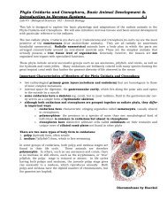
<strong>Phylum</strong> <strong>Echinodermata</strong>, Introduction to <strong>Phylum</strong> <strong>Chordata</strong>& More on Animal Development 14.2<strong>Lab</strong> <strong>#14</strong> -- Biological Sciences 102 – Animal BiologyClassification<strong>Phylum</strong> <strong>Echinodermata</strong>Class Crinoidea (cry-noy'de-a) (Gr. krinon, lily, + eidos, form, + ea, characterized by). About620 species. Sea lilies and feather stars. Aboral attachment stalk of dermal ossicles. Anus onoral surface; five branching arms with pinnules; ciliated ambulacral groove on oral surface withtentacle-like tube feet for food collecting; spines, madreporite, and pedicellariae absent.Examples: Antedon, FlorometraClass Asteroidea (as'ter-oy'de-a) (Gr. aster, star, + eidos, form, + ea, characterized by). About1600 species. Sea stars. Starshaped, with arms not sharply marked off from central disc;ambulacral grooves open, with tube feet on oral side; tube feet often with suckers; anus andmadreporite aboral; pedicellariae present. Examples: Asterias, PisasterClass Ophiuroidea (o'fe-u-roy'de-a) (Gr. ophi::,~ snake, + oura, tail, + eidos, form). About 2100species. Brittle stars and basket stars. Star-shaped, with arms sharply marked off fromcentral disc; ambulacral grooves closed, covered by ossicles; tube feet without suckers and notused for locomotion; pedicellariae absent. Examples: Ophiura, Gorgonocephalus, OphiodermaClass Echinoidea (ek'i-noy'de-a) (Gr. echinos, sea urchin, hedgehog, + eidos, form). About 950species. Sea urchins, heart urchins, and sand dollars. More or less globular or disc-shaped,with no arms; compact skeleton, or test, with closely fitting plates; movable spines; ambulacralgrooves closed and covered by ossicles; tube feet with suckers; pedicellariae present.Examples: Arbacia, Strongylocentrotus, Lytechinus, MellitaClass Holothuroidea (hol' o-thu-roy' de-a) (Gr. holothourion, sea cucumber, + eidos, form).About 1100 species. Sea cucumbers. Cucumber-shaped; with no arms; spines absent;microscopic ossicles embedded in thick, muscular wall; anus present; ambulacral groovesclosed; tube feet with suckers; circumoral tentacles (modified tube feet); pedicellariae absent;madreporite plate internal. Examples: Tbyone, parastichopus, CucumariaBilaterally symmetrical Auricularialarvae of a sea cucumber (x100)
<strong>Phylum</strong> <strong>Echinodermata</strong>, Introduction to <strong>Phylum</strong> <strong>Chordata</strong>& More on Animal Development 14.4<strong>Lab</strong> <strong>#14</strong> -- Biological Sciences 102 – Animal BiologyClassification<strong>Phylum</strong> <strong>Chordata</strong>Subphylum Tunicata (Urochordata) (u'ro-kor-da'ta) (Gr. oura, tail, + L. chorda, cord). About2000 species. Tunicates. Only larval forms have all chordate characteristics; adults sessile,without notochord and dorsal nerve cord; body enclosed in tunic.Example: Molgula, a sea squirtSubphylum Cephalochordata (sef'a-lo-kor-da'ta) (Gr. kephale, head, + L. chorda, cord). About22 species. Lancelets. Notochord and nerve cord persist throughout life; lance-shaped.Example: Branchiostoma (Amphioxus), a lanceletSubphylum Vertebrata (ver'te-bra'ta) (L. vertebratus, backboned). (Craniata). About 50,900species. Vertebrates. Enlarged brain enclosed in cranium; nerve cord surrounded by bony orcartilaginous vertebrae; notochord in all embryonic stages and persists in adults of somefishes; typical structures include two pairs of appendages and body plan of head, trunk, andpostanal tail.Subphylum Vertebrata is further divided into many classes and ordersthat we will be studying in upcoming labs and lectures.Animal Development – Terms Relevant to Classificationblastomere = an early cleavage cell in a developing embryoCleavage = early embryonic cell divisions (mitosis)determinate cleavage = the type of cleavage, usually spiral, in which the fate of theblastomeres is determined very early in development; mosaic cleavagemosaic cleavage = embryonic development characterized by independent differentiation ofeach part of the embryoindeterminate cleavage/development = the type of embryonic development in which the fateof blastomeres is not determined very early as to tissues or organs (cell differentiation);regulative cleavageregulative cleavage/development = embryonic development determined by interactionsamong neighboring cells; cell fates are not fixed early in developmentCoelom Formationschizocoelic coelom and mesoderm formation = formation of a coelom by splitting of theembryonic mesoderm; embryonic formation of the mesoderm as cords of cells betweenectoderm and endoderm; splitting of these cords results in the coelomic spaceenterocoelic coelom and mesoderm formation = formation of a coelom by outpouching ofthe mesodermal sac from the endoderm of the primitive gut; embryonic formation of themesoderm by a pouchlike outfolding from the archenteron, which then expands and obliteratesthe blastocoel, thus forming a large cavity, the coelom, lined with mesoderm
<strong>Phylum</strong> <strong>Echinodermata</strong>, Introduction to <strong>Phylum</strong> <strong>Chordata</strong>& More on Animal Development 14.5<strong>Lab</strong> <strong>#14</strong> -- Biological Sciences 102 – Animal Biologyarchenteron = the main cavity of an embryo in the gastrula stage; it is lined with endodermand represents the future digestive cavityProtostomiaA group of phyla in which cleavage is determinate, the coelom (in coelmate forms) is formed byproliferation of mesodermal bands (schizocoelic formation), the mesoderm is formed from aparticular blastomere (called 4d) and the mouth is derived from or near the blastopore;includes the phyla Annelida, Mollusca, Arthropoda and a number of minor phyla.Blastopore becomes the mouth.Ecdysozoan ProtostomesAny member of a clade within Protostomia whose members shed the cuticle as they grow;includes the phyla Arthropoda, Nematoda and several smaller phyla.Lophotrochozoan ProtostomesAny member of a clade within Protostomia whose members generally possess either atrochophore larva or a lophophore; includes the phyla Annelida, Mollusca and the ectoproctstrochophore larvae = a free-swimming ciliated marine larva characteristic of most molluscsand certain ectoprocts, brachiopods, and marine worms; an ovoid or pyriform body withpreoral circlet of cilia and sometimes a secondary circlet behind the mouth.lophophore = tentacle-bearing ridge or arm within which is an extension of the coelomic cavityin lophophorate animals.ectoproct = bryozoans (moss animals); psuedocoelomate animals that have their anus locatedwithin a crown of tentacles; about 4500 species; both freshwater and marine; few more than0.5 mm longDeuterostomiaA group of phyla in which cleavage is indeterminate (regulative) and primitively radial. Themesoderm arises by enterocoelic formation, and the mouth is derived away from theblastopore; includes the phyla <strong>Echinodermata</strong>, <strong>Chordata</strong> and Hemichordata.Blastopore becomes the anus.LABORATORY NOTES & DRAWINGS ON ANIMAL EMBRYOLOGY:
<strong>Phylum</strong> <strong>Echinodermata</strong>, Introduction to <strong>Phylum</strong> <strong>Chordata</strong>& More on Animal Development 14.6<strong>Lab</strong> <strong>#14</strong> -- Biological Sciences 102 – Animal BiologyLAB PROCEDURENAME:LAB SCORE:Observation of Living SpecimensClass Crinoidea (sea lilies and feather stars)‣ If available, observe the crinoid specimens on display in the lab.‣ Record the descriptive information requested at the end of the lab.‣ If unavailable, go to the following website to view a crinoid:Class Asteroidea (sea stars)‣ Observe the asteroid specimens on display in the lab.‣ Record the descriptive information requested at the end of the lab forPisaster giganteus.Class Ophiuroidea (brittle stars and basket stars)‣ Observe the ophiuroid specimens on display in the lab.‣ Record the descriptive information requested at the end of the lab for one brittle starspecies.Class Echinoidea (sea urchins and sand dollars)‣ Observe the echinoid specimens on display in the lab.‣ Record the descriptive information requested at the end ofthe lab for Stronglyocentrotus purpuratus orS. franciscanus..Class Holothuroidea (sea cucumbers)‣ Observe the holothuroid specimens on display in the lab.‣ Record the descriptive information requested at the end of the lab for one seacucumber species.Ophiodea by Haeckel
<strong>Phylum</strong> <strong>Echinodermata</strong>, Introduction to <strong>Phylum</strong> <strong>Chordata</strong>& More on Animal Development 14.8<strong>Lab</strong> <strong>#14</strong> -- Biological Sciences 102 – Animal BiologyDISSECTIONClass AsteroideaPisaster giganteus (sea star)Use a dissecting scope for your investigation.Obtain one sea star specimen for each two students.‣ Examine the external anatomy of the sea star on both the dorsal and ventral sides andbe able to identify the structures listed below. Note the pentaradial body plan (symmetry)and with five arms or rays and the oral-aboral flattening evident in this class.‣ External Structures‣ central disc‣ five rays (arms)‣ tube feet (podia)‣ mouth‣ anus (at center of central disc)‣ bivium = two arms nearest to the madreporite‣ trivium = three arms opposite the bivium‣ madreporite‣ ambulachral grooves (in each arm; contains tube feet)‣ calcareous spines on ciliated epidermis‣ dermal branchiae (fingerlike bulges in epidermis for respiration)‣ pedicellariae (two-jawed modified spines that capture tiny prey and protect dermalbranchiae from collecting sediment and small parasites)‣ sensory tentacles (elongated tube feet at end of each arm)‣ pigmented eyespots (on end of each arm; small red spot)‣ Examine the internal anatomy of the sea star from the dorsal side and be able to identifythe structures listed below. Place the specimen aboral side up (with the tube feet facing upon the dissecting tray) in a dissecting pan and cover with water. Select the three arms ofthe trivium and snip off their distal ends. Insert a scissors point under the body wall at thecut end of one of the arms. Carefully cut along the dorsolateral margins of at least twoarms to the central disc. Lift up the loosened wall and carefully free any clinging organs.Uncover the central disc, but cut around the madreporite plate, leaving it in place. Becareful not to damage the delicate tissue underneath. The tissue around the anal openingin the center of the disc will cling. Cut the very short intestine close to the aboral wallbefore lifting off the body wall.Internal Structures‣ stone canal leads to madreporite‣ ring canal‣ radial canals‣ ampullae of tube feet‣ gonads‣ pyloric stomach (more dorsal)‣ cardiac stomach (more ventral)‣ hepatic/pyloric cecae
<strong>Phylum</strong> <strong>Echinodermata</strong>, Introduction to <strong>Phylum</strong> <strong>Chordata</strong>& More on Animal Development 14.9<strong>Lab</strong> <strong>#14</strong> -- Biological Sciences 102 – Animal BiologyClass EchinoideaStudy of a Living Stronglyocentrotus purpuratus (sea urchin)‣ Each table of students should look at one of the living sea urchin specimens(3 specimens per lab or this may be done collectively as a class).‣ Obtain a sea urchin specimen and place it in a bowl of sea water.‣ Study the behavior and then examine it under the dissecting scope to reveal detail.‣ One group from the entire class will dissect the sea urchin and all students shouldreview its structure.Sea urchins are usually common on a rocky sea bottom, especially when there is a thickgrowth of algae. They occur most commonly from the low-tide mark to a depth of about 20fathoms, but they are also found at greater depths. They move slowly by means of ventralspines, and climb using their tube feet. The five large teeth in the jaw apparatus (Aristotle’slantern) are used for browsing on algae and on small encrusting animals. Breeding takesplace mainly in winter. Fertilization is external and each zygote develops into a pelagicechinopluteus larva which feeds on other planktonic organisms before going throughmetamorphosis and settling in the benthic habitat.‣ Place the sea urchin on its back in an aquarium and time its righting response.‣ How long does it take?‣ Briefly describe the techniques used in righting itself.‣ Briefly describe the feeding response of the sea urchin to small bits of kelp.‣ Describe or sketch a couple of different types of pedicellaria of the sea urchinStronglyocentrotus purpuratus as observed under the dissecting scope, from pictures shownby your instructor or from the textbook or Inter<strong>net</strong>.Generalized pedicellaria
<strong>Phylum</strong> <strong>Echinodermata</strong>, Introduction to <strong>Phylum</strong> <strong>Chordata</strong>& More on Animal Development 14.10<strong>Lab</strong> <strong>#14</strong> -- Biological Sciences 102 – Animal BiologyDISSECTIONClass EchinoideaStronglyocentrotus purpuratus (sea urchin)Use a dissecting scope for your investigation.Obtain one sea urchin specimen for dissection for the entire class to observe.‣ Examine the external anatomy of the sea urchin on both the dorsal and ventral sidesand be able to identify the structures listed below.‣ Note the pentaradial body plan (symmetry) – easily seen in the test (the urchin skeleton).External Structures‣ spines‣ five ambulacral regions (contain tube feet)‣ tube feet (podia)‣ mouth (with Aristotle’s lantern; teeth)‣ anus (not directly visible but on the center dorsal side)‣ madreporite‣ pedicellaria‣ periostomal gills (five pairs)‣ Examine the internal anatomy of the sea urchin and be able to identify the structureslisted below. Hold the sea urchin very gently when dissecting; the test is relatively fragile.Remove the thin “skin” around the mouth to get a better view of the teeth. Remove some ofthe spines around the “top” of the test to see the anus. Remove the spines from the test’sequator before making the midlateral cut. To make the cut, force the sharp point of thescissors through the test wall (don’t cut too deeply; you could damage the gonads andintestines) and cut completely around the test’s equator. Gently separate the 2 halves ofthe test to examine the internal structures. Try to keep all internal structures intact ifpossible.Internal Structures‣ Aristotle’s lantern‣ ampullae of tube feet‣ stomach‣ intestine‣ rectum‣ gonad
<strong>Phylum</strong> <strong>Echinodermata</strong>, Introduction to <strong>Phylum</strong> <strong>Chordata</strong>& More on Animal Development 14.11<strong>Lab</strong> <strong>#14</strong> -- Biological Sciences 102 – Animal BiologyObservation of Living Specimens<strong>Phylum</strong> <strong>Chordata</strong>Subphylum Tunicata (Urochordata) = sea squirts, tunicates, salps‣ Observe the tunicate specimens on display in the lab.‣ Record the descriptive information requested at the end of the lab.Subphylum UrochordataUrochordata (Gr. oura, tail, + L. chorda, cord) are commonly called tunicates because of theirleathery covering, or tunic. They are divided into three classes: Ascidiacea,sea squirts; Thaliacea, salpians; and Larvacea, appendicularians. The largest group is theascidians (Gr. askidion, leather bag or bottle), which are also the most generalized. They arecalled sea squirts because of their habit, when handled, of squirting water from the excurrentsiphon. Any of the small, translucent ascidians may be used for this exercise. Adult tunicatesare sessile, whereas the larvae undergo a brief free-swimming existence.Tunicates are found in all seas and at all depths. Most of them are sessile as adults, althoughsome are pelagic (found in the open ocean). Ciona intestinalis (Gr. Chione, demigoddess ofmythology) is a cosmopolitan species common in shallow water on wharf pilings, on anchoredand submerged objects, and on eelgrass. It grows to 15 cm in its largest dimension. Althoughsea squirts (ascidians of class Ascidiacea) are common, they also are commonly overlooked inthe marine environment. Nearly all shallow-water sea squirts are found attached to almost anyavailable rigid surface: wharf pilings, rocks, shells, and ship bottoms. One of the best places tosee sea squirts is on pilings, where they may cover the surface.Subphylum Urochordata‣ Describe the feeding mode for an adult tunicate, tracing the path of water flow throughthe body. Be sure to include the names and roles of all the structures (from the diagramsincluded in the lab) involved in food collection. You may use the Inter<strong>net</strong> to aid you in youranswer.
<strong>Phylum</strong> <strong>Echinodermata</strong>, Introduction to <strong>Phylum</strong> <strong>Chordata</strong>& More on Animal Development 14.12<strong>Lab</strong> <strong>#14</strong> -- Biological Sciences 102 – Animal Biology<strong>Phylum</strong> <strong>Chordata</strong>Subphylum Cephalochordata (lancelets)‣ Observe the cephalochordate Branchiostoma (Amphioxus) sp. (as available) ondisplay in the lab.‣ Record the descriptive information requested at the end of the lab.Subphylum CephalochordataVarious researchers have suggested that the structures of Amphioxus listed below arehomologous with those of vertebrates. In other words, they may be derived from the samestructure in a common ancestor. Since Amphioxus appears to be primitive in structure(meaning it has probably changed very little from ancestors which gave rise to vertebrates),study of Amphioxus may give us an idea of what the early vertebrates looked like. On the otherhand, some biologists (eg. Hyman) are critical of some of these interpretations.Study of Branchiostoma (Amphioxus) sp. (lancelet)‣ Obtain prepared slides of a whole mount and cross section of Branchiostoma(Amphioxus) sp. and study them under the 4X and the 10X objectives and identify thefollowing structures:Whole Mount Structures‣ oral hood‣ notochord‣ neural tube‣ pharynx‣ dorsal fin‣ ventral fin‣ caudal fin‣ atrium‣ atriopore‣ myotomes‣ hepatic cecum‣ buccal cirriCross Section Structures‣ atrial cavity‣ dorsal fin‣ neural tube‣ pharynx‣ myotomes‣ gill bars‣ dorsal aorta‣ metapleural folds‣ notocord‣ Which direction does the apex of the chevron-like myotome “V” point?Anteriorly or Posteriorly?‣ Describe the feeding mode for an adult lancelet, tracing the path of water flow throughthe body. Be sure to include the names and roles of all the structures (from the diagramsincluded in the lab) involved in food collection. You may use the Inter<strong>net</strong> to aid you in youranswer.
<strong>Phylum</strong> <strong>Echinodermata</strong>, Introduction to <strong>Phylum</strong> <strong>Chordata</strong>& More on Animal Development 14.13<strong>Lab</strong> <strong>#14</strong> -- Biological Sciences 102 – Animal BiologyLiving Specimens Data<strong>Phylum</strong> <strong>Echinodermata</strong>Class Asteroidea (sea stars)Scientific name: Pisaster giganteusCommon name:Notes & observations to help you remember and distinguish this class:<strong>Phylum</strong> <strong>Echinodermata</strong>Class Ophiurodea (brittle stars and basket stars)Scientific name:Common name:Notes & observations to help you remember and distinguish this class:
<strong>Phylum</strong> <strong>Echinodermata</strong>, Introduction to <strong>Phylum</strong> <strong>Chordata</strong>& More on Animal Development 14.14<strong>Lab</strong> <strong>#14</strong> -- Biological Sciences 102 – Animal Biology<strong>Phylum</strong> <strong>Echinodermata</strong>Class Echinoidea (sea urchins and sand dollars)Scientific name: Stronglyocentratus purpuratus or S. franciscanusCommon name:Notes & observations to help you remember and distinguish this class:<strong>Phylum</strong> <strong>Echinodermata</strong>Class Holothuroidea (sea cucumbers)Scientific name:Common name:Notes & observations to help you remember and distinguish this class:
<strong>Phylum</strong> <strong>Echinodermata</strong>, Introduction to <strong>Phylum</strong> <strong>Chordata</strong>& More on Animal Development 14.15<strong>Lab</strong> <strong>#14</strong> -- Biological Sciences 102 – Animal Biology<strong>Phylum</strong> <strong>Echinodermata</strong>Class Crinoidea (sea lilies and feather stars)Scientific name:Common name:Notes & observations to help you remember and distinguish this class:<strong>Phylum</strong> <strong>Chordata</strong>Subphylum Tunicata (sea squirts, tunicates, salps)Scientific name:Common name:Notes & observations to help you remember and distinguish this class:<strong>Phylum</strong> <strong>Chordata</strong>Subphylum Cephalochordata (lancelets)Scientific name: Branchiostoma (Amphioxus) sp.Common name:Notes & observations to help you remember and distinguish this class:
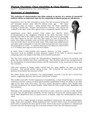
<strong>Phylum</strong> <strong>Echinodermata</strong>, Introduction to <strong>Phylum</strong> <strong>Chordata</strong>& More on Animal Development 14.16<strong>Lab</strong> <strong>#14</strong> -- Biological Sciences 102 – Animal BiologyLABORATORY NOTES:Asteridea by Haeckel