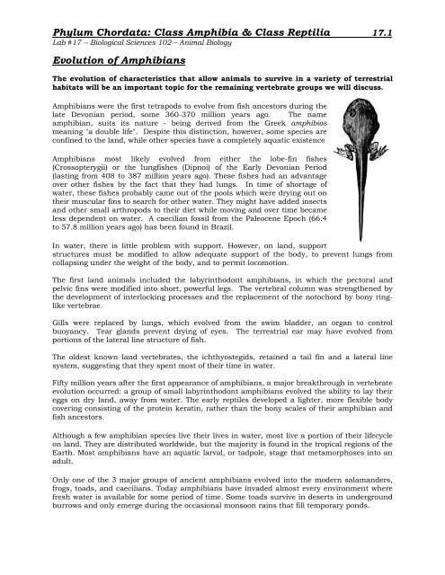
Class Amphibia & Class Reptilia Evolution of ... - Biosciweb.net
Class Amphibia & Class Reptilia Evolution of ... - Biosciweb.net
Class Amphibia & Class Reptilia Evolution of ... - Biosciweb.net

You also want an ePaper? Increase the reach of your titles
YUMPU automatically turns print PDFs into web optimized ePapers that Google loves.
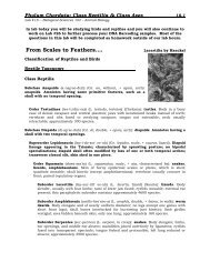
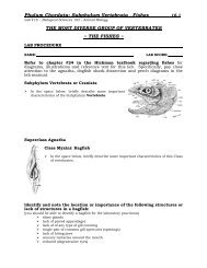
Phylum Chordata: <strong>Class</strong> <strong>Amphibia</strong> & <strong>Class</strong> <strong>Reptilia</strong> 17.5Lab #17 -- Biological Sciences 102 – Animal BiologyFROG DISSECTION<strong>Class</strong> <strong>Amphibia</strong>Order Anura (Salientia)Family RanidaeGenus Rana (or similar genus)AFTER YOU HAVE COMPLETED THE CARDIAC PHYSIOLOGY PORTION OF THELAB ON PAGES 17.10 TO 17.13 THEN YOU MAY COMPLETE THE DISSECTIONTO OBSERVE THE STRUCTURES LISTED BELOW. All students at each lab tableshould observe one live frog. You need not write out your observations, but youshould think about the importance <strong>of</strong> the various frog adaptations you observe.Your instructor will double-pith a frog for your dissection which you will do ingroups <strong>of</strong> 3 or 4 students.External Structures (entire frog)‣ head‣ trunk‣ sacral hump‣ cloacal opening‣ forelimbs (how many digits on each forefoot/hand?)‣ hindlimbs (how many digits on each hindfoot?)‣ eyes‣ nictating membrane‣ tympanic membrane‣ external naris (nares)Skeletal Structures(on frog skeleton and diagram in lab)‣ skull‣ axial skeleton‣ appendicular skeleton‣ nasal fossa‣ orbital fossa‣ auditory capsule‣ maxilla‣ dentary(middle portion <strong>of</strong> each side <strong>of</strong> the mandible)‣ vertebrae (ending in urostyle)‣ suprascapula and scapula‣ clavicle‣ humerus‣ radioulna‣ carpals‣ metacarpals‣ pelvis (urostyle, ischium and ilia;pubis is difficult to see)‣ femur‣ tibi<strong>of</strong>ibula‣ calcaneus (with astragalus = tarsals)‣ metatarsals‣ phalanges (on all forefeet and hindfeet)
Phylum Chordata: <strong>Class</strong> <strong>Amphibia</strong> & <strong>Class</strong> <strong>Reptilia</strong> 17.6Lab #17 -- Biological Sciences 102 – Animal BiologyMuscles (see lab diagrams - can be removed from the frog once identified)‣ pectoralis major (ventral chest)‣ rectus abdominis (ventral abdomen)‣ external oblique (abdomen)‣ sartorius (ventral thigh)‣ gracilis major (ventral thigh)‣ adductor magnus (ventral thigh)‣ gastrocnemius (leg/shank) & Achilles tendon‣ latissimus dorsi (dorsal thorax)‣ triceps femoris (dorsal thigh)‣ biceps femoris (dorsal thigh)Head - Internal Structures‣ internal naris (nares)‣ teeth‣ tongue‣ opening to esophagusThoracic & Abdominal Organs – Internal Structures‣ liver‣ gallbladder‣ stomach‣ small intestines (supported by mesenteries)‣ large intestine‣ pancreas (as visible)‣ spleen (as visible)‣ mesonephric kidneys‣ urinary bladder‣ cloaca‣ gonads (ovary or testes)‣ oviducts (female)‣ lungs‣ heart‣ pericardial sac (around heart)‣ atria <strong>of</strong> heart‣ ventricle <strong>of</strong> heart‣ dorsal aorta (as visible)‣ posterior vena cava (as visible)‣ remove heart to view sinus venosus and interior <strong>of</strong> heart chambers
Phylum Chordata: <strong>Class</strong> <strong>Amphibia</strong> & <strong>Class</strong> <strong>Reptilia</strong> 17.7Lab #17 -- Biological Sciences 102 – Animal Biology‣ Regarding the basic classification <strong>of</strong> amphibians, be sure you are aware <strong>of</strong> the majordistinguishing characteristics to each <strong>of</strong> the following orders as described above:‣ Order Gymnophiona‣ Order Urodela‣ Order Anura‣ Be sure you can identify a given amphibian to its correct order for thelaboratory practicum.‣ Observe and study the live and preserved amphibian specimens displayed in the lab.Provide the following information for 6 different amphibians displayed in the lab.1. Common NameScientific NameOrder2. Common NameScientific NameOrder3. Common NameScientific NameOrder4. Common NameScientific NameOrder5. Common NameScientific NameOrder6. Common NameScientific NameOrder
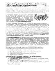
Phylum Chordata: <strong>Class</strong> <strong>Amphibia</strong> & <strong>Class</strong> <strong>Reptilia</strong> 17.8Lab #17 -- Biological Sciences 102 – Animal BiologyReview the dissections <strong>of</strong> the mudpuppy, snake, and turtle (as available).Be sure you can identify the various structures identified in these dissections.Answer the following questions using your lab manual, textbook, instructor feedback, theInter<strong>net</strong> and collective discussions with your classmates.‣ Regarding the dissections, list some major characteristics that differentiate a snake froma frog.‣ What skeletal structures form the shell <strong>of</strong> a turtle or tortoise?‣ Is the shell covered in skin? Is the shell covered with scales?‣ What causes the shell to have a specific color pattern?‣ What is the plastron <strong>of</strong> a turtle?‣ What characteristics allow a reptile to survive in a completely terrestrial environment?(Think about each organ system as you answer this question as thereare many terrestrial adaptations seen in reptiles).
Phylum Chordata: <strong>Class</strong> <strong>Amphibia</strong> & <strong>Class</strong> <strong>Reptilia</strong> 17.9Lab #17 -- Biological Sciences 102 – Animal Biology‣ Observe and study the live and preserved reptile specimens displayed in the lab.Provide the following information for 6 different reptiles displayed in the lab.1. Common NameScientific NameOrderSuborder2. Common NameScientific NameOrderSuborder3. Common NameScientific NameOrderSuborder4. Common NameScientific NameOrderSuborder5. Common NameScientific NameOrderSuborder6. Common NameScientific NameOrderSuborder
Phylum Chordata: <strong>Class</strong> <strong>Amphibia</strong> & <strong>Class</strong> <strong>Reptilia</strong> 17.10Lab #17 -- Biological Sciences 102 – Animal Biology‣ VERTEBRATE CARDIAC PHYSIOLOGYThe heart undergoes a constant cycle <strong>of</strong> contractions and relaxations called the cardiac cycle.The period <strong>of</strong> ventricular contraction is called systole, while the period <strong>of</strong> ventricular relaxationis called diastole. Diastole begins as the ventricles start to relax. Soon the pressures within theaorta and pulmonary artery exceed ventricular pressures, causing the semilunar valves toclose. As the ventricular pressure falls below the atrial pressure the AV valves open and theventricles fill with blood. The ventricles fill to about 80% <strong>of</strong> capacity prior to contraction <strong>of</strong> theatria, the last event in diastole. As the ventricles start to contract, the ventricular pressuresoon exceeds the atrial pressure, causing the AV valves to close. As the ventricles continue tocontract, the ventricular pressure exceeds the arterial pressures causing the semilunar valvesto open. Blood is forcefully ejected out <strong>of</strong> the ventricles and into the aorta and pulmonaryartery.The heart is autorhythmic, meaning it generates its own rhythmic action potentialsindependent <strong>of</strong> the nervous system. The rhythmic beating <strong>of</strong> the heart is controlled by a smallgroup <strong>of</strong> cells in the wall <strong>of</strong> the right atrium, collectively called the sinoatrial node (SA node).Since the SA node controls heart rate, it is called the pacemaker <strong>of</strong> the heart. The electricalimpulse that arises in the SA node travels through gap junctions to the atrial myocardiumand then to the atrioventricular (AV) node. The impulse is delayed slightly in the AV node (AVnodal delay) before traveling into the bundle <strong>of</strong> His. The impulse then travels through thebundle branches, into the purkinje fibers and terminates in the ventricular myocardiumwhere it stimulates muscle contraction. The specialized structures that conduct the electricalimpulse through the heart are collectively called the conduction system.‣ Draw a diagram <strong>of</strong> the conduction system within the heart and label its parts.
Phylum Chordata: <strong>Class</strong> <strong>Amphibia</strong> & <strong>Class</strong> <strong>Reptilia</strong> 17.11Lab #17 -- Biological Sciences 102 – Animal Biology‣ Drugs that Effect the Vertebrate HeartBoth branches <strong>of</strong> the autonomic nervous system enervate the SA node and modify its activity.Sympathetic input (norepinephrine and epinephrine) increases heart rate, whileparasympathetic input (acetylcholine) decreases heart rate. Also, certain drugs can alter therate and strength <strong>of</strong> cardiac contraction. In today’s experiment you will record a frog’s cardiaccycle on the Biopac equipment using an exposed frog heart. You will also test the effects <strong>of</strong> thefollowing drugs on heart rate.Epinephrine: Epinephrine is a hormone secreted by the adrenal medulla. Epinephrine worksin concert with norepinephrine (a neurotransmitter secreted by the sympathetic nervoussystem) to increase both the strength and rate <strong>of</strong> cardiac contractions.Pilocarpine: Pilocarpine stimulates the release <strong>of</strong> acetylcholine from parasympathetic neuronsthat innervate the SA node, thus causing a decrease in heart rateAcetylcholine: Acetylcholine is a neurotransmitter secreted by the parasympathetic nervoussystem at the SA node <strong>of</strong> the heart. It causes a decrease in cardiac rate.Atropine: Atropine is a competitive inhibitor <strong>of</strong> acetylcholine. Atropine outcompetesacetylcholine for binding to the acetylcholine receptor. Atropine blocks the effects <strong>of</strong>acetylcholine, thus inhibiting the parasympathetic activity <strong>of</strong> the heart.Eserine: Eserine is an inhibitor <strong>of</strong> Acetylcholinesterase, the enzyme responsible for thebreakdown <strong>of</strong> acetylcholine in the synaptic cleft <strong>of</strong> the neuromuscular junction.Potassium Chloride (KCl): The initiation and propagation <strong>of</strong> action potentials in the heart isdependent, in part, on the steep K+ concentration gradient between the ICF and ECF (higherconcentration <strong>of</strong> K+ on the inside <strong>of</strong> the cell than on the outside). An increase in extracellularK+ above normal (hyperkalemia) disrupts this gradient, causing a decrease in heart rate andcontractility. In extreme hyperkalemia, the conduction rate <strong>of</strong> action potentials may be sodepressed that ectopic pacemakers appear in the ventricles and fibrillation may develop, <strong>of</strong>tenwith fatal results.
Phylum Chordata: <strong>Class</strong> <strong>Amphibia</strong> & <strong>Class</strong> <strong>Reptilia</strong> 17.12Lab #17 -- Biological Sciences 102 – Animal BiologyFrog Heart Cardiac Drug ExposureExperimental Procedure:Biopac setup:1. Plug the force transducer into channel 1 <strong>of</strong> the mp40 unit2. Place the small hook in the 50gram slot <strong>of</strong> the forcetransducer.3. Turn the mp40 unit on.4. Open the bms108biopacdoohan folder on the desktopand click on the Frog Heart file.Frog Preparation:1. Obtain a double-pithed frog from your instructor and place itventral side up on a wax dissecting pan.2. Pin the frog securely to the pan placing pins through all four appendages.3. Make an incision through the skin and cut a circular pattern to remove a section roughlythe size <strong>of</strong> a silver dollar. The center <strong>of</strong> this circle should be the sternal xiphoid process.4. After the skin is removed, cut a similar pattern through the musculature and rib cage inorder to expose the heart. Try to avoid cutting any large visible blood vessels.5. Lift the pericardium with forceps and cut through it so as to expose the heart. BECAREFUL TO NOT CUT THE HEART ITSELF!6. Insert the cardiac hook through the apex <strong>of</strong> the ventricle, but do not enter the ventricleitself. Use the thread to lift the heart to a vertical position. Now gently snip the frenulum,which is on the dorsal side <strong>of</strong> the heart. Be sure you do not cut the vena cava.7. Attach the thread from the heart hook to the lever <strong>of</strong> the force transducer. Adjust thetransducer so that the thread is perfectly vertical and there is adequate tension on theheart. The heart should now be in a vertical position.8. IMPORTANT! Keep heart constantly moist with Ringer’s solution throughout theexperiment.9. Before you begin recording, be certain you have all <strong>of</strong> the drugs required for theexperiment.Recording the Cardiac Cycle <strong>of</strong> the Frog Heart1. Click the blue “start” button in the bottom right corner <strong>of</strong> your screen to begin recording.You should now be seeing the tracing <strong>of</strong> a cardiac cycle. If not, then alert the instructor.• Records 20 cardiac cycles then click the stop button.2. Slowly drip 5 drops <strong>of</strong> epinephrine directly on to the heart.• Wait 1 minute before recording the effect.• Place an event marker on the screen (F9) to indicate a change in conditions.• Record 20 cycles then click stop.• Note the heart rate for this region <strong>of</strong> the data.• If there is no change then add more drug. If there is a significant change then goto the next step.
Phylum Chordata: <strong>Class</strong> <strong>Amphibia</strong> & <strong>Class</strong> <strong>Reptilia</strong> 17.13Lab #17 -- Biological Sciences 102 – Animal Biology3. Thoroughly rinse the heart with ringer solution and allow it to rest for 2 minutes beforeapplying the next drug.4. Repeat steps 2 and 3 for the remainder <strong>of</strong> the drugs. Be certain to go in the followingorder:• Pilocarpine• Acetylcholine• Atropine• Eserine• Potassium chlorideAnalysis <strong>of</strong> the Frog Heart Data1. You should now have 7 segments <strong>of</strong> data; the initial segment minus drug and data foreach drug condition. You can view all <strong>of</strong> these segments on one screen by clicking thehorizontal autoscale button in the top tool bar.2. You will now determine the average beats per minute for each <strong>of</strong> these segments andenter that data in table 1. Your instructions are detailed below.3. Using the magnifying tool, adjust the screen for optimal viewing <strong>of</strong> the last 10 cycles <strong>of</strong>segment 1 (no drug).4. Determine the average heart rate for this region. Using the I beam cursor, highlight thearea between 2 systolic peaks and note the heart rate in bpm. Repeat this for severalpeaks in this segment in order to get an average heart rate. Log the data in the table onpage 4.5. Repeat steps 2-4 for each drug condition and log the data in Table 1.The Effects <strong>of</strong> Drugs on the Frog HeartDrug AdministeredHeart Rate (bpm)Ringers (buffered saline)EpinephrinePilocarpineAcetylcholineAtropineEserinePotassium Chloride
Phylum Chordata: <strong>Class</strong> <strong>Amphibia</strong> & <strong>Class</strong> <strong>Reptilia</strong> 17.14Lab #17 -- Biological Sciences 102 – Animal BiologyCardiac Physiology Homework Questions1. For each <strong>of</strong> the following conditions, state whether the condition occurs during diastole orsystole <strong>of</strong> the cardiac cycle.• Ventricular pressure exceeds aortic pressure Systole or Diastole (circle one)• AV valves are open Systole or Diastole• Semilunar valves have just closed Systole or Diastole• Atrial pressure exceeds ventricular pressure Systole or Diastole• Blood is flowing into the pulmonary artery Systole or Diastole• The atria are contracting Systole or Diastole• The ventricles are contracting Systole or Diastole2. What components <strong>of</strong> the nervous system innervate the SA node?3. How is acetylcholine normally removed from the synaptic cleft <strong>of</strong> the neuromuscularjunction?4. Is epinephrine a hormone or neurotransmitter? From where is it secreted?5. Epinephrine is predicted to cause an increase in heart rate. Did you observe thisphenomenon? Explain how epinephrine increases heart rate.
Phylum Chordata: <strong>Class</strong> <strong>Amphibia</strong> & <strong>Class</strong> <strong>Reptilia</strong> 17.15Lab #17 -- Biological Sciences 102 – Animal Biology6. Eserine is predicted to cause a decrease in heart rate. Explain the mechanism by whicheserine could have this effect.7. What is hyperkalemia and what effect does it have on human cardiac function?8. What effect did potassium chloride have <strong>of</strong> the frog heart? Provide a rationale for thisobservation.9. Some pesticides (carbamates and organophosphates) act as acetylcholinesterase inhibitors.Cholinergic stimulation caused by exposure to high levels <strong>of</strong> these pesticides can result inrespiratory paralysis. Which <strong>of</strong> the drugs discussed in this lab might be used as atreatment for this condition? Explain.Hint: Under normal condition, somatic stimulation <strong>of</strong> the diaphragm results in rhythmicbreathing.10. Pilocarpine is predicted to slow the heart rate. Provide an explanation for this effect.
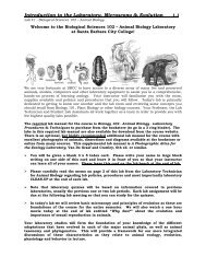
Phylum Chordata: <strong>Class</strong> <strong>Amphibia</strong> & <strong>Class</strong> <strong>Reptilia</strong> 17.16Lab #17 -- Biological Sciences 102 – Animal BiologyLABORATORY NOTES:Chelonia by Haeckel