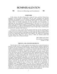
206 Young & HenriksenFigure 10. Holococcolith form. (A−C) Syracosphaera anthos holococcoliths (older name Periphyllophoramirabilis). (A) complete coccosphere; (B) detail <strong>of</strong> coccolith; (C) acid etched specimen (reproduced withpermission from Halldal and Markali (1955), showing organic coatings around the coccoliths. (D)Corisphaera sp. partially collapsed coccolith with similar wall fabric to that <strong>of</strong> S. anthos, showing that it isformed <strong>of</strong> rhombohedral crystallites. (E) Calyptrolithophora papillifera showing perforate hexagonalcrystallite arrangement. Again the constituent crystallites are rhombohedral. (F) Coccolithus pelagicusholococcoliths (older name Crystallolithus hyalinus), showing construction from rhombohedral crystallitesin rhombohedral assembly.
<strong>Biomineralization</strong> <strong>Within</strong> <strong>Vesicles</strong> 207Cytological observationsVery few holococcolithophore cultures have been maintained and only two specieshave been studied in cytological section. Coccolithus pelagicus was studied by Manton andLeedale (1963, 1969) and by Rowson et al. (1986) while Calyptrosphaera sphaeroidea wasstudied by Klaveness (1973). As with heterococcoliths these studies showed that theholococcoliths are underlain by organic base-plate scales and these scales could be seendeveloping in Golgi vesicles. However, despite numerous observations, no intracellularcalcification could be seen, nor have intracellular holococcoliths been seen in lightmicroscopy studies. Hence, it has been inferred that calcification occurs outside the cellmembrane, after exocytosis <strong>of</strong> the base-plate scale. This poses obvious problems forunderstanding how calcification is regulated. A partial solution is provided by observationsthat, in motile species, exocytosis <strong>of</strong> coccoliths and scales occurs at the flagellar pole, andthat, at least in the studied species, a delicate “skin” envelopes the coccosphere. <strong>The</strong>refore,even if calcification does occur outside the cell membrane, it is likely to occur in aprivileged and highly regulated environment. Alternatively, it is possible that calcificationoccurs just below the cell membrane but is a rapid process immediately precedingexocytosis and so has not been captured in cytological sections. Since holococcoliths instatu nascendi have not been observed, there is no evidence from cytology on the pattern <strong>of</strong>growth in holococcoliths.Biochemical observations<strong>The</strong>re have been no studies on the biochemistry <strong>of</strong> holococcolith biomineralization.This reflects the need for biochemical work to focus on a few case studies and the verysmall number <strong>of</strong> holococcolith cultures available for study. In addition, the two mostfrequently cultured coccolithophore species, Emiliania huxleyi and Pleurochrysiscarterae do not produce holococcoliths. <strong>The</strong> species have a similar haplo-diploid lifecycleto other coccolithophores, but the haploid phase cells do not calcify.Morphological observations<strong>The</strong> primary research carried out on holococcoliths is detailed study <strong>of</strong> morphologyin order to establish their taxonomy (e.g., Kleijne 1991, Cros et al. 2000, Cros andFortuño 2002). This has resulted in a rich archive <strong>of</strong> detailed morphological data. <strong>The</strong>primary data source is scanning electron microscopy; supplementary information comesfrom cross-polarized light microscopy, which has provided data on crystallographicorientation (Crudeli and Young 2003). Finally, invaluable observations <strong>of</strong> organiccoatings were made by early Norwegian researchers, notably Halldal and Markali (1955),who decalcified plankton samples using hydrochloric acid. This technique left the organiccoatings as ghost specimens. A representative image is included as Figure 10C. From thiswork several relevant observations can be made.1. Holococcolith shape is highly regulated. Separate species have unique and veryconsistent morphologies. In the case <strong>of</strong> Syracosphaera anthos holococcoliths,(Fig. 10A−C) consistent features in addition to the gross shape, include thepresence <strong>of</strong> a hole at the base <strong>of</strong> the leaf-like distal process and a double-layeredstructure to both the process and the main tube wall. In comparison toheterococcoliths, there is at least as much regulation <strong>of</strong> total coccolith shape.2. <strong>The</strong> entire structure is made up <strong>of</strong> minute equant crystallites. This is obvious as asurface texture, but also applies where holococcoliths form thick basal structures.In such cases, the fabric consists <strong>of</strong> numerous layers <strong>of</strong> separate crystallites,rather than elongated crystallites. Moreover, observations <strong>of</strong> disintegratingholococcoliths (e.g., Fig. 10D) suggest that crystallites are predominantly and