

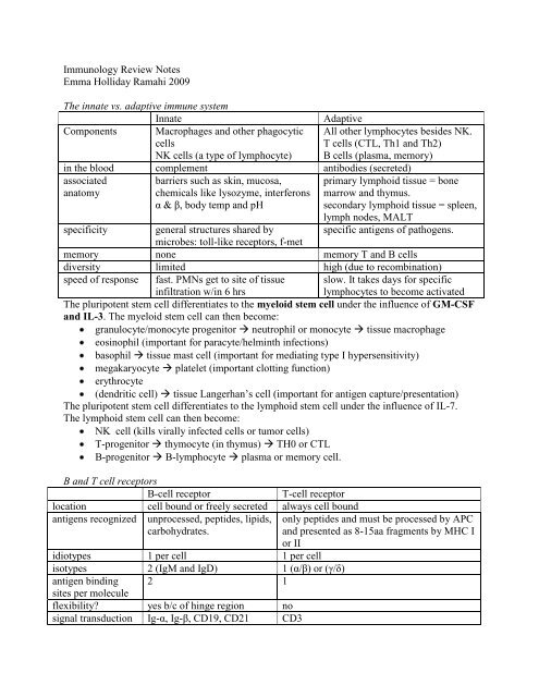
Immunology Review Notes
Immunology Review Notes
Immunology Review Notes

Create successful ePaper yourself
Turn your PDF publications into a flip-book with our unique Google optimized e-Paper software.
Immature T-cells leave the bone marrow and go to the thymus to differentiate further:Bone marrow Thymic Cortex Thymic Medulla Circulating T-cellsT-cellsTdt+ Tdt+“double positive” =CD4+ and CD8+express TCRexpress CD2/CD3based on whether they bindMHCI or II w/ higher affinity,choose either CD8+ or CD4+express TCRexpress CD2/CD3“single positive”either CD4 or CD8express TCRexpress CD2/CD3Positive Selection: cells that bind MHC are given the signal to divide and mature. If the TCRdoesn’t bind MHC, it is allowed to die by apoptosis.Negative Selection: cells that bind MHC too strongly are given a negative signal to die byapoptosis.Lymph Node Architecture:2 afferent lymphatic vessels bring antigen in from the tissues.Cortex contains primary follicles that are B-cell rich. Clones divide in the germinal centerParacortex contains T-cells (so B and T cells can interact)Medulla contains mature cells like plasma cellsMemory cells exit via the efferent lymphatic vessel.Spleen Architecture:Splenic artery brings in antigen from the blood.HEVs (high endothelial venules) bring in naïve lymphocytes L-selectins on lymphs bindto addressins on HEVs.Periarteriolar lymphoid sheaths (PALS) contain T-cellsWhite pulp = lymphoid follicles of lymphocytes and macrophagesRed pulp = sinusoids where blood collects before it leaves via the splenic vein.Antigen = something capable of inducing the formation of an antibodyImmunogen = something capable of generating an immune response. Requires that the moleculeto be recognized as foreign (different from self), be chemically complex, and have a MW of>5000Kd b/c B-cells must be crosslinked to be activated (need more than 1 identical epitope)Hapten = have only 1 epitope, so it can only bind one arm of the B-cell receptor.**Drug allergies: especially penicillin, streptomycin, aspirin, sulfa-drugs and succinylcholinecan induce an allergic response 7-14 days post-exposure showing mild symptoms.The next drug exposure life-threatening anaphylaxis.This can only happen b/c drugs act as haptens (MW
Arrest and Adhesion: Ig-CAMs on the endothelium bind integrins on the phagocyte tostabilize adhesion of the phagocyte to the endothelial cell.Transendothelial migration: phagocyte extends pseudopodia through vessel wall andextravasates into the tissues.Phagocytosis: extend pseudopodia to trap material in phagosome.Opsonization: enhances phagocytosis by 4,000x. IgG and C3b are main opsonins b/cphagocyte has Fc and C3b receptors that bind.Oxygen-dependent killing: “respiratory burst” = NADPH oxidase takes O2 to superoxide generates OH radical and H2O2 microbicidal. Myeloperoxidase takes H2O2 andCl to make hypochlorite (bleach) microbicidal.Oxygen-independent killing: lysozyme (digests cell wall of gram + bugs), defensins(punches holes in bacterial membrane), lactoferrin (chelates Fe so bugs can’t use it togrow), other hydrolytic enzymes.**Leukocyte Adhesion Deficiency (LAD): rare autosomal recessive inherited absence ofCD18 (the common β2) chain of several integrin molecules. Usually this is diagnosed when akiddo’s umbilical stump gets infected (omphalitis) more susceptible to bacterial but NOTviral infections. The kiddos have high WBC counts in their blood, (b/c WBCs areappropriately released from the marrow when infected), but the WBCs can’t get to theinfection no pus is formed. Diagnose w/ flow cytometry and treat w/ bone marrowtransplant.**Chronic Granulomatous Disease: inherited deficiency in one of the NADPH oxidasesubunits. Phagocytes cannot make superoxide, OH radical or H2O2. However,myeloperoxidase is still in tact, so if the bug is catalase negative myeloperoxidase canmake bleach from the bug’s own H2O2 biproducts. Kiddos present w/ increasedsusceptibility to catalase positive bacteria (staph aureus, klebsiella, and serratia) andfungus (aspergillus). Diagnose w/ negative (yellow) nitroblue tetrazolium test.MHC I and IIMHC Class IMHC Class IIHLA-A, B and CHLC-DP, DQ, DRpresent on all nucleated cells + platelets present on B-lymphocytes, macrophages and dendriticcells (+ activated endothelial cells)recognized by CD8+ cytotoxic T-cells recognized by CD4+ TH cellspresent endogenously synthesized present exogenously synthesized peptide(12-15aa) madepeptide (8-10aa) made from virus from bacteria (extracellular or intravacuolar pathogen),(intracytoplasmic pathogen), broken MHC II buds off in a vesicle plugged w/ invariant chain,down in proteosome enters ER via meets an acidic phagolysosome containing bug antigenTAP meets an MHCI and is acid degrades invariant chain, antigen binds MHCIItransported to plasma membrane and is transported to plasma membraneexpressed codominantly (contrast w/ TCR/BCR which do allelic exclusion), so all nucleated cellsexpress HLA A, B, and C from both mom and dad (6 total) and all APCs express HLA-DP, DQ,and DR from both mom and dad (6 total)made up of α heavy chain w/ 3 αdomains plus β2-microglobulin tosupport α in the membranemade up of 1 α and 1 β of equal length (looks like TCR).Antigen binding groove is at the N-terminus of bothchains.
1 st Signal: the CD4+ T-cell receptor binds to antigen-MHC complex on the APC (antigenspecificpart of the response).2 nd Signal: CD4 binds to non-antigen binding site on MHC-IILFA-1 (integrin) on T-cells binds to ICAM-1 on APCs to promote adherence.IgCAMs (CD2) on T-cells binds to LFA-3 (integrin) on APCs for adherence.CD28 on T-cells binds to B7 on APCs and triggers transcription of cytokine genes3 rd Signal: antigen binding promotes growth and proliferation of T-cells by stimulating bothsecretion of IL-2 from the T-cell and the upregulation of the IL-2 receptor on the SAME T-cell. IL-1, IL-6 and TNFα come from the macrophage to stimulate the T-cell. IFNγ comesfrom the T-cell to stimulate and activate the macrophage.**Superantigens like TSST-1 from staph aureus and pyrogenic exotoxin from strep: activatemany T-cells (as many as 10% of total number) by crosslinking β domain of TCR w/ αdomain of MHC II w/o the need for involvement of the antigen-binding site. This causespolyclonal activation of T-cells overproduction of IFNγ overactivation of macrophages overproduction of inflammatory cytokines IL-1, IL-6 and TNF-α systemic toxicity.**Bare Lymphocyte Syndrome: rare autosomal recessive inherited deficiency of MHC II.Kiddos present early w/ symptoms of mild SCID increased susceptibility to pyogenic andopportunistic infections. Can distinguish from SCID by treating w/ phytohemagglutinin (nonspecificT-cell mitogen) bare lymphocyte syndrome will show a response, but SCID won’t(b/c there are not T-cells to respond). These kiddos are deficient in CD4+ cells b/c they can’tdo positive selection in the thymus. Have hypogammaglobuminemia, but have CD8+ cells(But less functional b/c there are no Th1 chemokines to support them).Differentiation of TH0 cells into TH1 or TH2TH1 supports cell mediate immunity TH2 supports humoral immunityInduced By: intracellular pathogens producing astrong innate immune response(Listeria, mycobacteria, Leishmania) w/lots of IL-12 from macrophages andIFNγ from NK cells.extracellular pathogens whose antigen ispresent w/o much innate immunity (defaultsystem)IL-4 produced constituitively leads tomore IL-4 if no IL-12 around.Inhibited By: IL-4 and IL-10 from TH2 cells IFNγ from TH1 cellsCytokinesproducedIFNγ – enhances M0, enhancesexpression of MHC.IL-2 – induces proliferation and activityof T cells.TNFβ – has cytotoxic effects andenhances phagocyte’s activityIL-3 – supports growth anddifferentiation of myeloid cellsGM-CSF – induces proliferation ofgranulocyte precursorsIL-2 – induces proliferation and activity ofT-cells.IL-3 – supports growth and differentiationof myeloid cellsIL-4 – costimulates activation of B-cells,induces class switching to IgG1 and IgE.IL-5 – stimulates proliferation and inducesclass switching to IgA.IL-6 – stimulates Ab secretion, promotesterminal differentiation to plasma cells.IL-10 – suppresses cytokine production byTH1.GM-CSF – induces proliferation ofgranulocyte precursors.
**Tuberculoid Leprosy: mycobacterium leprae gets the strong TH1 response it needs to getrid of intracellular pathogen via granuloma formation. There is some skin and peripheralnerve damage, but the disease progresses slowly and the patient survives.**Lepromatous Leprosy: mycobacterium leprae gets an inappropriate TH2 response (andTH1 response is suppressed by reciprocal inhibition). The patient makes AB that don’t workagainst the bug and mycobacteria multiply w/in macrophages (10 10 bugs per gram of tissue) hypergammaglobulinemia and disseminated and disfiguring infection.Humoral Effector Mechanisms work against extracellular pathogens (microbes or toxins)Naïve B-cell is attracted to follicular areas of lymph nodes and spleenSignal 1: antigen binds and cross-links idiotypes of membrane receptorsSignal 2: if thymus-dependent antigen (most antigens in body) B-cell phagocytosesthe pathogen processes it and presents it on MHCII as well as expressing B7 CD+ THcell recognizes the MHC II and CD27 on T-cell binds to B7 on B-cell.CD40L on the T-cell binds to CD40 on the B-cell to give signal 2 for B-cell activation.Signal 3: cytokines released by TH2 cells (see above) induce class switching so the mostappropriate antibody can be made for the infection.*Thymus-independent antigens include stuff T-cells cannot respond to: lipids (like LPS fromgram negative cell envelope) and carbohydrate (like polysaccharide capsular antigen).B cells are directly stimulated or are activated as mitogens regardless of antigenic specificityto make antibodies (but can ONLY make IgM and cannot produce a memory response).The first AB type made by a B-cell is IgM (doesn’t need cytokine support from TH2)IgM = pentamer held together by J-chain. Big, bulky, can’t cross placenta, but has avalence of 10 so avidity is high. (2 binding sites for each of 5 IgM) so it canbind up antigen in tissue to effectively present it to lymphocytes. It is also mosteffective at activating complement, but cannot opsonize and cannot mediate ADCC(antibody dependent cytotoxicity). IgM is used to measure the extend of the primaryimmune response (to an acute infection).Cytokines from TH2 cells are needed to stimulate isotype switching. The idiotype (variableregion) is linked to another constant region downstream from M, and the DNA in the middle isexcised and degraded (ie, that B cell can never make IgM again).IgG (subclasses 1-4) = monomer than can cross the placental barrier (protects thefetus during gestation), activate complement, act as an opsonin, and mediate ADCC.IgA = dimer held together by J-chain and protects the mucosal surfaces of the bodyinhibiting toxins or bugs to the digestive, respiratory and urogenital surfaces. Itdoesn’t activate compliment or act as an opsinin, but it is in breast milk.IgE = binds directly to Fe receptors on mast cells and basophils first w/o bindingantigen. It triggers mast cell degranulation when crosslinked and protects againsthelminth parasites but also acts acts in allergic responses. (Type I HS).Somatic hypermutation: happens in germinal centers after the B-cell has encountered itsantigen and started proliferating. Single, point mutations are introduced to see if they can createbetter binding btwn antigen and BCR. The best fit wins and is clonally expanded. Affinitymaturation means that even though avidity is decreased (as we switch from IgM to other types),affinity is increased so the same amount of antigen can be bound.
**X-linked Hyper-IgM Syndrome: x-linked inherited disorder in the gene for CD40-ligand TH cells don’t have CD-40L so they can’t give signal 2 to B-cells B-cells can’t respond(proliferate or class switch) to thymus-dependent antigens. (Still see normal response to thymusindependentantigens). See high levels of IgM and a deficiency of IgG, IgA and IgE seeantibodies to neutrophils, platelets and RBCs, see a lack of germinal centers during humoralimmune response see recurrent infection w/ respiratory bugs esp Pneumocystis jiroveci.Complement:Alternative Pathway bacterial polysaccharide and LPS on the surface of pathogenstriggers cascade.Classical Pathway antigen-antibody complex (IgM or IgG) trigger cascade.1. C1 is triggered to cleave C42. C4b cleaves C23. C2a cleaves C3 (C3b binds on cell/particle surface to opsonize)4. C2a cleaves C5 (C5a is a important for chemoattractant activation)5. C5b binds to other complement factors to make MAC.**Complement Abnormalities:C3b deficiency immune complexes cannot be effectively cleared from the body.Hereditary angioedema uncontrolled complement activation at mucosal surfaces edema and pain.Paroxysmal nocturnal hemoglobinuria absence of regulatory proteins causeshemolysis of RBCs (esp at night when blood is relatively acidotic) hemoglobinuria.Cell Mediated Effector Mechanisms rid the body of antigenic stimuli inside the body’s cells(viruses, intracellular bacteria and some parasites). Th1 cells provide cytokine support to CD8+T-cells, NK cells (CD16+, CD56+ and CD2+) and macrophages (CD14+).TH1 cells release IFNγ to activate M0 cause tissue damage delayed typehypersensitivity. Can use DHT skin test to measure a person’s ability to mount CMI response.How CD8+ T-cells kill their target:Attachment: TCR binds antigen/MCH I complex. CD8 acts as coreceptor. LFA-1 andintegrin facilitate attachment.Activation: cytoskeleton rearranges to concentrate granulesExocytosis: CD8+ cell releases perforin (makes holes in the cell membrane) andgranzyme (serine proteases that activate caspases to carry out apoptosis). Cytokines likeIFNγ, TNFα, and TNFβ can also induce apoptosis. Fas-lignad on CD8+ T-cells can alsobind to Fas on the target cell to activate caspases and mediate apoptosis.Detachment: leaves to find another infected cell.How NK cells kill their target:NK cells don’t have TCR or CD3. They are CD16+ and CD56+If activating receptor binds lectins (common on pathogens) kill signalIf inhibiting receptor doesn’t bind to MHC I (b/c virus downregulated the expression ofMHC I) absence of no-kill signal (normal cells have normal MHC I no-kill signal)Kill by granzymes, perforin and enhances by IFNα, IFNβ and IL-12.
How Antibodies can kill targets (via antibody-dependent cell-mediated cytotoxicity):NK cells, macrophages, monocytes, neutrophils and eosinophils have a membrane Fcreceptor for the Fc part of IgG.IgG binds to target cell cytotoxic cell binds to IgG’s Fc lysis of target cell.Involves lytic enzymes, TNF and perforin.**Cytomegalovirus: CMV downregulates MHC I molecules, but produces a “decoy” MHC-Ilikemolecule. The decoy is different enough that is cannot activate CTLs, but similar enough soit can evade killing from NK cells. However, ADCC can still kill these CMV-infected cells.Immunologic Memory B and T memory cells are created when a pathogen is cleared from thebody. Memory cells have increased adhesion molecules and home to inflamed tissue.Memory B cells are terminally differentiated, but unlike plasma cells (who only live2wks), when can remain for months or years w/ IgG, IgE or IgA membrane Ig. This means afaster response if the body ever encounters the antigen again.Memory T cells are the T-cells that escape inactivation and apoptosis after theircytokines are no longer necessary (b/c infection is cleared).Primary Immune Responseresponse takes 5-10days after introductionpeak response is smallIgM, then IgG latervariable to low affinityinduced by all immunogensneed high dose antigen w/ adjuvantSecondary Immune Responsetakes 1-3 dayslarge peakIncreasing IgG, IgA or IgEhigh affinity (already did affinity maturation)induced only by protein antigenscan do w/ low dose w/o adjuvant**Military Vaccine against Adenovirus types 4 and 7: enteric-coated, live, non-attenuatedvirus produces asymptomatic infection in the intestine generates memory IgA cells protected against 2 nd adenovirus infection via aresol (would otherwise cause pneumonia).Types of Immunity and their clinical applicationsNatural Passive Immunity fetus gets maternal IgG from placental transferinfant gets maternal IgA through colostrum/breast milkNatural Active Immunity when you have an infection and recover, your memory B and T cellskeep you from getting the same infection again (HepB)Artificial Passive Immunity antitoxin given to someone who is very sick from black widowspider bite, botulism or diphtheriapooled immunoglobulin against hepA, hepB, measles, rabies ortetanus.monoclonal antibodies against RSVArtificial Active Immunity traditional immunizations:Component vaccine HepBToxoid vaccine diphtheria, tetanus, pertussusCapsular vaccine haemophilus, pneumococcus, meningococcusLive, inactivated polioLive, attenuated measles, mumps and rubella, also varicella.
**Never give a live viral vaccine to an immunocompromised person (AIDS or chemo)**Never give a live viral vaccine to an infant under 12mo b/c maternal IgG is still present andwill inactivate the vaccine before it can mount a useful immune response in the infant.**Passive immunotherapy has risks such as generation of IgE antibodies anaphylaxis,formation of compliment-activating immune complexes (type III HS), large amounts ofantibodies being given at one time can induce anti-allotype antibodies, and people w/ selectiveIgA-deficiency (1 in 700) can get a reaction if given infused IgA.*Can use killed vaccine for naked capsid viruses but need live vaccine for enveloped virus.*Adjuvant is a substance added to a vaccine to increase its immunogenicity.Aluminum potassium sulfate prolongs antigen’s persistenceMuramyl dipeptide enhances co-stimulatory signalAlum induces granuloma formationLPS and synthetic polyribonucleotides induce a non-specific lymphocyte proliferation.Important Immunodeficiency DiseasesDisease Defect Clinical ManifestationChronic GranulomatousDiseaseChediak-HigashiSyndromeGlucose-6-phosphatedehydrogenase deficiencyMyeloperoxidasedeficiencyLeukocyte adhesiondeficiencyBruton X-linkedhypogammaglobuminemiaTransient hypogammaglobuminemiaof infancyCommon variablehypogammaglobulinemiadef of NADPH oxidase (1of 4 proteins) failure tomake superoxide or otherO2 radicalsgranule structural defectdef enzy in hexosemonophosphate shuntgranule enzyme deficiencyabsence of CD18 common β chain forleukocyte integrinsdeficiency of tyrosinekinase that blocks B cellmaturationdelayed onset of IgGsynthesisunknown MOArecurrent infections w/ catalasepositive organisms (staph, klebsiella,serratia and aspergillus)recurrent infection w/ bacteria,chemotactic and degranulationdefecits, absent NK activity, partialalbanismsame as CGD but also w/ hemolyticanemiamild or none (b/c can still dorespiratory burst, just no bleach)recurrent/chronic infections, can’tform pus, umbilical stump won’t falloff.low IG of all classes, no circulatingB cells (can’t leave marrow),stopped at pre-B stage. Normal CMI5 th -6 th month of life but resolves by16-30mo. Incr susceptibility topyogenic bacteriaonset in late teens/20s, B cells are inperipheral blood but IG levelsdecrease and autoimmunity incrSelective IgA deficiency deficiency of IgA repeated sinopulmonary and GIinfectionsX-linked hyper IgMsyndromedeficiency of CD40-L onactivated T-cellshigh titers of IgM w/o other isotypes.Normal B and T cell numbers butincreased susceptibility toextracellular bugs and opportunists
Deficiency in classicpathway of complementDeficiency in alternativepathway of complementC3 deficiencyC1q, C1r, C1s, C4 or C2like factor B or properdinincreased immune complex disease,increased pyogenic bacteria infectionincreased Neisseria infectionsbacterial infections and immuneC5, C6, C7, C8 deficiency complex diseaserecurrent meningococcal andgonococcal infectionsHereditary angioedema deficient C1-INH overuse of C1, C4 and C2 edemaParoxysmal nocturnalhemoglobinuriaDiGeorge SyndromeMHC class I deficiencyWiskott-AldrichSyndromeAtaxia telangietctasiaX-linked SCIDautosomal recessive SCIDBare lymphocytesyndromedeficient complementdecay-activating factor3 rd and 4 th pharyngealpouches don’t develop get thymic aplasiaTAP can’t transportmolecules to ERdefect in cytoskeletalglycoprotein (can’t fusephagosome and lysosome)defect in a kinase involvedin the cell cycledefect in common γ chain ofIL-2, IL-4, IL-7, IL-9 andIL-15 receptorsadenosine deaminasedeficiency (toxic byproductsaccumulate)defect in signal transductionfrom T-cell IL-2 receptorsMHC class II deficiencyat mucosa surfacesRBCs are lysed by complement espat night when blood is more acidichypoparathyroidism, cardiacmalformations, depressed T-cellnumbers and no T-cell responsedeficient in CD8+ but normal CD4+T-cells. Get recurrent viral infectionscan respond to bacterialpolysaccharides, depressed IgM, lossof humoral and CMI responses,thrombocytopenia and eczema.Ataxia, telangiectasias (in the eye),deficiency of IgA and IgE.Chronic diarrhea, skin, mouth andthroat lesions, fungal opportunisticinfections, low levels of circulatinglymphocytes and cells areunresponsive to mitogensT-cells are present and will respondto mitogens, no GVHD anddeficiency of CD4+ T-cells w/hypogammaglobulinemiaAIDS: HIV is a D-type retrovirus that attaches to CD4+ cells (TH, macrophages and microglia) affects both innate and adapted immunity.Early: uses CCR5 chemokine co-receptor: prefers macrophagesLate: uses CXCR4 chemokine co-receptor: prefers CD4+ T-cellsIncreases viral load by multiplying inside activated lymphocytes and macrophagesEliminates CMI by exerting a cytopathic effect on lymphs and macrophages (decr CD4 count)Makes infected cells less susceptible to CMI by Nef gene product downregulating MHCIInhibition cytokine synthesis by Tat gene productgp120 undergoes antigenic drift evades antibody mediated effector mechanisms and exhaustsimmune capacitygp120 heavy glycosylation hides epitopes from immune recognition.
Type I Hypersensitivity:Mediated by IgE antibodies and mast cells called atopic or allergic responseManifested w/in minutes upon re-exposure to antigen1 st exposure to antigen TH2 released IL-4 to tell B-cells to make IgE.IgE binds Fc-down onto mast cells.2 nd exposure to antigen allergen cross-links IgE molecules on the mast cells openscalcium channels contents of mast cell granules are released.Mast cells contain:Histamine contracts smooth muscle and incr vascular permeabilityHeparin anticoagulantEosinophil chemotactic factor A attracts eosinophilsPGE2 (from AA via COX) incr pain and vascular permeabilityPGD2 (from AA via COX) incr smooth muscle contractions and vascularpermeabilityLTC4, LTD4. LTE4 (from AA via Lipoxygenase) same as PGD2LTB4 (from AA via lipoxygenase) chemotactic for PMNs.Eosinophils contain:Cationic granule proteins major basic proten kills parasitesEnzymes like eosinophil peroxidase tissue remodelin.Type I HS Disease Allergen Clinical FindingAllergic rhinitis trees, grass, dust, cats, edema, irritation, mucus in nasal mucosadogs, mitesFood allergy milk, eggs, fish, cereals, hives and GI problemsgrainsWheal and flare insect stings, in vivoallergy skin testinglocal skin edema, reddening and vasodilationof vesselsAsthma inhaled materials bronchial and tracheal constriction, edema,mucus production and massive inflammationSystemicanaphylaxisinsect stings, snake venomsand drug reactionsbronchial and tracheal constriction, completevasodilation and death!Type II Hypersensitivity:Mediated by antibodies (usually IgG) directed directly at the body’s tissues.Sometimes autoantibodies are produced when they are cross reactive w/ foreign antigenAuto-AB damage host tissues by: opsonizing host cells and activating complement,recruiting PMNs and macrophages that cause tissue damage, or binding normal cellreceptors and interfering w/ their function. ADCC can also be triggered (hemolytic dz)Type II HS Disease Target Antigen Mechanism Clinical PictureAutoimmunehemolytic anemiaAutoimmunethrombocytopenicpurpuraRBC membraneproteins (Rh, I,Ag)plateletmembraneproteinsRBC is opsonized,phagocytosed, and destroyedvia complementAb-mediated plateletdestruction throughopsonization and complementHemolytic anemia: highindirect bilirubin, jaundice,if infant kernicterus(basal ganglia)Bleeding d/o: menorrhagia,nosebleeds, normal PT andPTT but increased BT
GoodpasturesyndromeAcute RheumaticFever**NOT post-strepglomerulonephritis!!Noncollagenouspart ofbasementmembrane (IV)in kidney glomand lung alveoliAB againstStreptococcalcell wall Agcross-reacts w/myocardial AgComplement and Fc receptormediated inflammationinflammation and macrophageactivationMyasthenia Gravis Ach receptor Ab inhibits ACH from binding down-regulates receptorsGraves Disease TSH receptor Ab-mediated stimulation ofTSH receptor on the thyroidType II DiabetesMellitus (some)Pernicious AnemiaKidney: smooth, linear IgGfluorescence w/ symptomsof nephritic syndrome(hematuria, HTN). Type IIRPGN (crescent disease)Lung: hemoptysis usuallypreceeds kidney problemsMyocarditis and arthritis.Muscle weakness andparalysis, see first inextraoccular muscles, ptgets diplopia and ptosisthat gets worse late in day.Hyperthyroidism: heatintolerance, incr HR,weight loss despite incrappetite. Followed byhypothyroidism whenburnout occurs.hyperglycemia,Insulin Ab inhibits binding of insulinReceptorketoacidosisIntrinsic factor Neutralization of intrinsic Megaloblastic anemia w/of gastric factor decreased absorption hyperseg PMNs,parietal cells of B12neurologic symptoms**Hemolytic disease of the newborn happens when a Rh- mother gives birth to an Rh+ babyand the mother makes anti-Rh antibodies. If the mother gets pregnant a 2 nd time w/ an Rh+baby, the anti-Rh IgG can cross the placenta and produce hemolytic disease. Mother shouldbe treated w/ RhoGam = human anti-RhD IgD antibody at 28 wks gestation (with the firstRH+ pregnancy) and then w/ human anti-RhD IgG antibody w/in 72hr of birth. This preventsthe anti-Rh antibodies from being formed.Type III Hypersensitivity:Caused by immune complexes involving foreign or self antigens bound to antibodiesbeing deposited in places like the glomerulus of the kidney, or capillary bed of the skin.The site of damage does NOT reflect the site of origin usually causes systemicdamage.Disease Antigen Clinical PictureSystemic LupusErythmatosisdsDNA, Sm, otherneucleoproteins,Need 4 out of 11 criteria: malar rash, discoid rash, ANA,other Ig like dsDNA or Smith, oropharyngeal ulcers,neurologic d/o, serositis (pleuritis and pericarditis),hematologic d/o (antiphospholipid), arthritis, renal d/o,photosensitivity
RheumatoidArthritisPost-strepglomerulonephritisIgM against IgGFc region (calledRF)strep wall Agscoated w/ Absdeposited onglomerular BMSerum Sickness various proteins arthritis, vasculitis, nephritisArthus Rxn any injected local pain and edema.proteinInflammatory d/o affecting synovial joints w/ pannusformation in MCPs and PIPs but not DIPs. See rheumatoidnodules, morning stiffness improving w/ use, systemicsymptoms like fever, fatigue, pleuritis. Associated w/ HLA-DR4See “lumpy bumpy” Ig deposition on fluorescence,See subepithelial “humps” on EM.See enlarged, hypercellular gloms on LM.Clinically: nephritic syndrome (hematuria, HTN, azotemia)Type IV Hypersensitivity:Tissue injury is caused by T-cells (either released cytokines or direct killing)CD4+ TH1 cells and CD8+ cells release cytokines (IFNγ) that activate macrophages torelease TNF.The process can be auto-reactive or directed against foreing antigen that happens to bebound to host tissue.Disease T-Cell Specificity Clinical PictureType I DiabetesMellitusMultiple SclerosisContact DermatitisGuillain-Barre’syndromePeripheral neuritisHashimotoThyroiditisIslet-cell antigen, insulin,glutamic acid decarboxylase,other antigensMyelin basic protein,proteolipid proteinNickel (cheap jewelry), poisonivy/oak, catechols,hapten/carrierperipheral nerve myelin organgliosidesP2 protein of peripheral nervemyelinUnknown Ag in thyroid (TSHreceptor?)Usually kiddo
HLA types w/ associated diseases.Rheumatoid ArthritisType I Diabetes MellitusMultiple SclerosisSystemic Lupus ErythematosusAnkylosing SpondylitisCeliac DiseaseDR4DR3/DR4DR2DR2/DR3B27DQ2 or DQ8Transplants:Autograft/Autologous graft: tissue moved from one place to another (skin graft for burnpatient or CABG w/ saphenous vein)Syngeneic graft: transplantation between monozygotic twinsAllogenic graft: transplant between genetic different members of the same species(kidney, liver, heart, lung).Xenogeneic graft: transplant between different species (baboon heart into human)Because of HLA differences: any graft (besides autografts) will be recognized as foreign anddestroyed. As the graft is vascularized, CD4+ and CD8+ cells migrate into the graft and becomeexposed to the foreign antigen (different HLAs = foreign antigen).Rejection Type Time Course CauseHyperacute Minutes hours preformed anti-donor antibodies and complementAccelerated Days Reactivation of already sensitized T-cellsAcute Days weeks Primary activation of T-cellsChronic Months years Not clear: probably combo of antibodies, immunecomplexes, slow cellular reaction.**Graft-versus-Host Disease: happens in bone marrow transplantation b/c if bone marrowcontains some mature T-lymphocytes they can attack the host (who must beimmunocompromised to prevent rejection of the transplant). Symptoms = widespread epithelialcell death, rash, jaundice, diarrhea and GI hemorrhage.Before transplantation, can test for tissue compatibility:ABO blood typing: a person makes IgM against A/B antigens not present on self RBCs.Similar glycoprotein antigens are also found in intestinal flora. An ABO mismatch causesa hyperacute rejection reaction b/c IgM is already formed.HLA matching (tissue typing): the larger the number of matched alleles, the better thechances for graft survival. Routine testing is not done for heart, liver and lung (b/crecipients are in critical condition and the tissue won’t last long enough to get the resultsback anyway). HLA-A, B and DR are routine typing done b/c they are best predictors ofrejection.*Class I Microcytotoxicity testing: mix lymphs from donor or recipient w/different antisera for different antigens. If antisera recognizes a class I HLA it will bind,complement will lyse, and a special dye will be able to penetrate the broken cell membrane.*Class II Mixed Lympocyte reaction: lymphs from a potential donor are irradiatedso they can’t proliferate, but can still present antigen. The recipient’s cells are added to theculture and labled thymidine is measured to indicate cell proliferation. If the class II antigensare different proliferation will occur. No proliferation = good match.
labled anti-IG antibody. That way, if antibodies are present in the patient serum, they willbind to the antigen in the fake tissue, and the fluorescence labled anti-AB antibody willbind to those and show color.Radioimmunoassay and Enzyme-Linked Immunoabsorbent Assay (ELISA): are verysensitive and can pick up very small amounts of material. Usually used to test forhormones, drugs, antibiotics, serum proteins, infectious disease antigens and tumormarkers. *Used as the screening test for HIV w/ p24 capsid antigen on a microtiter plate,patient serum is added. If anti-HIV p24 antibodies are present in patient’s serum, theywill bind to the plate. Then, anti-human gamma globulin is added w/ an enzyme attached.This enzyme will change color when the enzyme substrate is added.Western Blot or Immunoblot: the test used to confirm HIV when the patient had apositive ELISA (ELISA has a high false positive rate). Virus antigens are blotted ontonitrocellulose paper then patient serum is added so if antibodies are present they can bind.Then, antihuman immunoglobulin is added conjugated to enzyme or radioactive labels.Flow Cytometry is used to analyze cell types in a complex mixture and sort them basedon their binding to different fluorescent dyes. Can analyze relative numbers of cells inthat tissue location. Computerized histiogram spits out w/ one dye on the x axis andanother dye on the y axis. “Double positives” = cells w/ high fluorescence from both dyes(ie, they carry both markers) will be in the top right quadrant.


