Canine calcinosis circumscripta â retrospective studies
Canine calcinosis circumscripta â retrospective studies
Canine calcinosis circumscripta â retrospective studies

Create successful ePaper yourself
Turn your PDF publications into a flip-book with our unique Google optimized e-Paper software.
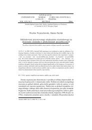
Medycyna Wet. 2008, 64 (12) 1399Fig. 3. Macroscopic view of <strong>calcinosis</strong> <strong>circumscripta</strong>Fig. 4. Microscopic view of <strong>calcinosis</strong> <strong>circumscripta</strong>. Note thevisible proliferating granulomatous inflammation in the surroundingsof the calcium masses. Stain HE, magnification 100 ×moderate pressure. The calcified masses were usually foundto be hypovascularized, occasionally supplied with small--sized blood vessels or more frequently, the absence of bloodvessels within their area was noted. The size of lesions excisedin the early stage of treatment averaged from 0.5 to 1.5 cm.The other pathological changes were variably sized even upto 3-5 cm in diameter. The consistency of resected formationsvaried from semi-liquid to solid, brittle mass of oval shapes.The cut section of the aggregates had chalky calcified foci(fig. 3). During the follow-up period the patients were providedwith an antibiotic cover.The surgically removed mineralized cutaneous masses weresubmitted to histopathologic evaluation. The collected segmentswere fixed in buffered formalin, embedded in paraffin,processed to sections and stained with hematoxylin and eosin.The microscopic picture of studied lesions revealed basophiliccalcified mass stained violet under hematoxylin and eosin.Quite frequently, the aggregates of calcification were surroundedby a visible inflammatory-granulomatous reactionoccurring as a proliferating fibroconnective tissue (fig. 4).Disease recurrence was reported in three cases, a short timeafter surgical treatment, i.e. 2 months post operation, in twodogs and a year after in one dog. The recurrent lesions weresmall-sized and painless. Only one animal had another surgeryperformed after two years.Results and discussionCalcinosis <strong>circumscripta</strong> (CC) is presented in literatureas a disorder affecting both humans and variousspecies of animals. In medicine CC defines smallmineralized foci formed within the subcutaneous tissue.Bigger lesions located in the connective tissue arecalled tumoral <strong>calcinosis</strong>, but both terms are equallyapplicable in veterinary medicine (1, 11, 14). However,in the literature this disorder has been identifiedby many names, such as cutaneous <strong>calcinosis</strong>, calciumgout, lipo<strong>calcinosis</strong>, apocrine cystic <strong>calcinosis</strong>, hipstone, Kalkgicht, which follows from the fact that thepathogenesis of this disease is not established (6, 14,15). Therefore, considering the potential etiology ofCC, different factors are taken into account, whichmakes it possible to classify the disorder into a fewcategories.Pathological processes that damage cutaneous tissues,impair proper blood supply and nutrient flow arelikely to induce calcium deposition. These processesinclude, among others, connective tissue diseases, trauma,neoplasms, degeneration as well as formation ofabscesses, hematomas and granulomas. This type of<strong>calcinosis</strong> is termed dystrophic and is not associatedwith calcium and phosphorus concentration in bloodserum (6, 14, 15). The same group may include iatrogenic<strong>calcinosis</strong> mentioned while discussing the surgicalprocedures performed earlier as well as combinedtreatment with medroxy-progesterone and proligestoneinjections (4, 5, 9, 12, 14-16). Calcinosis incidencehas been reported as a result of long-standinginflammations caused by a foreign body, among others,polydioxanone suture material, otitis externa, interdigitalspace inflammation, demodicosis, neoplasticand degenerative changes detected within apocrineglands as well as urate gout (6, 7, 11-16). Most <strong>calcinosis</strong><strong>circumscripta</strong> lesions described were formed incutaneous tissues close to the joints and over bony prominences,where tissue is especially susceptible to injury.That may support the theory of trauma being themechanism responsible for CC lesion development.In three cases reported (17%) calcification occurreddue to an apparent damage to tissues, whereas in theother reports no single cause was established. However,mechanical trauma can not be ruled out asa mechanism promoting or contributing to the formationof these pathological changes. None of the dogshad similar symptoms in the case history and noneof them had surgical procedures performed in theaffected region earlier. Only in one patient <strong>calcinosis</strong>circumcripta was not the only disease that occurred.A six-month-old West Highland White Terrier waspresented initially to the Clinic and Department ofAnimal Surgery for severe deformations of the mandibleregion that persisted for over 2 weeks. TheX-ray evaluation of the puppy’s head revealed thepresence of cranial mandibular osteopathy. A sub-