A Mechanistic View of the Formation of ... - CIC biomaGUNE
A Mechanistic View of the Formation of ... - CIC biomaGUNE
A Mechanistic View of the Formation of ... - CIC biomaGUNE

You also want an ePaper? Increase the reach of your titles
YUMPU automatically turns print PDFs into web optimized ePapers that Google loves.
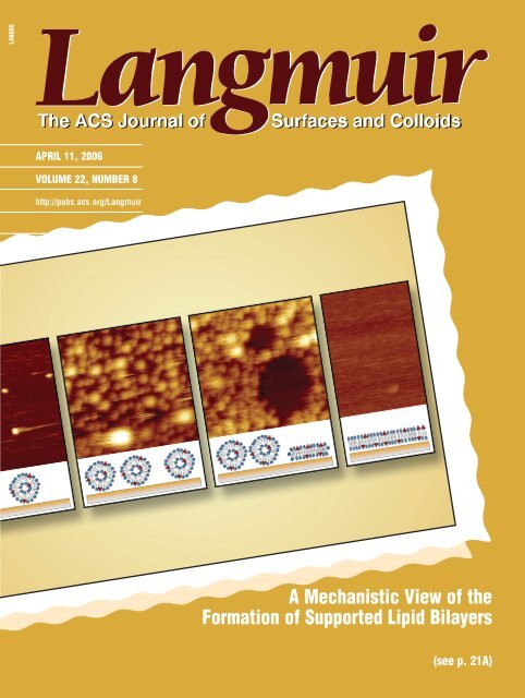
LANGD5APRIL 11, 2006VOLUME 22, NUMBER 8http://pubs.acs.org/LangmuirA <strong>Mechanistic</strong> <strong>View</strong> <strong>of</strong> <strong>the</strong><strong>Formation</strong> <strong>of</strong> Supported Lipid Bilayers(see p. 21A)
Langmuir 2006, 22, 3497-35053497InVited Feature Article<strong>Formation</strong> <strong>of</strong> Solid-Supported Lipid Bilayers: An Integrated <strong>View</strong>Ralf P. Richter,* ,† Rémi Bérat, and Alain R. BrissonLaboratoire d'Imagerie Moléculaire et Nanobiotechnologie, IECB, UMR-CNRS 5471, UniVersité BordeauxI, 2 Rue Robert Escarpit, 33607 Pessac Cedex, FranceReceiVed October 4, 2005. In Final Form: NoVember 22, 2005Supported lipid bilayers (SLBs) are popular models <strong>of</strong> cell membranes with potential bio-technological applications.A qualitative understanding <strong>of</strong> <strong>the</strong> process <strong>of</strong> SLB formation after exposure <strong>of</strong> small lipid vesicles to a hydrophilicsupport is now emerging. Recent studies have revealed a stunning variety <strong>of</strong> effects that can take place during thisself-organization process. The ensemble <strong>of</strong> results in our group has revealed unprecedented insight into intermediates<strong>of</strong> <strong>the</strong> SLB-formation process and has helped to identify a number <strong>of</strong> parameters that are determinant for <strong>the</strong> lipiddeposition on solid supports. The pathway <strong>of</strong> lipid deposition can be tuned by electrostatic interactions and by <strong>the</strong>presence <strong>of</strong> calcium. We emphasize <strong>the</strong> importance <strong>of</strong> <strong>the</strong> solid support in <strong>the</strong> SLB-formation process. Our resultssuggest that <strong>the</strong> molecular-level interaction between lipids and <strong>the</strong> solid support needs to be considered explicitly,to understand <strong>the</strong> rupture <strong>of</strong> vesicles and <strong>the</strong> formation <strong>of</strong> SLBs as well as to predict <strong>the</strong> properties <strong>of</strong> <strong>the</strong> resultingSLB. The impact <strong>of</strong> <strong>the</strong> SLB-formation process on <strong>the</strong> quality and <strong>the</strong> physical properties <strong>of</strong> <strong>the</strong> resulting SLB aswell as implications for o<strong>the</strong>r types <strong>of</strong> surface-confined lipid bilayers are discussed.IntroductionBiological membranes play key roles in cell life, controlling<strong>the</strong> transfer <strong>of</strong> information and <strong>the</strong> transport <strong>of</strong> ions and moleculesbetween <strong>the</strong> inside and outside cellular worlds and participatingin various intra- and extracellular processes. These highly complexand dynamic assemblies, only a few nanometers thick, consist<strong>of</strong> two main components: a two-dimensional space made <strong>of</strong>lipid molecules held toge<strong>the</strong>r by hydrophobic interactions andself-assembled as a continuous bilayer and proteins embeddedwithin <strong>the</strong> membrane or transiently associated with it.Our current knowledge <strong>of</strong> <strong>the</strong> molecular processes occurringat biological membranes is based on studies performed both onintegrated and on reconstituted systems using models <strong>of</strong> biologicalmembranes. The deposition <strong>of</strong> model membranes on solidsupports has become very popular, 1-3 both for studying basicmembrane processes and for possible biotechnological applications.4-16 The growing interest in confining lipid membranes on* Corresponding author. E-mail: ralf.richter@urz.uni-heidelberg.de.† Current address: Department <strong>of</strong> Biophysical Chemistry, Institute <strong>of</strong>Physical Chemistry, Heidelberg University, Im Neuenheimer Feld 253, 69120 Heidelberg, Germany.(1) Sackmann, E. Science 1996, 271, 43-48.(2) Boxer, S. G. Curr. Opin. Chem. Biol. 2000, 4, 704-709.(3) Knoll, W.; Frank, C. W.; Heibel, C.; Naumann, R.; Offenhäusser, A.;Rühe, J.; Schmidt, E. K.; Shen, W. W.; Sinner, A. ReV. Mol. Biotechnol. 2000,74, 137-158.(4) Bieri, C.; Ernst, O. P.; Heyse, S.; H<strong>of</strong>mann, K. P.; Vogel, H. Nat. Biotechnol.1999, 17, 1105-1108.(5) Kung, L. A.; Kam, L.; Hovis, J. S.; Boxer, S. G. Langmuir 2000, 16,6773-6776.(6) Sapuri, A. R.; Baksh, M. M.; Groves, J. T. Langmuir 2003, 19, 1606-1619.(7) Sackmann, E.; Tanaka, M. Trends Biotechnol. 2000, 18, 58-64.(8) Knoll, W.; Park, H.; Sinner, E. K.; Yao, D.; Yu, F. Surf. Sci. 2004, 570,30-42.(9) Reviakine, I.; Brisson, A. Langmuir 2001, 17, 8293-8299.(10) Kam, L.; Boxer, S. G. Langmuir 2003, 19, 1624-1631.(11) Larsson, C.; Rodahl, M.; Höök, F. Anal. Chem. 2003, 75, 5080-5087.(12) Milhiet, P. E.; Giocondi, M.-C.; Baghdadi, O.; Ronzon, F.; Roux, B.; leGrimellec, C. EMBO Rep. 2002, 3, 485-490.(13) Yip, C. M.; Darabie, A. A.; McLaurin, J. J. Mol. Biol. 2002, 318,97-107.surfaces has been nourished by <strong>the</strong> emergence <strong>of</strong> a multitude <strong>of</strong>surface-sensitive characterization techniques, 5,17-19 advancedsurface patterning methods, 5,20-24 and liquid handling systems(micr<strong>of</strong>luidics). 10During <strong>the</strong> past decade <strong>the</strong> conceptual base <strong>of</strong> surface-confinedmembrane systems has grown considerably. A large number <strong>of</strong>systems has been described, including solid-supported lipidbilayers, 9,11,15,25,26 polymer-cushioned lipid bilayers, 27-29 hybridbilayers, 30,31 te<strong>the</strong>red lipid bilayers, 32 suspended lipid bilayers, 33,34(14) Salafsky, J.; Groves, J. T.; Boxer, S. G. Biochemistry 1996, 35, 5, 14773-14781.(15) Reviakine, I.; Bergsma-Schutter, W.; Brisson, A. J. Struct. Biol. 1998,121, 356-361.(16) Watts, T. H.; Brian, A. A.; Kappler, J. W.; Marrack, P.; McConnell, H.M. Proc. Natl. Acad. Sci. U.S.A. 1984, 81, 7564-7568.(17) Shao, Z.; Mou, J.; Czajkowsky, D. M.; Yang, J.; Yuan, J.-Y. AdV. Phys.1996, 45, 1-86.(18) Knoll, W. Annu. ReV. Phys. Chem. 1998, 49, 569-638.(19) Höök, F.; Larsson, C.; Fant, C. In Encyclopedia <strong>of</strong> Surface and ColloidScience; Marcel Dekker: New York, 2002; pp 774-791.(20) Srinivasan, M. P.; Ratto, T. V.; Stroeve, P.; Longo, M. L. Langmuir 2001,17, 7951-7954.(21) Michel, R.; Reviakine, I.; Su<strong>the</strong>rland, D.; Fokas, C.; Csucs, G.; Danuser,G.; Spencer, N. D.; Textor, M. Langmuir 2002, 18, 8580-8586.(22) Svedhem, S.; Pfeiffer, I.; Larsson, C.; Wingren, C.; Borrebaeck, C.; Höök,F. ChemBioChem 2003, 4, 339-343.(23) Morigaki, K.; Kiyosue, K.; Taguchi, T. Langmuir 2004, 20, 7729-7735.(24) Jackson, B. L.; Groves, J. T. J. Am. Chem. Soc. 2004, 126, 13878-13879.(25) Brian, A. A.; McConnell, H. M. Proc. Natl. Acad. Sci. U.S.A. 1984, 81,6159-6163.(26) Lenz, P.; Ajo-Franklin, C. M.; Boxer, S. G. Langmuir 2004, 20, 11092-11099.(27) Goennenwein, S.; Tanaka, M.; Hu, B.; Moroder, L.; Sackmann, E. Biophys.J. 2003, 85, 646-655.(28) Wagner, M. L.; Tamm, L. K. Biophys. J. 2001, 81, 266-275.(29) Munro, J. C.; Frank, C. W. Langmuir 2004, 20, 10567-10575.(30) Silin, V. I.; Wieder, H.; Woodward, J. T.; Valincius, G.; Offenhausser,A.; Plant, A. L. J. Am. Chem. Soc. 2002, 124, 14676-14683.(31) Terrettaz, S.; Mayer, M.; Vogel, H. Langmuir 2003, 19, 5567-5569.(32) Purrucker, O.; Förtig, A.; Jordan, R.; Tanaka, M. ChemPhysChem 2004,5, 327-335.(33) Römer, W.; Lam, Y. H.; Fischer, D.; Watts, A.; Fischer, W. B.; Göring,P.; Wehrspohn, R. B.; Gösele, U.; Steinem, C. J. Am. Chem. Soc. 2004, 126,16267-16274.(34) Römer, W.; Steinem, C. Biophys. J. 2004, 86, 955-965.10.1021/la052687c CCC: $33.50 © 2006 American Chemical SocietyPublished on Web 01/14/2006
3498 Langmuir, Vol. 22, No. 8, 2006 Richter et al.Figure 1. Surface-confined membrane models: (A) solid-supportedlipid bilayer; (B) polymer-cushioned lipid bilayer; (C) hybrid bilayer,consisting <strong>of</strong> a self-assembled monolayer (e.g., thiols on Au or silaneson glass or silica) and a lipid monolayer; (D) te<strong>the</strong>red lipid bilayer;(E) freely suspended lipid bilayer; (F-G) supported vesicular layers.or supported vesicular layers 22,35 (Figure 1). In parallel, a multitude<strong>of</strong> methods has been proposed to create such biomimetic edifices,including Langmuir-type approaches (Langmuir-Blodgett orLangmuir-Schäfer deposition) 36-39 and <strong>the</strong> spreading <strong>of</strong> vesicleson various preconditioned supports. 20,38,40-44The spreading <strong>of</strong> small lipid vesicles on hydrophilic solidsupports, pioneered by McConnell et al., 25 presents an attractiveand simple route to form supported lipid bilayers (SLBs). Theone-step procedure allows creating SLBs <strong>of</strong> different lipidmixtures. 45,46 The fact that such SLBs form a fluid twodimensionalspace allowing free diffusion in translation androtation <strong>of</strong> lipid molecules and lipid-associated proteins makes<strong>the</strong>m well suited to analyze lipid domain formation, 13,47-54intermembrane interactions, 55-57 or membrane processes such(35) Yoshina-Ishii, C.; Boxer, S. G. J. Am. Chem. Soc. 2003, 125, 3696-3697.(36) Swart, R. M. In Langmuir-Blodgett Films; Roberts, G., Ed.; PlenumPress: New York, 1990; pp 273-316.(37) Grandbois, M.; Clausen-Schaumann, H.; Gaub, H. E. Biophys. J. 1998,74, 2398-2404.(38) Wong, J. Y.; Majewski, J.; Seitz, M.; Park, C. K.; Israelachvili, J.; Smith,G. S. Biophys. J. 1999, 77, 1445-1457.(39) Ross, M.; Steinem, C.; Galla, H.-J.; Jansh<strong>of</strong>f, A. Langmuir 2001, 17,2437-2445.(40) Steinem, C.; Jansh<strong>of</strong>f, A.; Ulrich, W.-P.; Sieber, M.; Galla, H.-J. Biochim.Biophys. Acta 1996, 1279, 169-180.(41) Keller, C. A.; Kasemo, B. Biophys. J. 1998, 75, 1397-1402.(42) Starr, T. E.; Thompson, N. L. Langmuir 2000, 16, 10301-10308.(43) Zhao, J.; Tamm, L. K. Langmuir 2003, 19, 1838-1846.(44) Baumgart, T.; Offenhäusser, A. Langmuir 2003, 19, 1730-1737.(45) Richter, R. P.; Mukhopadhyay, A.; Brisson, A. Biophys. J. 2003, 85,3035-3047.(46) Richter, R. P.; Brisson, A. Biophys. J. 2005, 88, 3422-3433.(47) Mou, J.; Yang, J.; Shao, Z. J. Mol. Biol. 1995, 248, 507-512.(48) Reviakine, I.; Simon, A.; Brisson, A. Langmuir 2000, 16, 1473-1477.(49) Schneider, J.; Dufrêne, Y. F.; Barger, W. R., Jr.; Lee, G. U. Biophys. J.2000, 79, 1107-1118.(50) Rinia, H. A.; Kruijff, B. d. FEBS Lett. 2001, 504, 194-199.(51) Lawrence, J. C.; Saslowsky, D. E.; Edwardson, J. M.; Henderson, R. M.Biophys. J. 2003, 84, 1827-1832.(52) Tokumasu, F.; Jin, A. J.; Feigenson, G. W.; Dvorak, J. A. Biophys. J.2003, 84, 2609-2618.(53) Giocondi, M.-C.; Milhiet, P. E.; Dosset, P.; le Grimellec, C. Biophys. J.2004, 86, 861-869.(54) Jansh<strong>of</strong>f, A.; Bong, D. T.; Steinem, C.; Johnson, J. E.; Ghadiri, M. R.Biochemistry 1999, 38, 5328-5336.(55) Kaizuka, Y.; Groves, J. T. Biophys. J. 2004, 86, 905-912.(56) Parthasarathy, R.; Groves, J. T. Proc. Natl. Acad. Sci. U.S.A. 2004, 101,12798-12803.as protein adsorption, 58,59 protein self-assembly, 13,48,60 proteinlocalization at lipid phase boundaries, 12 or protein function. 37The self-organization steps involved in this methodsvesicleadsorption, rupture, and spreading into planar membranesspresent fundamental interest in colloidal and interfacial science.Both <strong>the</strong>oretical 61-63 and experimental work 41,45,64-70 during <strong>the</strong>past decade have considerably improved <strong>the</strong> general understanding<strong>of</strong> this process, and a detailed image <strong>of</strong> <strong>the</strong> structuralintermediates and <strong>the</strong> driving forces is now emerging. Figure 2shows four archetypes <strong>of</strong> lipid deposition processes, as followedby quartz crystal microbalance with dissipation monitoring(QCM-D). The QCM-D technique has proven very valuable toscreen <strong>the</strong> overall properties <strong>of</strong> <strong>the</strong> lipid deposition, 41 thanks to<strong>the</strong> dissipation parameter that allows distinguishing betweenintact, adsorbed vesicles (high dissipation) and bilayer patches(low dissipation). As shown in <strong>the</strong> schemes in Figure 2, vesiclesei<strong>the</strong>r do not adsorb (Figure 2A), adsorb and remain intact, givingrise to a supported vesicular layer (SVL) (Figure 2B), or forman SLB (Figure 2, panels C and D). Notably, SLB formation canoccur via two scenarios with distinctly different kinetics. In onecase, vesicles rupture quickly upon interaction with <strong>the</strong> solidsupport (Figure 2D), whereas in <strong>the</strong> o<strong>the</strong>r, a large amount <strong>of</strong>intact vesicles is adsorbed at an intermediate state <strong>of</strong> <strong>the</strong> process(Figure 2C).Recent technical developments, combining QCM-D and atomicforce microscopy (AFM), have allowed us to characterize <strong>the</strong>intermediate states leading to SLB formation in unprecedenteddetail. Here we present an overview <strong>of</strong> work performed in ourgroup that sheds light on <strong>the</strong> mechanisms and critical parametersinvolved in <strong>the</strong> formation <strong>of</strong> SLBs as well as on <strong>the</strong> propertiesand <strong>the</strong> quality <strong>of</strong> <strong>the</strong> resulting SLB.Mechanism <strong>of</strong> SLB <strong>Formation</strong>To satisfactorily describe <strong>the</strong> mechanism <strong>of</strong> SLB formation,two critical steps in this process need to be understood: (i) <strong>the</strong>adhesion and rupture <strong>of</strong> vesicles on <strong>the</strong> support and (ii) <strong>the</strong>evolution <strong>of</strong> <strong>the</strong> supported bilayer patches thus formed into acomplete SLB. Figure 3 provides an overview <strong>of</strong> mechanisms<strong>of</strong> vesicle rupture that have been reported or suggested in <strong>the</strong>literature.Stability <strong>of</strong> Adsorbed Vesicles. A simple rationale to evaluate<strong>the</strong> binding and <strong>the</strong> stability <strong>of</strong> surface-bound vesicles wasprovided by <strong>the</strong> <strong>the</strong>ory <strong>of</strong> Seifert and Lipowsky. 62,71 In <strong>the</strong>ircontinuum approach, where <strong>the</strong> bilayer is treated as a thin twodimensionalsheet embedded in three-dimensional space, <strong>the</strong>balance between <strong>the</strong> gain in adhesion energy (as given by <strong>the</strong>(57) Fix, M.; Melia, T. J.; Jaiswal, J. K.; Rappoport, J. Z.; You, D.; Söllner,T. H.; Rothman, J. E.; Simon, S. M. Proc. Natl. Acad. Sci. U.S.A. 2004, 101,7311-7316.(58) Corsel, J. W.; Willems, G. M.; Kop, J. M. M.; Cuypers, P. A.; Hermens,W. T. J. Colloid Interface Sci. 1986, 111, 544-554.(59) Andree, H. A. M.; Stuart, M. C. A.; Hermens, W. T.; Reutelingsperger,C. P. M.; Hemker, H. C.; Frederik, P. M.; Willems, G. M. J. Biol. Chem. 1992,267, 17907-17912.(60) Czaikowsky, D. M.; Shao, Z. FEBS Lett. 1998, 430, 51-54.(61) Seifert, U.; Lipowsky, R. Phys. ReV. A1990, 42, 4768-4771.(62) Seifert, U. AdV. Phys. 1997, 46, 13-137.(63) Zhdanov, V. P.; Keller, C. A.; Glasmästar, K.; Kasemo, B. J. Chem. Phys.2000, 112, 900-909.(64) Rädler, J.; Strey, H.; Sackmann, E. Langmuir 1995, 11, 4539-4548.(65) Keller, C. A.; Glasmästar, K.; Zhdanov, V. P.; Kasemo, B. Phys. ReV.Lett. 2000, 84, 5443-5446.(66) Jass, J.; Tjärnhage, T.; Puu, G. Biophys. J. 2000, 79, 3153-3163.(67) Reviakine, I.; Brisson, A. Langmuir 2000, 16, 1806-1815.(68) Johnson, J. M.; Taekijp, H.; Chu, S.; Boxer, S. G. Biophys. J. 2002, 83,3371-3379.(69) Reimhult, E.; Höök, F.; Kasemo, B. Langmuir 2003, 19, 1681-1691.(70) Benes, M.; Billy, D.; Benda, A.; Speijer, H.; H<strong>of</strong>, M.; Hermens, W. T.Langmuir 2004, 20, 10129-10137.(71) Lipowsky, R.; Seifert, U. Mol. Crys. Liq. Cryst. 1991, 202, 17-25.
<strong>Formation</strong> <strong>of</strong> Solid-Supported Lipid Bilayers Langmuir, Vol. 22, No. 8, 2006 3499Figure 2. Lipid deposition pathways measured by QCM-D on silica. (A) Vesicles do not adsorb. (B) Vesicles adsorb and remain intact,forming a supported vesicular layer (SVL). (C) Vesicles adsorb and remain initially intact. At high vesicular coverage an SLB is formed.(D) Vesicles adsorb and rupture instantaneously, to form an SLB. The dissipation, ∆D, allows distinguishing between <strong>the</strong> morphologicalstate <strong>of</strong> <strong>the</strong> adsorbed lipids: intact vesicles exhibit high dissipation while bilayer (patches) show low dissipation. The legends indicate <strong>the</strong>lipids usedsdioleoyltrimethylammonium-propane (DOTAP), dioleoylphosphatidylcholine (DOPC), and dioleoylphosphatidylserine (DOPS)swith <strong>the</strong>ir molar mixing ratios. 45Figure 3. Mechanisms <strong>of</strong> vesicle rupture: (A) an isolated adsorbed vesicle ruptures spontaneously, driven by its support-induced deformation;(B) neighboring adsorbed vesicles fuse and eventually rupture; (C) <strong>the</strong> active edge <strong>of</strong> a supported bilayer patch induces <strong>the</strong> rupture <strong>of</strong> aneighboring vesicle; (D) <strong>the</strong> cooperative action <strong>of</strong> several neighboring vesicles leads to <strong>the</strong> rupture <strong>of</strong> a first vesicle (at <strong>the</strong> critical vesicularcoverage). The active edge <strong>the</strong>reby exposed triggers <strong>the</strong> rupture <strong>of</strong> adjacent vesicles.adhesion area) and <strong>the</strong> cost in <strong>the</strong> vesicles’ curvature energy (asgiven by <strong>the</strong> bilayer’s bending rigidity) is determinant for <strong>the</strong>adsorption, deformation, and rupture <strong>of</strong> vesicles. Initial data inour group provided support for this model for egg-PC on mica. 67The examples given in Figure 2, panels A, B, and D, exemplify<strong>the</strong> scenarios were vesicles do not adsorb, adsorb intact, andrupture spontaneously, respectively.However, recent experimental data have provided evidencethat this continuum approach does not convey <strong>the</strong> whole answerto <strong>the</strong> question <strong>of</strong> vesicular stability under conditions commonlyemployed for SLB formation. Cooperative effects <strong>of</strong> neighboringvesicles as well as <strong>the</strong> dynamic distribution <strong>of</strong> different lipidspecies in a vesicle have to be taken into account for a betterdescription <strong>of</strong> <strong>the</strong> rupture propensity <strong>of</strong> surface-bound vesicles.Critical Vesicular Coverage. An intriguing effect <strong>of</strong> <strong>the</strong>cooperative action <strong>of</strong> surface-bound vesicles was first reportedby Kasemo and co-workers. By combining measurements byQCM-D and surface plasmon resonance (SPR) 65 toge<strong>the</strong>r withcomputer simulations, 63 <strong>the</strong> group could show (i) that isolatedvesicles <strong>of</strong> egg-PC remain intact when bound to a silica supportand (ii) that a certain surface density <strong>of</strong> vesicles (henceforwarddenoted <strong>the</strong> critical vesicular coverage) is required to initiate <strong>the</strong>decomposition <strong>of</strong> surface-bound vesicles into bilayer patches.Zhdanov and Kasemo 72 proposed that <strong>the</strong> support-induced stress(or deformation) <strong>of</strong> an adsorbed vesicle is fur<strong>the</strong>r enhanced by<strong>the</strong> adsorption <strong>of</strong> vesicles in its vicinity. When a certainconfinement <strong>of</strong> neighboring vesicles, corresponding to <strong>the</strong> criticalcoverage, is reached, <strong>the</strong> stress on <strong>the</strong> vesicle becomes sufficientto induce its rupture (Figure 3D).Figure 2C exemplifies <strong>the</strong> response obtained by QCM-D, when<strong>the</strong> critical vesicular coverage is involved. As techniques suchas QCM-D and SPR give average information about <strong>the</strong> adsorbedmaterial, a small fraction <strong>of</strong> prematurely ruptured vesicles maypotentially go undetected. 73 Our images by atomic forcemicroscopy (AFM) provide direct evidence that silica waferscan indeed be covered with vesicles that remain stable for days,being devoid <strong>of</strong> bilayer patches over areas <strong>of</strong> several squaremicrometers (Figure 4). 45 These images also demonstrate <strong>the</strong>(72) Zhdanov, V. P.; Kasemo, B. Langmuir 2001, 17, 3518-3521.(73) Reimhult, E.; Höök, F.; Kasemo, B. J. Chem. Phys. 2002, 117, 7401-7404.
3500 Langmuir, Vol. 22, No. 8, 2006 Richter et al.Figure 4. Imaging an intermediate <strong>of</strong> <strong>the</strong> SLB-formation processby AFM. Vesicles made <strong>of</strong> DOPC/DOPS (4:1) were exposed to asilica wafer. Spherical objects, identified as vesicles, densely populate<strong>the</strong> surface. No bilayer patches are visible, indicating that <strong>the</strong> criticalvesicular coverage is not attained. Image size (z scale): 2 µm (50nm). Adapted from ref 45. Copyright 2003 Biophysical Society.Figure 5. Tracing vesicle rupture kinetics by QCM-D. Vesiclesinitially adsorb intact but rupture into bilayer patches over <strong>the</strong> timerange <strong>of</strong> 30 min and more, as indicated by <strong>the</strong> decrease in dissipation(- - -) and <strong>the</strong> increase in frequency (-O-), after rinsing away vesiclesin solution (arrow). The AFM image (inset, image size: 750 nm ×500 nm), taken after <strong>the</strong> QCM-D measurement, confirms <strong>the</strong>coexistence <strong>of</strong> vesicles and bilayer patches. Adapted from ref 46.Copyright 2005 Biophysical Society.strength <strong>of</strong> <strong>the</strong> AFM to resolve <strong>the</strong> local morphology <strong>of</strong> <strong>the</strong> lipidassemblies down to nanometer resolution.Long-Term Stability <strong>of</strong> Adsorbed Vesicles. We observed apeculiar effect for vesicles containing a mixture <strong>of</strong> DOPC andDOPS when exposed to mica in a calcium-containing solution:when adsorbing vesicles at low surface density (i.e., <strong>the</strong> interaction<strong>of</strong> neighboring vesicles is negligible), <strong>the</strong>y initially remainedintact but ruptured individually over a time range <strong>of</strong> minutes tohours (Figure 5). 74 This strongly contrasted our commonobservation on silica supports that isolated vesicles ei<strong>the</strong>r ruptureimmediately (i.e., within less than a second) after adsorption orremain intact for days.What is <strong>the</strong> origin <strong>of</strong> such particular rupture kinetics? It appearsreasonable that a support-induced reorganization <strong>of</strong> <strong>the</strong> two lipidspecies within <strong>the</strong> adsorbed vesicle may lead to dynamic changesin <strong>the</strong> vesicle-support interaction and in <strong>the</strong> stability <strong>of</strong> <strong>the</strong>vesicles. The observed time range for rupture is, however, much(74) Richter, R. P.; Brisson, A. Biophys. J. 2005, 88, 3422-3433.Figure 6. Tracking <strong>the</strong> propagation <strong>of</strong> bilayer patch formation byAFM. (A) All vesicles are intact. (B) Two vesicle segments areresolved (arrowheads) followed by an extended bilayer domain,indicating <strong>the</strong> rupture <strong>of</strong> vesicles. The cross sections <strong>of</strong> threesuccessive scan lines (right) reveals that <strong>the</strong> two vesicles do notrupture simultaneoulsy: The right vesicle (white arrowhead) rupturesfirst (between scan line 1 and 2), likely induced by <strong>the</strong> AFM-tip.At scan line 3, <strong>the</strong> left vesicle (black arrowhead) is ruptured, likelyinduced by <strong>the</strong> “active edge” <strong>of</strong> <strong>the</strong> bilayer patch that is formed from<strong>the</strong> right vesicle. (C) The rupture <strong>of</strong> a single vesicle induces <strong>the</strong>transformation <strong>of</strong> several adjacent vesicles into a stable bilayer patch.A small gap (a few nanometers) separates <strong>the</strong> patch edge fromneighboring intact vesicles. A part <strong>of</strong> <strong>the</strong> image is distorted because<strong>the</strong> tip was accidentally retracted from <strong>the</strong> surface. Image size: 250nm. The slow scan direction is indicated with white arrows. Adaptedfrom ref 45. Copyright 2003 Biophysical Society.slower than <strong>the</strong> time needed for lipids within a single lipid leafletto reorganize. 75 We <strong>the</strong>refore propose that <strong>the</strong> translocation <strong>of</strong>lipids between <strong>the</strong> two leaflets <strong>of</strong> <strong>the</strong> vesicle is <strong>the</strong> parameterresponsible for <strong>the</strong> slow vesicle rupture. The suggested rupturemechanism correlates with our observation that mica induces anasymmetric inter-leaflet lipid distribution in SLBs, 76 an issuethat will be discussed in detail later on.Growth and Coalescence <strong>of</strong> Supported Lipid Bilayers. Oncea vesicle has ruptured, <strong>the</strong> resulting bilayer patch exposes anedge. 77,78 These edges are energetically unfavorable and, at leastfrom a <strong>the</strong>rmodynamic perspective, expected to promote <strong>the</strong>interaction with adjacent lipid material, such as <strong>the</strong> rupture <strong>of</strong>surface-bound vesicles (Figure 3C) or vesicles from solution.Provided <strong>the</strong> density <strong>of</strong> adsorbed vesicles is sufficiently high,such a process can propagate in a cascade <strong>of</strong> rupture eventsacross several neighboring vesicles and leads to <strong>the</strong> formation<strong>of</strong> extended bilayer patches. 45,63 The intermediate steps in thisprocess can be traced by AFM, as illustrated in Figure 6, andsuggest that <strong>the</strong> propagation speed is in <strong>the</strong> range <strong>of</strong> seconds. 45Fur<strong>the</strong>rmore, adjacent bilayer patches usually coalesce in order(75) Bernard, A.-L.; Guedeau-Boudeville, M.-A.; Jullien, L.; de Meglio, J.-M.Langmuir 2000, 16, 6809-6820.(76) Richter, R. P.; Maury, N.; Brisson, A. Langmuir 2005, 21, 299-304.(77) Kasson, P. M.; Pande, V. S. Biophys. J. 2004, 86, 3744-3749.(78) Jiang, F. Y.; Bouret, Y.; Kindt, J. H. Biophys. J. 2004, 87, 182-192.
<strong>Formation</strong> <strong>of</strong> Solid-Supported Lipid Bilayers Langmuir, Vol. 22, No. 8, 2006 3501Figure 8. Possible scenarios <strong>of</strong> <strong>the</strong> mutual interaction <strong>of</strong> neighboringvesicles. The surface-induced flattening <strong>of</strong> a newly adsorbing vesicleinduces <strong>the</strong> deformation <strong>of</strong> a neighboring one. If sliding and rollingare inhibited (A), <strong>the</strong> deformation represents a persisting stress for<strong>the</strong> vesicles, and facilitates <strong>the</strong>ir rupture. Sliding or rolling along <strong>the</strong>surface can release <strong>the</strong> stress (B). Thus, if sliding or rolling is enabled,neighboring vesicles can only induce added stress (and <strong>the</strong>rebyrupture) when a high overall packing <strong>of</strong> vesicles on <strong>the</strong> surface isattained.Figure 7. (A) Incomplete SLB made from DOPC/DOPS (4:1)vesicles on mica: bilayer patches are predominantly circular,indicating that <strong>the</strong>y are laterally mobile. Image size: 1 µm. (B)Incomplete SLB made from DOTAP-vesicles on silica. Some bilayerpatches (arrowheads) exhibit stable, strongly noncircular shapes.Image size: 1 µm. (C-F) Sequential images <strong>of</strong> patch coalescenceinduced by dynamic changes in <strong>the</strong> shape <strong>of</strong> a bilayer patch. After<strong>the</strong> merger <strong>of</strong> patches 1-3 (C), induced by <strong>the</strong> AFM-tip, <strong>the</strong>coalescence with patches 4 (D), 5-6 (E), and 7 (F) is generated bymovements <strong>of</strong> <strong>the</strong> reshaping patch. Image size: 1.75 µm. Adaptedfrom ref 46. Copyright 2005 Biophysical Society.to minimize <strong>the</strong>ir edge length. 45,46,67 Taken toge<strong>the</strong>r, <strong>the</strong>se effectsincrease <strong>the</strong> size <strong>of</strong> individual bilayer patches and <strong>the</strong> overallbilayer coverage and will, in <strong>the</strong> ideal case, lead to a completeSLB. 45,46Some <strong>of</strong> <strong>the</strong> vesicles imaged in Figure 6 remain intact eventhough <strong>the</strong>y are situated as close as a few nanometers to <strong>the</strong> edge<strong>of</strong> a bilayer patch. 45 This suggests that <strong>the</strong> edge almost needs tocontact a vesicle to induce its rupture and illustrates that <strong>the</strong>efficiency <strong>of</strong> edge-induced processes relies strongly on <strong>the</strong> spatialarrangement <strong>of</strong> vesicles and bilayer patches.Lateral Mobility <strong>of</strong> Vesicles and Bilayer Patches. Lipidassemblies as a whole can be laterally mobile and undergocollective shape changes, an effect not to be confused with <strong>the</strong>lateral diffusion <strong>of</strong> individual lipid molecules. The shape <strong>of</strong> bilayerpatches on <strong>the</strong> solid support provides a first indication about<strong>the</strong>ir mobility. Laterally mobile patches tend to reshape intocircular patches to minimize <strong>the</strong>ir line tension, an effect that weobserved on mica surfaces (Figure 7A). 46 In contrast, bilayerpatches on silica frequently retained a strongly noncircular shape(Figure 7B), providing evidence for <strong>the</strong> lack <strong>of</strong> mobility. 45It is instructive to compare our observations on <strong>the</strong> mobility<strong>of</strong> lipid assemblies on mica and on silica with data previouslyreported by Rädler et al. 64,79,80 With reflection interference contrastmicroscopy (RICM), <strong>the</strong>y observed that lipid bilayers, continuouslyformed from a deposited blob <strong>of</strong> concentrated DOPC inwater, easily slide over surfaces <strong>of</strong> both types <strong>of</strong> support. Thekinetics <strong>of</strong> <strong>the</strong> sliding motion on mica could be describedquantitatively by <strong>the</strong> shear flow <strong>of</strong> a thin water film that issandwiched between <strong>the</strong> solid support and <strong>the</strong> bilayer 81,82 andra<strong>the</strong>r high spreading coefficients <strong>of</strong> up to 40 µm 2 /s wereobtained. 79In light <strong>of</strong> <strong>the</strong> results by Rädler and co-workers, it was surprisingthat we found lipid assemblies to be immobile on silica. Also,<strong>the</strong> shape changes observed by us and o<strong>the</strong>rs 83 on mica seemconsiderably slower than postulated from <strong>the</strong> action <strong>of</strong> alubricating water film. 83 Variations in <strong>the</strong> employed experimentalconditions, in particular <strong>the</strong> presence <strong>of</strong> divalent ions 45,74,83 versuspure water, 64,79 may well be at <strong>the</strong> origin <strong>of</strong> <strong>the</strong> observeddifferences. However, <strong>the</strong> large range <strong>of</strong> variations in mobilityremains intriguing and points toward a current lack in understanding<strong>the</strong> coupling between <strong>the</strong> bilayer and <strong>the</strong> solid support.What kind <strong>of</strong> effects can be induced by <strong>the</strong> lateral mobility<strong>of</strong> lipid assemblies? The series <strong>of</strong> AFM images in Figure 7C-Fdemonstrates how dynamic changes <strong>of</strong> <strong>the</strong> patch shape canenhance <strong>the</strong> coalescence <strong>of</strong> neighboring bilayer patches. 46Similarly, vesicles are expected to rupture, induced by <strong>the</strong> activeedge <strong>of</strong> an approaching bilayer patch.The mobility <strong>of</strong> surface-bound vesicles also has importantimplications for <strong>the</strong> nature <strong>of</strong> <strong>the</strong> critical vesicular coverage.Mobile vesicles can avoid stress from neighboring vesicles bydisplacement along <strong>the</strong> surface (Figure 8B). Consequently, stressdue to intervesicle interactions can build up only when <strong>the</strong> oVerallvesicular coverage is high enough to force <strong>the</strong> vesicles to interact.The critical vesicular coverage is thus directly determined by <strong>the</strong>overall density <strong>of</strong> adsorbed vesicles. Given that intervesicleinteractions are commonly short-ranged, this implies that <strong>the</strong>critical coverage must be elevated. 72 In contrast, <strong>the</strong> shaperelaxation <strong>of</strong> immobile vesicles is constrained to <strong>the</strong> local(79) Nissen, J.; Gritsch, S.; Wiegand, G.; Rädler, J. O. Eur. Phys. J. B 1999,10, 335-344.(80) Nissen, J.; Jacobs, K.; Rädler, J. O. Phys. ReV. Lett. 2001, 86, 1904-1907.(81) Bayerl, T. M.; Bloom, M. Biophys. J. 1990, 58, 357-362.(82) Johnson, S. J.; Bayerl, T. M.; McDermott, D. C.; Adam, W. A.; Rennie,A. R.; Thomas, R. K.; Sackmann, E. Biophys. J. 1991, 59, 289-294.(83) Muresan, A. S.; Lee, K. Y. C. J. Phys. Chem. B 2001, 105, 852-855.
3502 Langmuir, Vol. 22, No. 8, 2006 Richter et al.environment (Figure 8A), and hence, <strong>the</strong> critical coverageoriginates in a local effect that involves a limited number <strong>of</strong>neighboring vesicles. In <strong>the</strong> minimal configuration, two immobilizedneighboring vesicles may be sufficient to inducerupture. Due to <strong>the</strong> statistical distribution <strong>of</strong> vesicles over <strong>the</strong>surface, <strong>the</strong> local effect, however, translates into an apparentcritical coverage <strong>of</strong> <strong>the</strong> ensemble. In this case <strong>the</strong> critical vesicularcoverage can thus span from very low to very high values, aswe could demonstrate for silica surfaces. 45Parameters that Govern SLB <strong>Formation</strong>Which pathway <strong>of</strong> vesicle deposition will be taken is essentiallydetermined by <strong>the</strong> interplay <strong>of</strong> bilayer-support, interbilayer, andintrabilayer interactions. In principle, <strong>the</strong> relative contribution<strong>of</strong> <strong>the</strong>se interactions will be susceptible to <strong>the</strong> nature <strong>of</strong> <strong>the</strong> support(its surface charge, chemical composition and roughness), <strong>the</strong>lipid vesicles (<strong>the</strong>ir composition, charge, size, and physical state),as well as <strong>the</strong> aqueous environment (its composition, pH andionic strength). In <strong>the</strong> following, we will outline some essentialexperimental parameters that appear to control SLB formation.Electrostatic Interactions. Several studies have pointed out<strong>the</strong> influence <strong>of</strong> <strong>the</strong> charge <strong>of</strong> support and lipids as well as <strong>the</strong>ionic strength <strong>of</strong> <strong>the</strong> solution on <strong>the</strong> adsorption <strong>of</strong> vesicles. 64,84-86In systematic studies on silica 45 and mica, 46 we provided evidencethat all four pathways <strong>of</strong> vesicle deposition outlined in Figure2 can actually be generated by varying one experimental parameteronly: <strong>the</strong> vesicle charge. These studies demonstrate that <strong>the</strong>SLB-formation process will be strongly influenced by electrostaticinteractions. Consequently, adjustments in <strong>the</strong> pH or in <strong>the</strong> ionicstrength are expected to constitute relatively simple means tooptimize <strong>the</strong> formation <strong>of</strong> SLBs for a given surface and a givenlipid composition. 85Calcium Ions. The influence <strong>of</strong> divalent ions in general andcalcium in particular appears notoriously surprising. The ions donot only participate in <strong>the</strong> screening <strong>of</strong> charges, <strong>the</strong>reby modifying<strong>the</strong> electrostatic interactions, but <strong>the</strong>y also directly interact, in<strong>of</strong>ten subtle ways, with surfaces and lipids. 48,87 As a generaltrend, calcium was found to promote <strong>the</strong> adsorption and rupture<strong>of</strong> vesicles and SLB formation. 45,67,84,88 Effects are particularlystrong on mica. 46,67,70 Often minor concentrations (mM and below)<strong>of</strong> <strong>the</strong> ion are sufficient to generate significant effects.Solid Support. The role <strong>of</strong> <strong>the</strong> solid support in <strong>the</strong> process<strong>of</strong> SLB formation cannot be underestimated. It is probably <strong>the</strong>most complex and still <strong>the</strong> most enigmatic parameter.Work on different supports has pointed out that hydrophilicityis a necessary 26 but not a sufficient condition to promote <strong>the</strong>rupture <strong>of</strong> vesicles and subsequent SLB formation. A number <strong>of</strong>reports has actually revealed difficulties to form SLBs on surfacessuch as gold, 41 SrTiO 2 , 42 TiO 2 , 42,73 or platinum, 69 leaving micaand silicon-based materials, such as glass, Si 3 N 4 , or silica, as <strong>the</strong>most common surfaces used for <strong>the</strong> preparation <strong>of</strong> SLBs. Progresson TiO 2 has though recently been reported, 89 again confirming<strong>the</strong> importance <strong>of</strong> electrostatic interactions and calcium.Although surface roughness in general was reported to haveconsiderable effects on <strong>the</strong> spreading <strong>of</strong> bilayers on solidsupports, 64,85 we experienced that SLB formation is only little(84) Nollert, P.; Kiefer, H.; Jähnig, F. Biophys. J. 1995, 69, 1447-1455.(85) Cremer, P. S.; Boxer, S. G. J. Phys. Chem. B 1999, 103, 2554-2559.(86) Hennesthal, C.; Steinem, C. J. Am. Chem. Soc. 2000, 122, 8085-8086.(87) Wilschut, J.; Hoekstra, D. Trends Biochem. Sci. 1984, 479-483.(88) Ekeroth, J.; Konradsson, P.; Höök, F. Langmuir 2002, 18, 7923-7929.(89) Rossetti, F. F.; Bally, M.; Michel, R.; Textor, M.; Reviakine, I. Langmuir2005, 21, 6443-6450.Figure 9. Transmission electron cryo-microscopy image <strong>of</strong> a silicananoparticlecovered by a nanoSLB. The silica nanoparticle appearsas a sphere <strong>of</strong> uneven density with a rough surface. The surroundingring <strong>of</strong> electron-dense material (green circle) corresponds to <strong>the</strong>outer lipid layer <strong>of</strong> an SLB covering <strong>the</strong> particle surface. The SLBtightly follows <strong>the</strong> particle’s corrugations (arrow) and does not leavesolvent-rich pockets. Scale bar: 20 nm. Adapted from ref 91 withpermission. Copyright 2005 American Chemical Society.affected by roughness in <strong>the</strong> nanometer range. 45,90,91 It isremarkable that SLBs can even be formed on silica filmsexhibiting extreme roughness and porosity at <strong>the</strong> nanoscale, suchas aerogels or xerogels, 92 even though <strong>the</strong> kinetics <strong>of</strong> SLBformation and <strong>the</strong> quality <strong>of</strong> <strong>the</strong> final bilayer seem substantiallyaffected under such conditions. An obvious question is to whatextent <strong>the</strong> SLB follows <strong>the</strong> support’s corrugations. An answerto this question has recently been given by imaging, bytransmission electron cryo-microscopy, <strong>of</strong> lipid vesicles adsorbedto and lipid bilayers surrounding silica nanoparticles. Membranecoatednanoparticles (Figure 9) illustrate that <strong>the</strong> lipid membranefollows very intimately <strong>the</strong> topography <strong>of</strong> <strong>the</strong> underlyingsupport. 91 The attraction between <strong>the</strong> solid support and <strong>the</strong> lipidmembrane is obviously strong enough to overcome <strong>the</strong> bilayer’sbending energy, inhibiting <strong>the</strong> formation <strong>of</strong> solvent-rich pocketsbetween SLB and support.Although relatively little acknowledged in <strong>the</strong> literature, <strong>the</strong>surface preparation may considerably influence <strong>the</strong> kinetics <strong>of</strong>lipid deposition and <strong>the</strong> nature <strong>of</strong> <strong>the</strong> lipid assembly that isultimately formed. 45 The hydroxylation state <strong>of</strong> silica surfaces,for example, can vary considerably, as a function <strong>of</strong> <strong>the</strong>manufacturing procedure, exposure to high temperature or tobasic solutions 93 and thus influence <strong>the</strong> charge 94 and o<strong>the</strong>rphysicochemical properties <strong>of</strong> <strong>the</strong> support. Apart from effects on<strong>the</strong> physicochemical state <strong>of</strong> <strong>the</strong> surface as a whole, surfacemanufacturing and preparation are susceptible to creating lateralheterogeneities in <strong>the</strong> surface properties. Some responses in <strong>the</strong>SLB formation have indeed been attributed to surface defects(“hot spots”). 68 The AFM images in Figure 10 present someexamples <strong>of</strong> lipid deposits on several glass surfaces provided bydifferent manufacturers. Even though all surfaces are essentiallysilica-like, substantial variability in <strong>the</strong> morphology <strong>of</strong> <strong>the</strong> lipiddeposits can be observed.The interaction between lipids and solid support can alsostrongly affect <strong>the</strong> properties 95,96 and <strong>the</strong> quality <strong>of</strong> <strong>the</strong> finalSLB. Some aspects will be discussed below.(90) Richter, R. P.; Brisson, A. Langmuir 2003, 19, 1632-1640.(91) Mornet, S.; Lambert, O.; Duguet, E.; Brisson, A. Nano Lett. 2005, 5,281-285.(92) Weng, K. C.; Stålgren, J. J. R.; Duval, D. J.; Risbud, S. H.; Frank, C. W.Langmuir 2004, 20, 7232-7239.(93) Iler, R. K. The Chemistry <strong>of</strong> Silica. Solubility, Polymerization, Colloidand Surface Properties, and Biochemistry; Wiley-Interscience: New York, 1979.(94) Toikka, G.; Hayes, R. A. J. Colloid Interface Sci. 1997, 191, 102-109.
<strong>Formation</strong> <strong>of</strong> Solid-Supported Lipid Bilayers Langmuir, Vol. 22, No. 8, 2006 3503Figure 10. Quality <strong>of</strong> <strong>the</strong> final SLB on different supports as imaged by AFM. (A) Glass cover slip (Deckgläser Nr. 1, Marienfeld GmbH,Lauda-Königsh<strong>of</strong>en, Germany): islands <strong>of</strong> many adsorbed vesicles remain stable for hours within <strong>the</strong> SLB. (B) Glass slide (Objektträgermit Mattrand, Knittel Gläser GmbH, Braunschweig, Germany): many individual vesicles remain intact within <strong>the</strong> SLB. (C) Silicon wafer:<strong>the</strong> SLB is homogeneous over areas <strong>of</strong> several µm 2 . Only a few defects, trapped vesicles, are visible (arrowheads). Surfaces, after cleaningwith SDS and UV/ozone, 45 were incubated with DOPC/DOPS (4:1)-vesicles at 2 mM CaCl 2 . Scale bar: 400 nm. The insets show respectivesurfaces prior to vesicle exposure at <strong>the</strong> same magnification. Imaging was performed as described in ref 46.Table 1. Amount <strong>of</strong> DOPS in <strong>the</strong> Bulk-Facing Leaflet <strong>of</strong> SLBsMade <strong>of</strong> DOPC and DOPSDOPS-content in <strong>the</strong>nominal DOPSbulk-facing leaflet (%)content (%) a on SiOb 2 on mica c on TiOd 20 0 0 010 10 3 ( 1 3380 >60 >60aAs determined by <strong>the</strong> mixing ratio <strong>of</strong> DOPS and DOPC in <strong>the</strong>vesicles. b From ref 97. c From ref 76. d From Figure 11.Interleaflet Distribution <strong>of</strong> Lipids in <strong>the</strong> SLBsLet us consider SLBs that are formed from vesicles containinga mixture <strong>of</strong> different lipid species. How are lipids distributedbetween <strong>the</strong> two SLB leaflets? This question, even though highlyrelevant for many applications, has until recently received ra<strong>the</strong>rlittle consideration. The interleaflet distribution is commonlyassumed to be symmetrical.We have investigated <strong>the</strong> adsorption behavior <strong>of</strong> prothrombinand annexin A5, two proteins that bind specifically toDOPS, to quantify <strong>the</strong> amount <strong>of</strong> DOPS in <strong>the</strong> bulk-facing lipidleaflet <strong>of</strong> SLBs containing both DOPC and DOPS. The ensemble<strong>of</strong> our results 76,97 provides evidence for a substantial degree <strong>of</strong>asymmetry in <strong>the</strong> interleaflet distribution <strong>of</strong> DOPS on mica. Forexample, an SLB that is formed from vesicles containing 20%DOPS exhibits a DOPS content in <strong>the</strong> bulk-facing leaflet <strong>of</strong>only 7% (Table 1). In contrast, we found <strong>the</strong> distribution <strong>of</strong>DOPS on silica to be symmetrical, within experimental error.The asymmetry on mica was suggested to originate from a specificcalcium-mediated interaction between <strong>the</strong> support andDOPS. 46,76Such an interaction is not restricted to mica. Recent studieson titanium oxide provide evidence for a similar, yet even strongerasymmetry in <strong>the</strong> distribution <strong>of</strong> DOPS (Figure 11, Table 1).These results suggest that an asymmetrical lipid distribution(95) Hetzer, M.; Heinz, S.; Grage, S.; Bayerl, T. M. Langmuir 1998, 14, 982-984.(96) Feng, Z. V.; Spurlin, T. A.; Gewirth, A. A. Biophys. J. 2005, 88, 2154-2164.(97) Richter, R. P.; Lai Kee Him, J.; Tessier, B.; Tessier, C.; Brisson, A.Biophys. J. 2005, 89, 3372-3385.Figure 11. Adsorbed amounts <strong>of</strong> annexin A5, a protein that bindsto DOPS in a calcium-dependent manner, to SLBs made <strong>of</strong> differentratios <strong>of</strong> DOPS and DOPC on silica (-0-), mica (-×-) and titaniumoxide (-]-). Annexin A5 was incubated at 2 mM CaCl 2 . Theresponses, given by <strong>the</strong> shifts, ∆f e , in QCM-D frequency, indicate<strong>the</strong> amounts that remain bound after removing excess annexin A5from solution. The sigmoidal curves are shifted toward higher nominalDOPS contents for mica and titanium oxide, indicating that lessDOPS is accessible in <strong>the</strong> SLB’s bulk-facing leaflets on <strong>the</strong>sesupports.<strong>of</strong> lipids in SLBs may be more prominent than commonlyappreciated.Integrity <strong>of</strong> <strong>the</strong> Final SLBsDirect or indirect evidence for <strong>the</strong> presence <strong>of</strong> defects in SLBshas frequently been reported. Defects were attributed to <strong>the</strong> choice<strong>of</strong> <strong>the</strong> employed lipids 45 and <strong>the</strong>ir mixture, 9 <strong>the</strong> preparation <strong>of</strong><strong>the</strong> liposomes, or <strong>the</strong> preparation <strong>of</strong> <strong>the</strong> solid support. The AFMimages on glass samples in Figure 10, panels A and B, illustratethat <strong>the</strong> formation <strong>of</strong> ideal SLBs cannot be taken for granted andthat <strong>the</strong> integrity <strong>of</strong> <strong>the</strong> final SLB needs to be validated.The importance <strong>of</strong> defects in an SLB will depend on <strong>the</strong>envisaged application. The action <strong>of</strong> lipases (i.e., lipid digestingenzymes), for example, was proposed to be triggered by <strong>the</strong>presence <strong>of</strong> point-defects in <strong>the</strong> membrane. 37 A few such defects,even though <strong>the</strong>y cover much less than one percent <strong>of</strong> <strong>the</strong> surface,may thus considerably affect <strong>the</strong> lipase activity. On <strong>the</strong> o<strong>the</strong>rhand, membranes that contain discontinuities that cover a fewpercent <strong>of</strong> <strong>the</strong> surface may be acceptable for o<strong>the</strong>r applications,such as protein adsorption studies.Methods to Characterize Defects. Whatever <strong>the</strong> application,appropriate characterization methods are required to determine<strong>the</strong> density and <strong>the</strong> nature <strong>of</strong> <strong>the</strong> defects in <strong>the</strong> SLB. In this
3504 Langmuir, Vol. 22, No. 8, 2006 Richter et al.Figure 12. “Trapped” vesicle. The surface-bound vesicle is locatedsufficiently far away to remain unaffected by <strong>the</strong> bilayer edges thoughclose enough to prevent <strong>the</strong> edge-induced rupture <strong>of</strong> o<strong>the</strong>r vesiclesfrom solution. We propose that such an arrangement inhibits <strong>the</strong>fur<strong>the</strong>r propagation <strong>of</strong> bilayer growth, leaving trapped vesicles asdefects.context, bulk methods such as QCM-D or ellipsometry can providean overall characterization <strong>of</strong> <strong>the</strong> state <strong>of</strong> <strong>the</strong> SLB and <strong>the</strong>intermediates in <strong>the</strong> SLB-formation process. However, localdefects that cover less than a few percent <strong>of</strong> <strong>the</strong> surface aredifficult to detect. A similar statement holds true for fluorescencerecovery after photobleaching (FRAP): <strong>the</strong> immobile fraction<strong>of</strong> molecules can rarely be determined to better than 1% and <strong>the</strong>detection <strong>of</strong> defects by fluorescence microscopy is limited by<strong>the</strong> optical resolution which exceeds <strong>the</strong> size <strong>of</strong> vesicles commonlyused to form SLBs. Only AFM appears capable <strong>of</strong> directlyvisualizing defects such as single holes or intact vesicles witha resolution in <strong>the</strong> range <strong>of</strong> a few nanometers. We note, however,that a careful control <strong>of</strong> <strong>the</strong> imaging conditions is required inorder to obtain reliable results, as AFM images may erroneouslypresent ideal bilayers, due to imaging artifacts. 74,90Apart from <strong>the</strong> effects that are due to <strong>the</strong> preparation <strong>of</strong> lipidsand support, one may wonder whe<strong>the</strong>r some <strong>of</strong> <strong>the</strong> abovediscussedmechanisms, which underlie <strong>the</strong> SLB formation, couldinherently be insufficient to generate a complete defect-free SLB.The AFM image in Figure 10C demonstrates that SLBs <strong>of</strong> highquality can be created on solid supports via <strong>the</strong> pathway in whichvesicle rupture is triggered by <strong>the</strong> critical vesicular coverage.The same is considered likely for <strong>the</strong> pathway in which vesiclesrupture individually, though conclusive evidence is yet lackingas our measurements were obstructed by contamination <strong>of</strong> <strong>the</strong>lipid sample.Investigations by Rädler et al. gave rise to <strong>the</strong> idea that slidingpromoting(or “self-healing”) surfaces should be ideal for <strong>the</strong>formation <strong>of</strong> defect-free SLBs. 1,64 However, we found SLBs <strong>of</strong>high quality under conditions where bilayer patches and vesicleswere virtually pinned to <strong>the</strong> surface. 45 This indicates that <strong>the</strong>mobility <strong>of</strong> lipid assemblies is not strictly necessary to formclose to ideal SLBs.Notwithstanding <strong>the</strong> indications that close to ideal SLBs canbe formed via <strong>the</strong> pathway <strong>of</strong> critical vesicular coverage, <strong>the</strong>spatial arrangement <strong>of</strong> surface bound vesicles and bilayer patchesmay in some cases inhibit fur<strong>the</strong>r propagation <strong>of</strong> bilayer growth.For example, a vesicle that is located sufficiently distant froman edge to remain undisturbed may still prevent <strong>the</strong> encounter<strong>of</strong> o<strong>the</strong>r vesicles from <strong>the</strong> solution with <strong>the</strong> edge (Figure 12).Such a vesicle, trapped in a bilayer hole, will thus stop bilayergrowth. From simple geometrical considerations, such an effectwould be expected to be more pronounced for larger vesicles.Indeed, a considerable amount <strong>of</strong> residual vesicles has beenreported in <strong>the</strong> case <strong>of</strong> larger vesicles on silica. 73 However, SLBformation from <strong>the</strong> smallest available vesicles seems to be largelydevoid <strong>of</strong> this effect.One application for which <strong>the</strong> quality <strong>of</strong> supported lipidmembranes has been a matter <strong>of</strong> recurrent discussion may bementioned here: <strong>the</strong> action <strong>of</strong> a few defects in <strong>the</strong> membranepotentially creates short circuits that disturb <strong>the</strong> measurement <strong>of</strong>ion transport through membranes or membrane-incorporatedproteins by electrosensing methods. 31,98 Despite frequentlyreported problems with <strong>the</strong> electrical properties <strong>of</strong> surfaceconfinedmembranes, no study has to <strong>the</strong> best <strong>of</strong> <strong>the</strong> authors’knowledge been undertaken to characterize <strong>the</strong> nature <strong>of</strong> <strong>the</strong>defects in detail. To date it remains <strong>the</strong>refore unclear how far<strong>the</strong> supported lipid bilayers used in <strong>the</strong> relevant studies correspondto <strong>the</strong> quality <strong>of</strong> <strong>the</strong> bilayers that we have reported here. Combinedapproaches with AFM, QCM-D, and electrosensing methodsmay provide valuable insight, to what extent SLBs can constitutesuitable membrane-mimics for <strong>the</strong> investigation <strong>of</strong> <strong>the</strong> channelproperties <strong>of</strong> membrane proteins.Conclusions and PerspectivesWe have described recent advancements in understanding <strong>the</strong>process <strong>of</strong> self-organization that leads from small vesicles inaqueous solution to a solid-supported lipid bilayer. Systematicstudies have allowed a number <strong>of</strong> mechanisms underlying SLBformation to be elucidated. Important insight in <strong>the</strong> involvedinteractions on <strong>the</strong> mesoscopic level has been gained, andparameters that are critical for <strong>the</strong> SLB-formation process havebeen identified.AFM and QCM-D, <strong>the</strong> main techniques employed in ourstudies, provide topographical information at <strong>the</strong> nanometer leveland quantitative physicochemical characterization <strong>of</strong> surfaceconfinedlipid assemblies, respectively. The combination <strong>of</strong> bothtechniques on identical supports has allowed for a considerableimprovement <strong>of</strong> our qualitative understanding <strong>of</strong> <strong>the</strong> SLBformation and opens up for a more quantitative assessment <strong>of</strong>this process.AFM, QCM-D, o<strong>the</strong>r methods, such as ellipsometry, SPR,fluorescence, electrosensing methods, and combinations <strong>the</strong>re<strong>of</strong>,constitute now an established toolbox for <strong>the</strong> detailed characterization<strong>of</strong> SLB formation. These tools may help to elucidatea number <strong>of</strong> apparently simple, but still debated questions,including <strong>the</strong> orientation <strong>of</strong> <strong>the</strong> lipid layers after vesicle ruptureas well as <strong>the</strong> role <strong>of</strong> vesicle fusion (Figure 3B) in <strong>the</strong> SLBformationprocess. AFM has emerged as a unique tool toinvestigate defects in SLBs down to <strong>the</strong> nanometer level. Adetailed and yet quick characterization <strong>of</strong> <strong>the</strong> quality <strong>of</strong> SLBs,however, remains a challenge.The experimental approaches described here and <strong>the</strong> presentunderstanding <strong>of</strong> <strong>the</strong> mechanisms involved in SLB formation onsolid supports can easily be extended to more complex systems,such as <strong>the</strong> formation <strong>of</strong> SLBs from protein-containing liposomesas well as polymer-cushioned, te<strong>the</strong>red, or pore-spanning lipidbilayers. It is hoped that, thanks to <strong>the</strong> recent maturation inunderstanding <strong>the</strong> SLB-formation process, formerly ra<strong>the</strong>r“artistic” approaches to SLB formation will be replaced by awell-controlled technology, <strong>the</strong>reby extending <strong>the</strong> applicability<strong>of</strong> surface-confined lipid membranes.We have pointed out <strong>the</strong> important role <strong>of</strong> <strong>the</strong> solid supportin <strong>the</strong> SLB-formation process. The interaction between lipidsand support appears complex and a good understanding on <strong>the</strong>molecular level is still lacking. It is intriguing that <strong>the</strong> solidsupport does not only affect <strong>the</strong> properties <strong>of</strong> <strong>the</strong> SLBs but also<strong>the</strong> two-dimensional organization <strong>of</strong> proteins bound to it. 90,97Future work will need to elucidate <strong>the</strong> nature <strong>of</strong> <strong>the</strong> thin solventlayer that separates <strong>the</strong> lipid bilayer from <strong>the</strong> solid support 81,82and its effect on <strong>the</strong> diffusion <strong>of</strong> lipid molecules in each <strong>of</strong> <strong>the</strong>two bilayer leaflets.(98) Naumann, R.; Schiller, S. M.; Giess, F.; Grohe, B.; Hartmann, K. B.;Kärcher, I.; Köper, I.; Lübben, J.; Vasilev, K.; Knoll, W. Langmuir 2003, 19,5435-5443.
<strong>Formation</strong> <strong>of</strong> Solid-Supported Lipid Bilayers Langmuir, Vol. 22, No. 8, 2006 3505Acknowledgment. The authors acknowledge <strong>the</strong> contributions<strong>of</strong> <strong>the</strong>ir collaborators in Bordeaux and A.R.B.’s formergroup in Groningen. We thank Aleš Benda (Heyrovský Institute,Prague, Czech Republic) for discussions and providing <strong>the</strong> glassslides (Figure 10). This research was supported by <strong>the</strong> ConseilRégional d’Aquitaine (France), <strong>the</strong> Fonds EuropéendeDéveloppementRégional, and European Community Grant FP6-NMP4-CT2003-505868 “Nanocues”.LA052687C