evaluating blood films evaluating blood films - Hungarovet
evaluating blood films evaluating blood films - Hungarovet
evaluating blood films evaluating blood films - Hungarovet
- No tags were found...

Create successful ePaper yourself
Turn your PDF publications into a flip-book with our unique Google optimized e-Paper software.
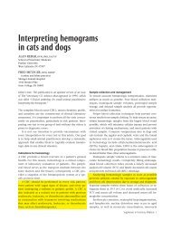
FIGURE 1Good staining technique is criticalto identifying polychromatophilson a peripheral <strong>blood</strong> film.1. The steps in preparing a <strong>blood</strong> film. A small dropof <strong>blood</strong> is placed near the end of a slide (top). Aspreader slide is drawn back into the <strong>blood</strong> dropat a 30-degree angle (middle). Then the spreaderslide is pushed away from the <strong>blood</strong> drop,creating a uniform film across the slide (bottom).Producing high-quality <strong>blood</strong> <strong>films</strong> begins byusing clean, new slides. Used slides frequentlyhave imperfections such as scratchesand other physical defects. Slides must befree of fingerprints, dust, alcohol, detergents,and debris. Using a microhematocrit tube,place a small drop (2 to 3 mm in diameter)of well-mixed EDTA <strong>blood</strong> about 1 to 1.5 cmfrom the end of the slide. Next, draw aspreader slide back into the <strong>blood</strong> drop atabout a 30-degree angle until the spreaderslide edge contacts the sample drop and capillaryaction disperses the sample along theedge. Then, using a smooth steady motion,push the spreader slide away from the <strong>blood</strong>drop, creating a uniform film that coversnearly the entire length of the slide (Figure1). Changing the angle of the spreader slidewill change the length and thickness of the<strong>blood</strong> film. For <strong>blood</strong> samples with lowhematocrits (severe anemia), you may needto decrease the angle of the spreader slide tomake a good-quality slide. In contrast, forsamples with high hematocrits (severe dehydrationand polycythemia due to a variety ofconditions), you may need to increase theangle of the spreader slide.Drying is an important step in the productionof good-quality <strong>blood</strong> <strong>films</strong>. Allow the<strong>blood</strong> film to thoroughly air-dry before applyingstain, or use a heat block (at the low setting)or a hair dryer to hasten the drying time,thus minimizing refractile markings that candistort erythrocyte morphology. In addition,keep formalin and formalin-containing containersaway from all <strong>blood</strong> and cytologysmears to prevent staining artifacts such as increasedcytoplasmic granularity and basophilia.Staining the <strong>blood</strong> filmMost veterinary practices use a modifiedWright’s stain (Romanowsky stain) for bothhematology and cytology. Modified Wright’sstains are available as either three- or two-solutionkits. We prefer kits with three separatesolutions: an alcohol fixative, an eosinophilicstaining solution, and a dark-blue stainingsolution. Veterinary practices should havetwo separate Coplin stain jar sets—one forhematology and cytology samples and onefor contaminated samples such as those collectedfor ear and fecal cytology. For optimalresults, replace staining solutions regularly.Recommended <strong>blood</strong> staining protocolincludes dipping the air-dried slide five to10 times in each solution while blotting oneedge briefly in between each solution. Rinsethe slide with distilled water after the finalstaining step, and allow the <strong>blood</strong> film to airdrybefore examination. Good staining techniqueis critical to identifying polychromatophilson a peripheral <strong>blood</strong> film. If thestaining is done improperly, many of thequick Romanowsky-type stains will not givethe needed tinctorial differences between matureRBCs (orange-red) and polychromatophils(bluish-pink). Typically, poor staining withthe various quick stains results in all RBCs havinga bluish or muddy tint.Evaluating the <strong>blood</strong> filmThe microscopeA high-quality microscope is essential forhematology. We recommend a binocular microscopewith a minimum of 10, 20, and100 (oil-immersion) objectives and widefield10 oculars. When <strong>evaluating</strong> the slide,make sure maximum light reaches the <strong>blood</strong>film. This means positioning the substage condenseras close as possible to the stage withthe iris wide open. The light source should beequipped with a variable rheostat to allowmaximal control of light intensity. To ensureoptimal focused light for the microscopic evaluation,the microscope should be in Köhler il-Feathered edgeMonolayer MonolayerBody of <strong>blood</strong> filmApplication point2. The components of the peripheral <strong>blood</strong> film slide.FIGURE 2Label4