How to Approach an Antibody Identification Panel
How to Approach an Antibody Identification Panel
How to Approach an Antibody Identification Panel

You also want an ePaper? Increase the reach of your titles
YUMPU automatically turns print PDFs into web optimized ePapers that Google loves.
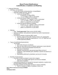
<strong>How</strong> <strong>to</strong> <strong>Approach</strong> <strong>Antibody</strong> <strong>Identification</strong>Sh<strong>an</strong> Yu<strong>an</strong>, MD(Last Updated 3/15/2011)Overview<strong>Antibody</strong> screen/identification are mainly performed on:o Potential tr<strong>an</strong>sfusion recipients <strong>to</strong> prevent hemolytic tr<strong>an</strong>sfusion reactionso Pregn<strong>an</strong>t women <strong>to</strong> evaluate the risk for HDFNThe screen:o Patient’s serum/plasma is mixed with a set of (2-3) reagent red blood cellso Reagent RBCs chosen such that various signific<strong>an</strong>t <strong>an</strong>tigens are expressed <strong>an</strong>drepresented, so that commonly formed allo<strong>an</strong>tibodies c<strong>an</strong> be detected.o Reagent RBCs are group O <strong>to</strong> avoid “interference” by ABO <strong>an</strong>tibodies.o Look for agglutination or hemolysis at three phases,• Immediate spin (IS) at room temp- this is often optional, because thisusually detects IgM, non-ABO <strong>an</strong>tibodies that are reactive at room temp,the majority of them are clinically insignific<strong>an</strong>t• After incubation at 37C, this facilitates agglutination by warm reactive<strong>an</strong>tibody, which are often IgG• After adding AHG (<strong>an</strong>ti-hum<strong>an</strong> IgG) <strong>to</strong> further enh<strong>an</strong>ce agglutination byIgG <strong>an</strong>tibodieso If screen positive, a more exp<strong>an</strong>ded p<strong>an</strong>el is done with 10-12 reagent cells,representing various <strong>an</strong>tigens in the same m<strong>an</strong>ner, <strong>to</strong> allow determination of<strong>an</strong>tibody specificity.o Au<strong>to</strong>logous control (patient’s own RBCs) done in parallel: <strong>to</strong> pick upau<strong>to</strong><strong>an</strong>tibodies.What makes <strong>an</strong> allo<strong>an</strong>tibody clinically signific<strong>an</strong>t? An <strong>an</strong>tibody is considered signific<strong>an</strong>t if its specificity has been associated witho Hemolytic disease of the fetus <strong>an</strong>d newborn (HDFN)o Hemolytic tr<strong>an</strong>sfusion reactionso Notable decreased survival of tr<strong>an</strong>sfused red cells. Some specificities are well known <strong>to</strong> be clinically signific<strong>an</strong>t, for e.g., Anti-Rh, Kidd,Kell, Duffy, S,-s For less well known ones, you c<strong>an</strong> consult the Antigen Facts Book by Marion <strong>an</strong>dReid, or check the current literature.Generally… An <strong>an</strong>tibody that reacts at the 37C or the AHG phase is more likely <strong>to</strong> be clinicallysignific<strong>an</strong>t –usually a warm reactive IgG <strong>an</strong>tibody If the <strong>an</strong>tibody is only reactive at the room temperature /immediate spin (IS) phase,then it is not very likely <strong>to</strong> be signific<strong>an</strong>t, usually a “cold” reactive, non-ABO, IgM<strong>an</strong>tibody (Part of the reason that some labs have ab<strong>an</strong>doned the IS phase of <strong>an</strong>tibodyscreen/identification.)In summary:1
For the boards, you c<strong>an</strong> probably ignore the high freq or low freq <strong>an</strong>tigens5. Use positive cells <strong>to</strong> try <strong>to</strong> fit a single <strong>an</strong>tibody (if all warm or all cold reactive), if one<strong>an</strong>tibody won’t explain all the reactions, or there seems <strong>to</strong> be a mixture of warm <strong>an</strong>dcold-reactive <strong>an</strong>tibodies, move on <strong>to</strong> two, three, etc.6. Special techniques that c<strong>an</strong> be used: Enzymes (include bromelin, papain, ficin, trypsin): destroy or expose<strong>an</strong>tigens, therefore c<strong>an</strong> enh<strong>an</strong>ce or weaken certain <strong>an</strong>tibody-RBC <strong>an</strong>tigenreactions:1. Enh<strong>an</strong>ces: Lewis P Is A Rhotten Kidd! And others.2. Destroys: M/N, Duffy, Luther<strong>an</strong> (Mnemonic: My Dog Luther<strong>an</strong>!)etc3. Does not affect: Kell Chemical modifications:1. Reducing reagents (DTT) treated serum <strong>to</strong> distinguish IgG fromIgM: DTT cleaves the bonds between IgM subunits <strong>an</strong>d inactivatedIgM. Useful when distinguishing warm reactive IgG from warmreactive IgM (rare)2. Chemical reagents (DTT, chloroquine) c<strong>an</strong> destroy certain <strong>an</strong>tigenson RBCa. DTT destroys Kell <strong>an</strong>tigen, by reducing the disulfide bonds<strong>to</strong> –SH groupsb. Chloroquine weakens Bg Phenotype the patient: by definition, you c<strong>an</strong>’t form <strong>an</strong> allo against <strong>an</strong>tigenyou have. Caveat is that patient should not have been tr<strong>an</strong>sfused recently.Question: <strong>How</strong> do you phenotype the RBC’s if the DAT is positive? The boundIgG will “block” the <strong>an</strong>tigens from the monoclonal <strong>an</strong>tibodies used inphenotyping:Answer: Elute <strong>an</strong>tibodies with choloroquine diphosphate, this will leave RBC <strong>an</strong>tigensrelatively intact <strong>an</strong>d accessibly <strong>to</strong> typing monoclonal <strong>an</strong>tibodies. Acid elution c<strong>an</strong> also be done, butthis might be <strong>to</strong>o harsh.Neutralizing or inhibi<strong>to</strong>r subst<strong>an</strong>ces: will eliminate reactivity of <strong>an</strong>tibodieswith certain specificities. This c<strong>an</strong> be used <strong>to</strong> confirm the suspectedspecificity of some allo<strong>an</strong>tibodies. Some examples are:1. saliva from secre<strong>to</strong>rs: <strong>an</strong>ti-Lewis2. hydatid cyst fluid: <strong>an</strong>ti-P13. serum: <strong>an</strong>ti-Chido, Rodgers4. urine from “super Sid” individuals, guinea pig urine : <strong>an</strong>ti-Sd a5. saliva: <strong>an</strong>ti-HAdsorption: Use patient’s own or specific RBC’s with known phenotype <strong>to</strong>remove cold or warm au<strong>to</strong>, or allogeneic cells with certain specificity fromthe serum <strong>to</strong> see if there is something else3
Acid elution (with glycine acid, digi<strong>to</strong>nin): Remove (“elute”) <strong>an</strong>tibodies fromDAT positive RBCs by acid, heat, or other org<strong>an</strong>ic solvent. The resulting“eluate” will give you a more concentrated solution of <strong>an</strong>tibodies <strong>an</strong>d mayallow you <strong>to</strong> finally identify the specificity when the serum reactivity is notstrong enough.7. Final rule in: positive reaction with three different cells positive for the specific<strong>an</strong>tigen <strong>to</strong> confirm the specificityPattern recognition: Always know the following1. Warm au<strong>to</strong>2. Cold au<strong>to</strong>3. HTLA <strong>an</strong>tibodies vs. <strong>an</strong>ti-high freqHTLA (Hi titer, low avidity) Specificities: Chido/Ridgers, John Mil<strong>to</strong>n Hagen, Knops, McCoy York, Cost-Sterling, Bg, Sda, Limited clinical signific<strong>an</strong>ce Weak reactivity (at most 1+), hence low avidity , but high titer, <strong>an</strong>d reactivity persistsafter serial dilutions4