

Cornea - ARVO
Cornea - ARVO
Cornea - ARVO

You also want an ePaper? Increase the reach of your titles
YUMPU automatically turns print PDFs into web optimized ePapers that Google loves.
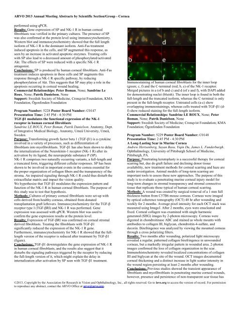
<strong>ARVO</strong> 2013 Annual Meeting Abstracts by Scientific Section/Group - <strong>Cornea</strong>performed using qPCR.Results: Gene expression of SP and NK-1 R in human cornealfibroblasts was verified in the primary cultures. The presence of SPwas also confirmed at the protein level using immunocytochemistry.Western blot and immunocytochemistry showed that the full lengthisoform of NK-1 R is the dominant isoform. Anti-Fas treatmentinduced apoptosis in the cells, and SP augmented this response, asseen by an increase in activated apoptotic enzymes. Treating cellswith SP also lead to a decreased amount of phosphorylated/activatedAkt. The effects of SP were reduced with a specific NK-1 Rantagonist.Conclusions: SP is produced by human corneal fibroblasts. Anti-Fastreatment induces apoptosis in these cells and SP augments thisresponse through a NK-1 R specific pathway, by reducingphosphorylation of Akt. This suggests that SP may play a role in theapoptosis occurring in corneal wound healing.Commercial Relationships: Peter Boman, None; Sandrine LeRoux, None; Patrik Danielson, NoneSupport: Swedish Society of Medicine, Cronqvist Foundation, KMAFoundation, Ögonfonden FoundationProgram Number: 5228 Poster Board Number: C0147Presentation Time: 2:45 PM - 4:30 PMTGF-β1 modulates the functional expression of the NK-1receptor in human corneal fibroblastsSandrine LE ROUX, Peter Boman, Patrik Danielson. Anatomy, Dept.of Integrative Medical Biology, Anatomy, Umeå University, Umeå,Sweden.Purpose: Transforming growth factor beta 1 (TGF-β1) is a cytokineinvolved in a variety of processes, such as differentiation offibroblasts into myofibroblasts. TGF-β1 has also been shown to delaythe internalization of the Neurokinin-1 receptor (NK-1 R) after itsactivation by its ligand, the neuropeptide substance P (SP).NK-1 R comprises two naturally occurring variants, a full-length anda truncated form, triggering different cellular responses. SP has beenshown to be involved in important events in the cornea essential forthe proper organization of collagen fibers and the transparency of thestroma. An impaired signaling through NK-1 R could thus disturb theextracellular matrix and impact the vision quality.We hypothesize that TGF-β1 modulates the expression pattern andfunction of the NK-1 R in human corneal fibroblasts. The purpose ofthis study was to test that hypothesis.Methods: Cultures of primary corneal fibroblasts were set-up withcells derived from healthy corneas, obtained from donatedtransplantation graft leftovers. Immunocytochemistry for the TGF-βreceptor type I (TGF-βRI) and NK-1 R was performed. Geneexpression was assessed with qPCR. Western blot was used toconfirm the gene expression results at the protein level.Results: Expression of TGF-βRI was confirmed on corneal stromalderived fibroblasts. Treating the fibroblasts with TGF-β1significantly reduced the expression of the NK-1 R gene.Furthermore, immunocytochemistry for NK-1 R showed that the fulllengthversion of the receptor is reduced after treatment by TGF-β1(figure).Conclusions: TGF-β1 downregulates the gene expression of NK-1 Rin human corneal fibroblasts, and the results also suggest that itdisturbs the signaling pathways triggered by the receptor by reducingthe full-length version of it, which might explain the delay ininternalization after activation by SP seen with TGF-β1 treatment.Immunostaining of human corneal fibroblasts for the inner loop(green; c, f) and the C-terminal (red; b, e) of the NK-1 receptor.Merged pictures in a (of b and c) and d (of e and f), with DAPI addedfor demonstrating nuclei (bluish). The inner loop is found in both thefull-length and the truncated isoform, whereas the C-terminal is onlypresent in the full-length receptor. Untreated cells (a-c) showoverlapping immunostainings, whereas cells treated with TGF-β1 (df)show reduced staining for the full-length isoform.Commercial Relationships: Sandrine LE ROUX, None; PeterBoman, None; Patrik Danielson, NoneSupport: Swedish Society of Medicine; Cronqvist Foundation; KMAFoundation; Ögonfonden FoundationProgram Number: 5229 Poster Board Number: C0148Presentation Time: 2:45 PM - 4:30 PMA Long-Lasting Scar in Murine <strong>Cornea</strong>Andrew Hertsenberg, Sayan Basu, Yiqin Du, James L. Funderburgh.Ophthalmology, University of Pittsburgh School of Medicine,Pittsburgh, PA.Purpose: Penetrating keratoplasty is a successful therapy for cornealscarring but, due do graft failure and declining donor tissueavailability, new treatment methods for corneal scarring and haze areunder investigation. Animal models of long-term scarring areimportant tools to assess these new approaches. The purpose of thisstudy is to evaluate a penetrating murine corneal injury model forlong-term changes in stromal transparency and stromal connectivetissue that replicate those typical of human corneal scarring.Methods: A wound was created by surgical removal of a 1 mm fullthickness button from C57B6 mouse corneas. <strong>Cornea</strong>s were imagedby optical coherence tomography (OCT) 48 hr after wounding andweekly for 2 months. Average pixel intensity for each OCT stack wasmeasured using ImageJ. After 2 months, eyes were enucleated andfixed. <strong>Cornea</strong>l collagen was examined with single harmonicgenerated (SHG) images by 2-photon microscopy. <strong>Cornea</strong>s weredigested in chondroitinase ABC and stained as whole mounts withantibodies to collagen III, biglycan, chondroitin-4-sulfate, anddecorin. Birefringence was analyzed by viewing the mounted corneasthrough a cross polarizing filters.Results: Two months after wounding, polarized light microscopyrevealed a regular, patterned collagen birefringence in unwoundedcorneas, but a markedly irregular pattern in wounded area. 2-photonimages confirmed the loss of collagen organization in the scar.Immunohistochemistry revealed localized concentrations of collagenIII and biglycan at the site of the wound. OCT images documentedcorneal thickening and a distinct increase in light scatter intensity inthe wound region persisting at least 2 months after wounding.Conclusions: Previous studies showed the transient appearance offibroblasts and myofibroblasts in penetrating murine corneal wounds,however, presence and persistence of non-transparent scar tissue has©2013, Copyright by the Association for Research in Vision and Ophthalmology, Inc., all rights reserved. Go to iovs.org to access the version of record. For permissionto reproduce any abstract, contact the <strong>ARVO</strong> Office at arvo@arvo.org.


