

Cornea - ARVO
Cornea - ARVO
Cornea - ARVO

You also want an ePaper? Increase the reach of your titles
YUMPU automatically turns print PDFs into web optimized ePapers that Google loves.
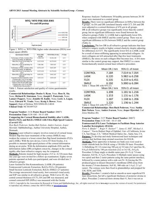
<strong>ARVO</strong> 2013 Annual Meeting Abstracts by Scientific Section/Group - <strong>Cornea</strong>Figure 1. WFG vs. WFO PRK higher order aberrations (HOA) rootmean square (RMS)Response Analyser. Furthermore 30 healthy persons between 20-30years were measured as a control group.Results: There were no significant differences in IOPcc between the4 groups. As DA and DR correlated linearly with CCT, DA and DRwere adjusted to a corneal thickness of 476 µm in all groups.DR in all refractive groups was significantly lower than the controlgroup, but no significant differences were found between therefractive groups (Table 1). LASIK had a significantly lower DA,when compared with SMILE and the control group. There was nosignificant difference in DA between SMILE and the control group(Table 2).Conclusions: The low DR in all refractive groups indicates that laserrefractive surgery results in higher corneal elasticity despite adjustingfor differences in CCT. The significantly lower DA in LASIK couldbe a result of stress-stiffening appearing in untouched collagens. Asmore collagens are left untouched after flap-free laser treatments(SMILE), the stress on each collagen fiber becomes less. A DA moresimilar to the control group may support, that SMILE is a morebiomechanically neutral corneal laser refractive procedure.Table 1. Patient satisfaction and quality of vision questionnaireresults.Commercial Relationships: Denise S. Ryan, None; Rose K. Sia,None; Richard D. Stutzman, None; Joseph F. Pasternak, None;Lamarr Peppers, None; Jennifer B. Eaddy, None; Lorie A. Logan,None; Edward W. Trudo, None; Kraig S. Bower, NoneSupport: Dept of Defense W81XWH-09-2-0018Clinical Trial: NCT01097525Program Number: 3136 Poster Board Number: D0071Presentation Time: 8:30 AM - 10:15 AMComparing the <strong>Cornea</strong>l Biomechanical Stability after LASIK,ReLEx FLEx and ReLEx SMILE with Ultra High Speed Camera(Corvis® ST)Iben Bach Pedersen, Sashia Bak-Nielsen, Anders Ivarsen, JesperHjortdal. Ophthalmology, Aarhus University Hospital, Aarhus,Denmark.Purpose: Laser refractive surgery involves removal of corneal tissue.Modern flap-free laser treatments of the cornea (SMILE) mayweaken the cornea to a lesser extent than flap based treatments (FLExand LASIK). With the new device, Corvis ST from Oculus, it ispossible to measure high-speed pictures of the corneal deformationduring an air-pulse. With the deformation amplitude (DA) and thedeformation radius (DR) at highest concavity, changes in the cornealrigidity after laser refractive surgery may be compared.Methods: Patients treated for high myopia (-9,5 to -4,5 D) more thanone year ago were invited for a follow up examination. Eighty-sevenpatients operated on both eyes participated, and were divided into 3refractive groups:Laser-Assisted In Situ Keratomileusis, LASIK (30 patients)Femtosecond Lenticule Extraction, ReLEx-FLEx (30 patients)Small Incision Lenticule Extraction, ReLEx-SMILE (27 patients)The average uncorrected visual acuity, best corrected visual acuityand IOP was similar in all refractive groups. With Corvis ST, thecentral corneal thickness (CCT), DA and DR was measured, andcorneal corrected IOP (IOPcc) was measured with the OcularTable 1: Mean DR with 95% CI.Table 2: Mean DA with 95% CI.Commercial Relationships: Iben Bach Pedersen, None; SashiaBak-Nielsen, None; Anders Ivarsen, None; Jesper Hjortdal, CarlZeiss Meditec (R)Program Number: 3137 Poster Board Number: D0072Presentation Time: 8:30 AM - 10:15 AMFemtosecond (FS) Laser Techniques to Facilitate Deep AnteriorLamellar Keratoplasty (DALK)Perry S. Binder 1, 2 , Roger F. Steinert 1, 2 , James E. Hill 2 , Michael A.Campos 2 . 1 Gavin Herbert Dept of Ophthal, Univ of California, IrvineCA, San Diego, CA; 2 Abbott Medical Optics Inc., Santa Ana, CA.Purpose: To develop and study femtosecond laser techniques andsettings that facilitate deep stromal resections for DALK.Methods: Three approches in human eye bank eyes were used tocreate smooth beds for DALK using a 150 kHz FS laser: Procedure1) Debulking (N=12) removing 300 µm @ 9.0 mm (3x3 spot/line SL,0.6 µJ) followed by 100 µm resection (2x2 or 3x3 SL, 0.6 µJ);Procedure 2) Deep single or double raster pass (N=3) @ 7.0 mmdiameter, 8x8 SL, 0.3 µJ); Procedure 3) Multiple raster passes usingtwo spiral and then 2 raster patterns using the same patient interface,followed by a raster pattern with a side cut (N= 8) forcing the FSproducedgases to dissect Descemet’s membrane (DM) away fromthe stroma (7.0 mm diameter, 8x8 SL, 900 side cut 1.6 µJ, 2.1 µJraster). The corneas were then fixed in glutaraldehye and prepped forLM, SEM, and TEM.Results: Procedure 1 created a bed as smooth as more superficial FSlaser dissections, but left a significant thickness of posterior stroma inplace. Procedure 2 removed more stroma than #1, but it was not as©2013, Copyright by the Association for Research in Vision and Ophthalmology, Inc., all rights reserved. Go to iovs.org to access the version of record. For permissionto reproduce any abstract, contact the <strong>ARVO</strong> Office at arvo@arvo.org.


