Guidelines - Haematology Association of Ireland
Guidelines - Haematology Association of Ireland
Guidelines - Haematology Association of Ireland
You also want an ePaper? Increase the reach of your titles
YUMPU automatically turns print PDFs into web optimized ePapers that Google loves.
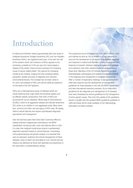
IntroductionIn <strong>Ireland</strong> and Northern <strong>Ireland</strong> approximately 800 new cases <strong>of</strong>malignant lymphoma, Hodgkin lymphoma (HL) and non-Hodgkinlymphoma (NHL), are registered each year. In line with the rest<strong>of</strong> the western world, the incidence <strong>of</strong> NHLs appears to beincreasing, probably by 3-5% per year and, being largely adisease <strong>of</strong> the elderly, these tumours represent an increasingcause <strong>of</strong> cancer-related death. The reasons for increasingincidence are multiple, ranging from the increasing elderlypopulation, greater accuracy <strong>of</strong> diagnosis and unknownenvironmental factors. The increase has not been noted inHL and in all subtypes <strong>of</strong> NHL and cannot solely be explainedon the basis <strong>of</strong> the HIV epidemic.NHLs are a heterogeneous group <strong>of</strong> diseases which aremainly linked by their origin within the lymphoid system andits different cellular components. Over 60% <strong>of</strong> NHLs areaccounted for by two diseases, diffuse large B-cell lymphoma(DLBCL) which is an aggressive disease and follicular lymphoma(FL) which is an indolent or non-aggressive entity. Many otherless- common and <strong>of</strong>ten rare sub-types <strong>of</strong> NHL exist. All displaydistinct natural histories and require sophisticated diagnosticapproaches and management.The Lymphoma Forum <strong>of</strong> <strong>Ireland</strong> (LFI) first met in October 2002and outlined its aims as: the promotion <strong>of</strong> a high standard <strong>of</strong>care and the development <strong>of</strong> all aspects <strong>of</strong> lymphoma diagnosisand treatment in <strong>Ireland</strong> and Northern <strong>Ireland</strong>, development andfostering <strong>of</strong> research initiatives relating to malignant lymphomasand interaction with other relevant bodies towards achievingthese aims. Members <strong>of</strong> the LFI include medical oncologists,haematologists, pathologists and radiation oncologists involvedin the diagnosis and management <strong>of</strong> malignant lymphomas.After a number <strong>of</strong> exploratory meetings, it was agreed that theinitial step required was the development <strong>of</strong> managementguidelines for these diseases in line with the WHO classificationand best international treatment practices. As an initial effort,guidelines for the diagnosis and management <strong>of</strong> 22 diseaseshave been developed as well as guidelines for the management<strong>of</strong> some generic issues. This is the first update <strong>of</strong> the guidelinesissued in parallel with the updated WHO guidelines published in2008 and these will be made available on the <strong>Haematology</strong><strong>Association</strong> <strong>of</strong> <strong>Ireland</strong> website.Over the last forty years there have been numerous efforts toclassify lymphoid malignancies culminating in the WHOclassification introduced after much international effort in 2001.It is clear that malignant lymphomas require a sophisticateddiagnostic approach based on clinical features, morphology,immunophenotyping and genetic analysis. It is essential thatsuch an approach underpins the clinical management <strong>of</strong> thesediseases, many <strong>of</strong> which are amenable to cure. Clinical care tooneeds to be delivered by those with expertise and experience inthe area within a multidisciplinary setting.1
Standards In Diagnosis<strong>of</strong> LymphomaTissue collectionInvestigations prior to biopsyA full blood count (FBC) and film (with flow cytometry ifappropriate), should be carried out before a node biopsy toavoid biopsying patients with CLL or acute leukaemia.Monospot: in patients < 30 years with lymphadenopathy.Epithelial carcinoma should be considered in patients >40with head and neck adenopathy, who should have an ENTexamination and FNA.Designated surgeons should perform all lymphnode biopsies in lymphoma diagnosisA designated surgeon ensures appropriate and uniformspecimen collection and prompt referral <strong>of</strong> patients to thelymphoma service. The preliminary biopsy report should beavailable to the multidisciplinary team (MDT) within 2 weeks <strong>of</strong>the patient’s hospital referral.An excision lymph node biopsy is preferablefor diagnosisAn excision biopsy allows detailed assessment <strong>of</strong> architecture,which is a key feature in lymphoma diagnosis. Needle biopsiesare more prone to artefact and may be inadequate for all thediagnostic investigations. A lymph node biopsy is preferable to abiopsy <strong>of</strong> an extra-nodal site.Approach to diagnosis <strong>of</strong> a patient withlymphadenopathy■ FBC with film (and cell marker studieswhere appropriate)■ Monospot in patients < 30 years■ Consider ENT examination and FNA toexclude epithelial malignancy <strong>of</strong> the head andneck in patients >40■ Designated surgeon(s)■ Excision biopsy preferred method; trucut biopsyif node not accessible■ Node biopsy – send unfixed to laboratoryLymph node biopsies should be sent freshto the laboratoryThis requires local arrangements for the prompt and safetransport <strong>of</strong> the specimen. Fresh material is essential for goodquality histology and facilitates the use <strong>of</strong> new diagnostictechniques. See Royal College <strong>of</strong> Pathologists minimum datasetfor lymphoma reports.Laboratory diagnosisSample handlingIn the laboratory, the lymph node should be sliced and imprintpreparations made. Thin slices should be placed in formalin for24 hours before processing as paraffin blocks. This is essentialfor high-quality morphology and reproducible results with markerstudies performed on paraffin sections. The remaining tissue maybe snap frozen and disaggregated into a single-cell suspension.2
Classification systemThe World Health Organization (WHO) classification <strong>of</strong>neoplastic diseasesTumours <strong>of</strong> the haematopoietic and lymphoidtissues, 4th edition 2008 should be used.Diagnostic requirements forhaematopathology diagnosisThe diagnosis <strong>of</strong> lymphoma should be made, or reviewed, in alaboratory with the necessary specialist expertise and facilities.A pathology laboratory diagnosing lymphoma requires access tothe following resources:a. Morphological expertise: Pathologists/haematologistsinvolved in lymphoma diagnosis should have the necessarytraining to undertake this work.b. Immunophenotyping: All marker studies should becarried out using panels designed to test the validity <strong>of</strong> themorphological diagnosis and to demonstrate key prognosticvariables. Marker studies should be carried out using flowcytometry and immunohistochemistry. An appropriate panelfor the lymphoma sub-types is included in the lymphomaspecificsections in this document.c. Molecular techniques: The two main techniques arepolymerase chain reaction (PCR) to detect monoclonalityand some translocations, and fluorescence in situhybridisation (FISH) techniques for translocations.These techniques should be used to confirm a provisionaldiagnosis and identify prognostic factors. Formal links witha molecular/cytogenetics service are required.d. Integrated reporting: Most patients withlymphoproliferative disorders have different specimens takenduring their clinical course. Departments should have amechanism for correlating results from lymph node biopsies,bone marrow aspirates and biopsies as well as differentanalyses <strong>of</strong> a single sample.ReportingA preliminary report should be available 5 working days after thespecimen is received. This interim report should state specificoutstanding investigations and be followed by a definitive report.Quality assurance and auditThe main component <strong>of</strong> quality assurance is access to a robustand timely diagnostic process. An audit system designed to testthe quality <strong>of</strong> the service should be in place. Laboratories shouldbe able to provide users <strong>of</strong> the laboratory with details <strong>of</strong> theirdiagnostic criteria and technical methods.Laboratories should participate in the relevant quality assuranceschemes for immunocytochemistry, flow cytometry and otherdiagnostic methods. Individual histopathologists should haveaccess to a lymphoma review panel.Diagnosis –laboratory procedures and standards■ Unfixed node biopsy imprint preparation –formalin preparation <strong>of</strong> material, snap freezing anddisaggregation into single-cell suspension■ WHO classification■ Access to immunophenotyping, moleculartechniques and molecular genetic techniques■ Preliminary report within 5 working days■ Systems <strong>of</strong> quality assurance in place: StandardOperating Procedures (SOPs); Lab Accreditation;National Quality Assurance Scheme■ Access to review panelMultidisciplinary team (MDT) workingMDT meetings are a desirable part <strong>of</strong> the diagnosis andmanagement <strong>of</strong> lymphoma. The arrangement will vary with localcircumstances but it is essential that diagnostic pathology andstaging radiology be reviewed in both new and relapsed patientsbefore making a management plan. This plan should be clearlydocumented in patients’ notes.3
Standards in Staging<strong>of</strong> LymphomaAnn Arbor staging classification for NHLThe International Prognostic Index (IPI)StageIIEIIIIEIIIIIIEIIISArea <strong>of</strong> involvementOne lymph node regionOne extralymphatic (E) organ or siteTwo or more lymph node regions on thesame side <strong>of</strong> the diaphragmOne extralymphatic organ or site (localised)in addition to criteria for stage IILymph node regions on both sides <strong>of</strong> the diaphragmOne extralymphatic organ or site (localised)in addition to criteria for stage IIISpleen (S) in addition to criteria for stage IIIThe IPI is a prognostic model based on 5 parameters■ Age (< 60 vs > 60 years)■■■Ann Arbor stage (I/II vs III/IV)Serum LDH (normal vs elevated)Extra-nodal involvement (≤ 1 site vs ≥ 1 site)■ Performance status (0,1 vs 2–4)Based on these factors, patients with DLBCL can be divided into4 prognostic categories as summarised below:Number <strong>of</strong> risk factorsIIISEIVSpleen and one extralymphatic organ or site(localised) in addition to criteria for stage IIIOne or more extralymphatic organs with or withoutassociated lymph node involvement (diffuse ordisseminated); involved organs should be designatedby subscript letters (P, lung; H, liver; M, bone marrow)A = asymptomatic;B = symptomatic; unexplained fever <strong>of</strong> ≥ 38ºC;unexplained drenching night sweats; or loss <strong>of</strong> > 10%body weight within the previous 6 months).IPI risk groupAll patientsLow-risk 0,1Low/intermediate-risk 2High/intermediate-risk 3High risk 4, 5The IPI describes a predictive model for patients with DLBCLat presentation. It has been adjusted for use in FL (FLIPI) and isless useful in ALCL, mediastinal B cell lymphoma and T-NHL. Itshould not used in Burkitt lymphoma or lymphoblastic lymphoma.4
ECOG performance statusGrade ECOG0 Fully active, able to carry on all pre-diseaseperformance without restriction1 Restricted in physically strenuous activitybut ambulatory and able to carry out work<strong>of</strong> a light or sedentary nature, e.g., lighthouse work, <strong>of</strong>fice work2 Ambulatory and capable <strong>of</strong> all selfcare butunable to carry out any work activities. Up andabout more than 50% <strong>of</strong> waking hours3 Capable <strong>of</strong> only limited selfcare, confined tobed or chair more than 50% <strong>of</strong> waking hours4 Completely disabled. Cannot carry on anyselfcare. Totally confined to bed or chair5 DeadStaging procedures-all patientsClinical■ Clinical history with reference to B symptomsand family history■ Physical examination with particular attention to node-bearingareas, Waldeyer’s ring, and size <strong>of</strong> liver and spleen■ Performance status (ECOG) including co-morbidityRadiology■ CXR■ Chest and abdominopelvic computed tomography (CT)<strong>Haematology</strong>■ FBC, differential and film■ Bone marrow aspirate and trephine■ Immunophenotyping <strong>of</strong> marrow +/- blood in low gradelymphomas and any other lymphomas with morphologicalevidence <strong>of</strong> marrow/blood involvementBiochemistry■ LDH, urea and electrolyte, creatinine, albumin, aspartatetransaminase (AST), bilirubin, alkaline phosphatase,serum calcium, uric acid■ Pregnancy test in females <strong>of</strong> child-bearing age5Serology■■Hepatitis B and CHIV statusStaging procedures (sometimes indicated)Radiology■■■Plain bone X-ray and bone scintigraphyNeck CTHead CT or magnetic resonance imaging (MRI)■ PET scan<strong>Haematology</strong>■ Coagulation screen■ ESR■ DCTBiochemistry■ Serum immunoglobulins/electrophoresis■ Beta 2 microglobulin■ CRP■ Tissue transglutaminase test (tTG) to exclude coeliac diseaseSerology■ EBV, HTLV serologyMolecular genetics■ FISH or PCR on involved marrow/blood for specificlymphoma-associated translocations■ IgH and TCR rearrangements on marrow/blood if molecularstaging clinically indicatedOthers■ ECHO and PFTs■ Lumbar puncture if lymphomatous meningitis is suspected orif indications for prophylactic treatment are present. CNSprophylaxis is currently used in patients with BurkittLymphoma, lymphoblastic lymphoma, HIV-related lymphoma,HTLV-1-related lymphoma and post-transplantlymphoproliferative disease. About 5% <strong>of</strong> patients withDLBCL develop CNS disease but there is no consensusabout which patients need to have a diagnostic lp.
Pathologic Diagnosis<strong>of</strong> LymphomaGeneral comments1. Primary diagnosisa. Complete lymph node excision in the presence <strong>of</strong> nodaldisease is the optimum diagnostic material and should beutilised wherever possible. *b. The complete lymph node excision should be transportedimmediately in a fresh state to the laboratory.c. The fresh lymph node should be handled as per algorithmon page 15.d. *Where full lymph node excision is not possible e.g.inaccessible mass or extranodal disease, core biopsy is thenext best option for accurate primary diagnosis unless aresection has been or will be undertaken. Multiple largecores should ideally be submitted fresh to the laboratory onsaline soaked gauze otherwise they should be submitted in10% buffered formalin.e. All cases must be received with relevant clinical information.2. The role <strong>of</strong> Flow Cytometry (FCM)a. FCM is useful for accurate diagnosis in all small cell /follicular pattern lesions.b. FCM on aspirated material may provide a reasonablealternative to biopsy in recurrent disease where tissue isnot easily obtainable e.g. elderly/frail patient, inaccessiblesite. Such FNA samples require co-ordination with thelaboratory as special fixation (RPMI) and immediateprocessing is required3. Optimum handling <strong>of</strong> fresh lymph node excisiona. Ensure immediate transfer from the theatre to laboratory.b. Bisect node and perform touch preparations.i. Air dried x 2 – Giemsa stainii. Air dried X6-8 FISH studies if requirediii. Fixed x 2 – H & E stainc. Sample portion <strong>of</strong> tissue for:i. Freezing – 1 portion in “RNA later”, 1 portion freshfrozen (this will preserve material for molecular studyshould this be required).ii. RPMI for FCM (this will preserve the cells and allowtransport to nearest centre <strong>of</strong>fering FCM and/orCytogenetics service).iii. Formalin for routine fixation and processing.4. Classification and gradinga. Use WHO classification 4th edition 2008b. Where applicable use WHO grading e.g.Follicular Lymphoma (see page 29)5. The role <strong>of</strong> FCM/Cytogenetics/Gene expression pr<strong>of</strong>ilingMaterial should be preserved in RPMI and frozenfor availability for cytogenetic analysis and otherrelevant evaluation6
Immunohistochemistry (IHC)Hodgkin Lymphoma (HL)BASIC IHC PANEL ON FFPET (Formalin fixed, paraffin-embedded tissue)- CD45- CD3- CD20- CD15-CD30- EMA (EMA should be negative in classic HL; positive in NLPHL)IF H&E MORPHOLOGY IS TYPICALCD45CD3CD20CD15CD30CD3CD20CD15CD30}}.......................NEGATIVE.......................POSITIVE}.......................NEGATIVE}.......................NEGATIVE.......................POSITIVE}.....................HLConsider ALCLPerform ALK1If ALK 1 NEGIf ALK 1 POS................................................................HL................................................................ALCLIF H&E MORPHOLOGY IS ATYPICAL ADD THE FOLLOWING TO THE INITIAL BASIC IHC PANELCD79ACD5CD10ALK 1Myeloperoxidase – to exclude myeloid origin.MCT – to exclude mast cell origin.Non-lymphoid markers as appropriate e.g. to exclude – Germ Cell tumoursMalignant melanomaPoorly differentiated carcinoma7
Hodgkin lymphoma and its differential diagnosisCD20 CD79a T-Cell CD4 CD30 CD15 EMAantigen CD8Nodular lymphocytepredominant HL + + - - -/+ - +Classical HL -/+ -/+ - - + + -/+T-cell rich large B-cell lymphoma + + - - - - -Anaplastic large cell lymphoma - - +/- CD8>CD4> + - +CD4&8-veKEY +/- The lymphoma cells are commonly but not always positive-/+ The lymphoma cells are usually but not always negativePotential diagnostic pitfalls in Hodgkin lymphomaWith “Typical” H&E Morphology■ ALCL■ T cell rich B cell lymphoma■ Diffuse large B cell lymphomaWith “Atypical” H&E Morphology■ ALCL■■■■■■T cell rich B cell lymphomaDiffuse large B cell lymphomaIntermediate CHL/DLBCL (Grey zone lymphoma)Myeloid originMast cell originNon-lymphoid neoplasms8
Large/Intermediate Cell Morphology on H&EBASIC IHC PANEL ON FFPETCLACD20CD3CD5 + Cyclin D1 (To exclude Blastic Mantle Cell Lymphoma)CD30Ki67BCL2BCL6CD10IRF4/MUM1}To provide additional prognostic/therapeutic information to clinicianCLA}+CD20 +CD79a.........................Pr<strong>of</strong>ile <strong>of</strong> Diffuse large B Cell lymphomaCD3 -CD30 -CD5 -IF CD20+ /CD79a + but proliferative index(Ki67) is high (>90%)■ Consider BURKITT (look for “starry sky” morphology;BCL2 NEG) or Intermediate BL/DLBCL Confirm withMolecular testing for MYC , BCL2, BCL6■ Consider Lymphoblastic – Do TdTIF CD20 - Do CD79A and CD138IF CD79a +■ Consider MYELOMA (CD138 +)■ Consider Myeloid neoplasms (MPO, CAE)IF CLA –■ Consider ALCL (CD30+, ALK +) orNon Lymphoid MalignancyT CELL■■■■IF CD20- / CD79A-/CD3+T-Cell LymphomaDo CD30, ALK-1CD56, CD4, CD89
Potential diagnostic pitfalls forlarge/intermediate cell morphologyNon-lymphoid malignancy■ Germ Cell tumour■ Carcinoma■ Melanoma■ SarcomaBurkitt lymphoma is missed – (If proliferative index(Ki67) is high and BCL2 is negative – think Burkitt)Confirm with Molecular genetic studies for MYC andBCL2 translocationsB Cell Lymphoblastic lesions - (include TdT in panel)Myeloid lesions If CD20 is negative but CD79A positivePlasma Cell lesions consider these“Blastic” Mantle Cell lymphoma – include CD5 ininitial panel. Confirm with Cyclin D1 or molecular geneticstudies for BCL1 translocation.10
Nodular/Follicular patterna. Nodular/follicular pattern with small cell morphologyFollicular LymphomaMantle Cell LymphomaMarginal Zone LymphomaSLL/CLLBASIC IHC PANEL ON FFPETCD20CD3CD5CD10CD23Cyclin D1BCL 2Ki67FCMCytogeneticsb. Nodular/follicular pattern with larger cells or “atypical” morphology consider -NLPHLLymphocyte Rich HLNSHLAnd handle as per HLc. Non-nodular small cell morphologyInvestigate as per basic IHC panelConsider Lymphoplasmacytic lymphomaPotential diagnostic pitfalls for small cell morphology■Lymphoplasmacytic lesions11
Summary <strong>of</strong> the usual Immunostaining Pattern <strong>of</strong> B-cell NeoplasmsPrecursor B-cell neoplasmsCD20 CD79 CD5 CD23 CD10 CD30 CD15 CyclinD1Precursor B-lymphoblasticleukaemia/lymphoma - +/- - - + - - -Mature B-cell neoplasmsB-cell chronic lymphocyticleukaemia/lymphoma + + + + - - - -B-cell prolymphocytic leukaemia + + - +/- - - - -/+Lymphoplasmacytic lymphoma + + - -/+ - - - -Mantle Cell lymphoma + + + - - - - +Follicular lymphoma + + - -/+ + - - -Marginal zone B-cell lymphoma<strong>of</strong> mucosa associated lymphoidtissue type + + - - - - - -Nodal marginal zone lymphoma +/-(monocytoid B-cells) + + - - - - - -Splenic marginal zone lymphoma + + - - - - - -Hairy cell leukaemia + + - - - - - -Plasmacytoma - + - - - -/+ - -Plasma cell myeloma - +/- - - - -/+ - -Diffuse large B-cell lymphoma + + -/+ -/+ -/+ -/+ - -Mediastinal (thymic) + + - +/- -/+ -/+ -/+ -Intravascular + + -/+ - -/+ -/+ - -Primary effusion lymphoma - + - - - + - -Burkitt lymphoma + + - - + - -KEY +/- The lymphoma cells are commonly but not always positive-/+ The lymphoma cells are usually but not always negativeNote that for T-cell and putative NK-cell neoplasms, immunostaining is complex and variable.12
Pathological diagnosis <strong>of</strong> suspected lymphoid diseaseReceive whole Lymph Node FRESH*Freeze Sample1-2mmWhole & "RNA Later" mediumRPMI Sample1-2mm(Flow cytometry & cytogenetics)Divide NodeFormalin fixationand wax embeddingH&E and touchprep evaluationTouch preparationsAir Dried - GiemsaFixed - H&E? Reactive Lymphoma ? OtherMalignancyLarge CellSmall CellHodgkin-likeBasic IHCCD3, CD20Ki 67Kappa/LambdaBcl-2Basic IHCCLA, CD3,CD5, CD20,CD30, CD79a,ALK-1, Ki 67Basic IHCCD3, CD5,CD10, CD19CD20, CD23CD79a, Bcl-2Ki 67, CyclinD1Kappa/LambdaBasic IHCCD3, CD15CD20, CD30EMABasic IHCCytokeratinS100VimentinFurther IHCTdT, MPOFurther IHCCD15, CD30Further IHCCD5, CD10, CD57CD79a, ALK-1MPO, MCT* For extranodal disease receive resection or core biopsy freshIssue Report using WHO Classification13
Mature B-Cell NeoplasmsChronic Lymphocytic Leukaemia/Small Lymphocytic LymphomaDefinition and Incidence:Chronic lymphocytic leukaemia / small lymphocytic lymphoma(CLL/SLL) is a neoplasm <strong>of</strong> monomorphic small, round B-lymphocytes in the peripheral blood, bone marrow and lymphnodes admixed with prolymphocytes and paraimmunoblastsexpressing CD5 and CD23. The term SLL is restricted to caseswith the tissue morphology and immunophenotype <strong>of</strong> CLL butwithout a leukaemic component. CLL comprises 90% <strong>of</strong> chronicleukaemias in the USA and Europe and 7% <strong>of</strong> NHLs present asCLL/SLL. The majority <strong>of</strong> patients are >50 years old (medianage 65) And the M: F ratio is 2:1. The incidence is 0.72cases/100,000 per year.ICD – O Codes: CLL 9823/3B-SLL 9670/3Clinical presentationMost patients with CLL are asymptomatic and the disease isdiagnosed incidentally on routine full blood count. Presentingfeatures can include lymphadenopathy, fatigue, auto-immunehaemolytic anaemia, infection, or evidence <strong>of</strong> bone marrow failure.PathologyThe lymphoid infiltrate effaces normal lymphoid architecturewith a pseudo-follicular pattern <strong>of</strong> regularly distributedpale areas containing larger cells in a dark background <strong>of</strong> smallcells The cells are slightly larger than normal lymphocytes, havea high nuclear-cytoplasmic ratio, round nucleus and occasionalsmall nucleolus. The pseud<strong>of</strong>ollicles contain a continuum <strong>of</strong>small, medium and large cells i.e. lymphocytes, prolymphocytesand paraimmunoblasts. The size <strong>of</strong> the pseud<strong>of</strong>ollicles and thenumber <strong>of</strong> paraimmunoblasts vary but there is no welldocumentedcorrelation between histological findings andclinical outcome.Cell morphology can vary and may be confused with mantle celllymphoma (MCL). Plamacytoid differentiation may also be present.In the blood and bone marrow similar small lymphocytes arefound and smudge, (smear) or basket cells are typically seen onblood films. Prolymphocytes, which are larger cells with aprominent nucleolus, usually account for
ImmunophenotypeThe tumour cells express pan B markers CD19 and 20 (weak),surface IgM and IgD (weak) and CD5 and CD23. Thelymphocytes do not express CD10, cyclin D1, FMC7 and CD79b.StagingThere are two clinical staging systems for CLL, the Rai (0-IV) andBinet (A-C) classifications.Recommended investigationsClinical:History and examination withreference to any family historyDiagnosticImaging:CXR, CT thorax abdomen and pelvis.Abdominal Ultrasound is acceptablefor patients being treated with lowintensity treatment or on a watchand wait policyPrognostic factors and GeneticsConventional bad prognostic indicators are; male gender,lymphocyte doubling time <strong>of</strong>
TreatmentApproximately 25% <strong>of</strong> patients with CLL (ie stable CLL , Binetstage A) do not require treatment and are managed with a watchand wait policy. Indications for treatment are progressive stage Adisease or stage B or C CLL.Monotherapy in CLLTraditionally CLL was treated with Chlorambucil with symptomaticintent to control progressive disease or B symptoms. Fludarabinemonotherapy results in an increased complete remission rate andlonger treatment free intervals.Combination treatmentsCombined Fludarabine and Cyclophosphamide (FC), increasesthe complete remission rate, treatment free interval and survival ata cost <strong>of</strong> increased toxicity.Refractory/ poor risk CLLPatients who are refractory to Fludarabine, have p53 dysfunctionor deletion <strong>of</strong> 17p have a median survival <strong>of</strong>
Lymphoplasmacytic LymphomaDefinition and IncidenceLymphoplasmacytic lymphoma (LPL) is composed <strong>of</strong> anadmixture <strong>of</strong> small B lymphocytes, lymphoplasmacytoid cellsand plasma cells usually involving the bone marrow, lymphnodes and spleen and usually associated with a serumparaprotein <strong>of</strong> IgM subtype.LPL is rare and comprises
TreatmentThis is an indolent lymphoma, which is incurable withconventional treatment. Patients with a hyperviscosity syndromeshould be treated as an emergency with plasma exchange untilplasma viscosity has normalised. Plasma exchange should becontinued regularly until IgM production has been controlledsufficiently to prevent further episodes <strong>of</strong> hyperviscosity.Oral Chlorambucil and Fludarabine are the most commonlyused agents in LPL, usually for 6 months.Combination immunochemotherapy can be used as first linetherapy in young patients or as second line therapy.Combinations include Rituximab with Fludarabine, orCycophosphamide, or using all three drugs in FCR. Bortezomibis active in LPL and combining it with Dexamethasone andFudarabine results in an ORR <strong>of</strong> 96% and CR in >20% <strong>of</strong>patients. Vincristine and possibly anthracyclines may not be activein LPL. Rituximab is active in LPL but should only be used whenthe paraprotein is low (eg after plasma exchange) because <strong>of</strong> therisk <strong>of</strong> aggravating hyperviscosity and a high incidence <strong>of</strong> severeinfusional reactions.Response EvaluationNormalisation <strong>of</strong> blood count, total, reduction <strong>of</strong> igM withplateau levels <strong>of</strong> the protein and a normal viscosity. The viscosityshould be checked weekly until normalisation and then monthlywhile on treatmentFollow UpTwo monthly for 1 year, 3 monthly for second year, followed bylong term follow up between 6 – 12 monthly with FBC,biochemistry pr<strong>of</strong>ile, paraprotein level and viscosity.19
Splenic MarginalZone LymphomaDefinition and IncidenceSMZL is an indolent B-cell lymphoma usually involving spleen,bone marrow and blood. Patients usually present withsplenomegaly and/or anaemia. Splenic hilar lymph nodes may beinvolved, although other lymphadenopathy is rare. Bone marrowand peripheral blood involvement is common. The disease is rare,comprising
Potential Pitfalls■ Failure to establish a diagnosis in the patient presentingwith isolated, unexplained splenomegaly – the decisionto proceed with diagnostic splenectomy may be difficult,especially in elderly patients.■■Failure to distinguish from other CD5-negative indolentB cell lymphomas.Failure to distinguish from nodal marginal zone lymphoma.TreatmentMost behave in an indolent fashion, and patients should betreated as for low-grade/follicular lymphoma. Median survival is10-13 years. Splenectomy may be followed by haematologicalresponses and prolonged survival, and is the treatment <strong>of</strong> choicefor fit patients. Other treatment options include splenic irradiation,alkylating agents, purine analogues or anti CD20 antibody.Transformation to large cell lymphoma may occur.Response Evaluation and Follow UpAs for other indolent lymphomas.21
Extra-nodal Marginal Zone B-CellLymphoma (Malt-Lymphoma)Definition and IncidenceExtranodal Marginal Zone B-cell Lymphoma <strong>of</strong> MucosaAssociated Lymphoid Tissue (MALT Lymphoma) is an extranodallymphoma comprising morphologically heterogeneous small B-cells. The gastrointestinal tract is the commonest site <strong>of</strong>development <strong>of</strong> MALT lymphoma, and the stomach is the mostcommon location (85%). Gastric MALT lymphoma is considered tobe derived from MALT acquired as a result <strong>of</strong> Helicobacter pyloriinfection. Incidence <strong>of</strong> 0.6 new cases / 100,000 population peryear, median age 60yrs, sex ratio shows a slight female excess.ICD-O Code 9699/3Clinical PresentationMost patients have a history <strong>of</strong> chronic inflammatory, secondaryto autoimmune disorders or low grade infections which result inaccumulation <strong>of</strong> extranodal lymphoid tissue. Examples includeHelicobacter pylori associated chronic gastritis, Sjogren’sSyndrome or Hashimoto’s thyroiditis. Helicobacter pylori isdetectable in most cases <strong>of</strong> gastric MALT lymphoma. Patientswith Sjogren’s syndrome and lymphoepithelioid sialadenitis have a40-fold increased risk <strong>of</strong> developing lymphoma, and most <strong>of</strong>these are MALT lymphomas. Patients with Hashimoto’s thyroiditishave a 3-fold increased risk <strong>of</strong> lymphoma development. Mostpatients present with Stage I or II disease, but 20% <strong>of</strong> patientshave bone marrow involvement. Multiple extranodal sites arepresent in 10% <strong>of</strong> patients at presentation, with 30% becomingdisseminated over time, and some transforming to DLBCL. The 5-years overall survival is >80.Pathology and GeneticsThe lymphoma cells infiltrate around reactive B-cell follicles,external to a preserved follicle mantle, in a marginal zonedistribution, and spread out to form larger confluent areaswhich eventually overrun some or most <strong>of</strong> the follicles. Thecharacteristic marginal zone B cells have small to medium sized,slightly irregular nuclei with moderately dispersed chromatin andinconspicuous nuclei, resembling those <strong>of</strong> centrocytes; they haverelatively abundant, pale cytoplasm. Plasmacytic differentiation ispresent in approximately one-third <strong>of</strong> gastric MALT-typelymphomas. Lymphoepithelioid lesions are usually present.Phenotype:CD19+ CD20+ CD22+ CD79a+ Slg+ Cd11c± CD43± CD5-CD10- CD23-. The tumour cells typically express IgM, and less<strong>of</strong>ten IgA or IgG, and show light chain restriction.Genetics:Trisomy 3 is found in 60% <strong>of</strong> cases, and the t(11,18)(q21;q21)in 25-50% and is not found in other lymphomas.StagingAs for other indolent lymphomas22
Recommended InvestigationsAs for other indolent B-cell lymphomas. Additional investigationsrecommended for patients with Gastric MALT lymphomas are:■■Biopsy with H pylori stain and culture(H pylori serology may be useful if infection is notconfirmed histologically)Breath testEndoscopic Ultrasound may be useful but is not universallyavailable. The t(11;18) status should be established.Prognostic Factors / IndexThe clinical course is typically indolent, and these lymphomasare slow to disseminate. Involvement <strong>of</strong> multiple extranodalsites and bone marrow involvement do not appear to confer aworse prognosis.Adverse prognostic factors identified for Gastric MALTlymphomas include:■H.pylori negative■■Failure to distinguish from other conditions. The differentialdiagnosis includes reactive processes (Helicobacter pylorigastritis, lymphoepithelial sialadenitis, Hashimoto’s thyroiditis),and other small B-cell lymphomas (follicular lymphoma,mantle-cell lymphoma, small lymphocytic lymphoma).Ann Arbour staging is misleading – for example, involvement<strong>of</strong> multiple extranodal sites, especially within the same organ(e.g., salivary gland, skin, GI tract) does not indicatedisseminated disease.TreatmentGastric MALT Lymphoma:Limited disease:For disease confined to the mucosa or submucosa, H. Pylorieradication produces complete remission rates <strong>of</strong> approximately70%. There is no clinical trial evidence to support the use <strong>of</strong>consolidation Chlorambucil therapy for patients with successfulH.pylori eradication■■■■Tumour invasion beyond the submucosat(1;14)(p22;q21)t(11;18)(q21;q21) - this translocation does notrespond to H pylori eradicationBcl-10 nuclear expressionVarious effective regimens for H Pylori Eradication are available.One is:■■■Omeprazole 20mg po bid x 1 weekAmoxycillin 1g po bid x 7 daysClarithromycin 500mg po bid x 7 daysPotential Pitfalls■ Failure to properly identify diffuse large B cell lymphomain the presence <strong>of</strong> an accompanying MALT lymphomaor to distinguish from de novo gastric diffuse largeB-cell lymphoma.■Metronidazole 400 mg po bid x 7 days23
Persistent / progressive disease ordisease requiring specific anti-lymphomatreatment at diagnosis:Additional treatment is needed for persistant or progressivedisease, infiltration <strong>of</strong> the muscularis mucosa, nodal involvementor presence <strong>of</strong> t(11;18).Management includes chemotherapy +/- Rituximab, withChlorambucil being the commonest therapy used in gastricMALT lymphomas at a dose <strong>of</strong> 6mg/m2/day for 14 days q 28days for 6-12 cycles (2 cycles beyond CR). Rituximab therapyis active in this lymphoma, though there is no consensusabout when it should be used. Loco-regioal RT <strong>of</strong> 30Gy(20 fractions) RT to stomach and adjacent lymph nodes hasbeen advocated as second line therapy.Response Evaluation and Follow UpFollow-up is essential following HP eradication in early stagedisease. Serial endoscopy with biopsy as follows is recommendedto ensure eradication <strong>of</strong> HP and disappearance <strong>of</strong> lymphoma:■■■2-3 months after antibiotic therapyTwice annually for 2 years at leastAnnually thereafterIf H.pylori has not been eradicated by 2 months, alternativesecond line antibiotic therapy should be given. If there istumour progression at any stage, chemotherapy +/-radiotherapyshould be given.Patients who are well and showing stable disease or partialresponses should not be deemed 'failures' until 1 year aftertreatment as responses can be slow, unless the patient has poorprognostic features (tumour invasion beyond the submucosa, H.pylori negative patients, t(1;14)(p22;q21), t(11;18)(q21;q21),Bcl-10 nuclear expression). These patients should be deemed'failures' to H.pylori eradication if there is no PR at 2 months orCR at 6 months.Non-gastric MALT lymphomas behave in an indolentfashion and should be treated in the same way as GastricMALT Lymphomas but without H. pylori eradication. Stage Iand II disease may be treated with observation post surgicalresection, chemotherapy or locoregional radiotherapy. Stage IIIand IV disease (uncommon) should be treated as for otherindolent lymphomas, unless transformation to high-gradehistology is demonstrated.24
Nodal Marginal ZoneB-Cell LymphomaDefinition and IncidenceNodal Marginal Zone Lymphoma is a primary nodal B-cellneoplasm that morphologically resembles lymph nodes involvedby marginal zone lymphoma <strong>of</strong> extranodal or splenic types, butwithout evidence <strong>of</strong> extranodal or splenic disease. Monocytoid B-cells may be prominent. The disease is rare, comprising
Follicular LymphomaDefinition and incidenceFollicular Lymphoma (FL) is a neoplasm <strong>of</strong> follicle centreB cells (centrocytes/ cleaved follicle centre cells (FCC) andcentroblasts/ non cleaved FCC).Worldwide FL is the second most frequent subtype <strong>of</strong> nodallymphoid malignancies. The incidence <strong>of</strong> this disease hasbeen increasing steadily during recent decades rising from 5-6cases/ 100,000/ year in the 1950s to about 15/ 100,000/ yearaccording to recent US figures. It accounts for about a third <strong>of</strong>cases <strong>of</strong> adults with NHL in the USA and 22% worldwide.The incidence <strong>of</strong> FL is lower in Asia and in underdevelopedcountries. FL accounts for 70% <strong>of</strong> cases <strong>of</strong> indolent lymphomasenrolled in clinical trials in the USA. The median age <strong>of</strong> diagnosisis 59 years with a male: female ratio 1:1.7. It rarely affects peopleunder the age <strong>of</strong> 20.ICD-O CodesFollicular Lymphoma 9690/3Grade 1 9691/3Grade 2 9695/3Grade 3 9698/3Clinical PresentationFL predominantly involves lymph nodes but also involves spleen,bone marrow, peripheral blood and Waldeyer’s ring. Involvement<strong>of</strong> non- haematopoietic extra-nodal sites such as skin,gastrointestinal tract, and s<strong>of</strong>t tissues is usually in the context <strong>of</strong>widespread nodal disease. Primary follicular lymphoma <strong>of</strong> theskin (cutaneous follicular lymphoma) is rare but is one <strong>of</strong> thecommonest cutaneous B- cell lymphomas.Most patients have widespread disease at diagnosis withperipheral and central (abdominal and thoracic) nodalenlargement as well as splenomegaly. Bone marrow involvementis found in 40% at diagnosis and only 30% will have stage I/IIdisease. Most patients are asymptomatic apart from lymph nodeswelling, despite widespread disease. The disease ischaracterised by a recurring and remitting course (usually inresponse to treatment) over several years with increasingresistance to chemotherapy and radiation over time. Deathusually occurs due to bulky, resistant disease or high gradetransformation to diffuse large B-cell lymphoma (DLBCL)Pathology and GeneticsFour growth patterns can be found in FL (1) follicular (>75%follicular), follicular and diffuse (25-75% follicular) and minimallyfollicular (< 25% follicular) and diffuse (0% follicular). FL iscomposed <strong>of</strong> two cell types normally found in the germinalcentre; centrocytes or cleaved follicle centre cells and the largertransformed centroblasts.26
FL is graded on the proportion <strong>of</strong> centroblasts present and theWHO classification describes three grades based on countingthe absolute number <strong>of</strong> centroblasts present per: 40 x highpowermicroscopic field/hpf. It is recognosed that distinctionbetween grades 1 and 2 is not clinically useful and the use <strong>of</strong> aGrade 1-2 (low grade) category is encouraged.■■■■■■Grade 1-2 (low grade) 0-15 centroblasts/hpfGrade 1 cases have 0-5 centroblasts/hpfGrade 2 cases have 6-15 centroblasts/hpfGrade 3 cases have >15 centroblasts/hpfGrade 3A centrocytes still presentGrade 3B solid sheets <strong>of</strong> centroblastsIn bone marrow, FL characteristically localises to theparatrabecular region but can involve the interstitial areas.Rare FLs have a completely diffuse growth pattern, but musthave either a typical FL immunophenotype or a t(14;18)before this diagnosis can be made. The cells must resemblecentrocytes with only a minor component <strong>of</strong> centroblasts.If there are >15 centroblasts/hpf, in a diffuse area, then thisshould be diagnosed as diffuse large B cell lymphoma.‘In situ’ FL is another rare variant in which there is colonization<strong>of</strong> lymphoid follicles with bcl2-overexpressing FL cells.Its’ clinical significance is unclear but it may represent aprecursor lesion to true FL.ImmunophenotypeImmunophenotype: FL cells express the B-cell antigens CD 19,CD 20, CD 22 and CD 79a and are usually Slg +, BCL 2+,CD10+, CD5-. The nuclear protein BCL6 is usually expressed.Cutaneous FL is typically BCL 2-ve.GeneticsFL is characterised by the t(14;18)(q32;q21) which involvesjuxtaposition <strong>of</strong> the BCL 2 gene and the immunoglobulin heavychainlocus and is present in 70- 95% <strong>of</strong> cases leading to upregulation<strong>of</strong> the anti-apoptotic BCL-2 gene. The translocationcan be detected by PCR technology in 85% <strong>of</strong> cases.StagingStaging <strong>of</strong> disease is reported according to the Ann Arborstaging classification. Nodal areas are defined as follows:Cervical:Mediastinal:AxillaryPara-aortic:Mesenteric:Inguinal:Other:pre-auricular, cervical, supraclavicular.paratracheal, mediastinal, hilar, retrocrural.Para-aortic, common iliac, external iliacCoeliac, splenic, portal, mesentericInguinal , femoraleg trochlearRecommended investigationsGeneric See page 2Specific:Immunoglobulins and electrophoresisPrognostic factors/ FLIPI indexPrognostic factors: Age > 60 vs 4 vs< 4 (see staging)LDH, high vs normalRisk Group: Adverse % patients 10yr OSFactorsLow Risk 0-1 36% 70%Intermediate Risk 2 37% 50%High Risk ≥3 27% 35%27
Potential Pitfallsa. Failure to differentiate from reactive diseaseb. Failure to differentiate from mantle cell lymphomac. Failure to grade and especially to distinguish grade 3bd. Failure to recognise as a component <strong>of</strong> DLBCL(diffuse large B-cell lymphoma) when FL hasundergone high- grade transformationTreatmentStage I /IIRadiation therapy with curative intent is the treatment <strong>of</strong>choice. Extended field radiation using 30-40 Gy results in a10 year DFS <strong>of</strong> 70%, however involved field radiotherapywould now be recommended.Stage III/IVConventional treatment is not curative and there is no evidencethat early treatment <strong>of</strong> asymptomatic patients improves overallsurvival. Treatment should be delayed until disease becomessymptomatic, leads to critical organ impairment or undergoeshigh- grade transformation. Spontaneous regression may occurin 10- 20% <strong>of</strong> patients being observed.The least toxic, effective treatment should be used to avoid longterm effects in patients who may have a prolonged survival.Patients should be treated to a stable or asymptomatic diseasestatus and then observed until disease progression. At this stagere-evaluation is undertaken (tissue sampling and re-staging) andfurther treatment planned. Disease which has not progressed for>2 years may be managed without escalated therapy. Thisapproach results in a median survival <strong>of</strong> 8-13 years with patientsreceiving an average <strong>of</strong> 3 courses <strong>of</strong> treatment. Younger patientswith good performance status at first or second progressionshould be considered for potentially “curative approaches” usingstem-cell transplantation.Single agent chemotherapyChlorambucil: can be used as pulse therapy (10mg/m2) for5 days every 28 days or continuous low dose therapy, usuallyfor 6 months resulting in an ORR <strong>of</strong> about 80%. Chlorambucil isstem cell toxic and should be avoided if a stem cell harvest isbeing considered.Fludarabine: Results in an ORR rate <strong>of</strong> 30-60% in relapseddisease and should be avoided if a stem cell harvest isbeing envisagedRituximab: Active in >80% as a single agent in de novo patientsand 65% <strong>of</strong> previously treated/refractory patients. No datasupports its use as a first line single agent, but it may be useful inpatients with compromised marrow or poor performance status.Combination chemotherapyR-CVP has an ORR 81% and EFS <strong>of</strong> 32 months after 8 cycles.R-CVP is not stem cell toxic and is useful first line therapy inpatients 65 years.R-combination (anthracycline-containing): R-CHOP is indicatedfor rapid disease control, in disease refractory to non-anthracyclinecontaining therapy, in patients with grade 3 disease or as secondline therapy. The use <strong>of</strong> RCHOP as primary chemotherapy isassociated with a lower rate <strong>of</strong> high grade transformation.Rituximab maintenance can be given in various schedules <strong>of</strong>which the commonest is Rituximab 375mg/m2 every 3 monthsfor 2 years. A meta-analysis confirms the survival advantage <strong>of</strong>RM as part <strong>of</strong> the primary therapy and at relapse.28
Radioimmunoconjugates such as ibritumomab-tiuxetan (Zevalin)are available for FL previously treated with rituximab. This agentis licensed for use in relapsed/ refractory disease followingrituximab therapy and gives a RR <strong>of</strong> 78% vs 46% for rituximab inthis setting. Zevalin has also been used as consolidation afterfirst line treatment and prolongs PFS by a median <strong>of</strong> 2 yearsStem cell transplantationHigh dose therapy (HDT) with autologous blood stem cell support(PBSCT) can be considered for patients
Mantle Cell LymphomaDefinition and incidenceMantle cell lymphoma (MCL) is a B-cell neoplasm composed <strong>of</strong>monomorphic small to medium sized lymphoid cells with irregularnuclei which most closely resemble centrocytes/ follicle centrecells but with less-irregular nuclei. MCL accounts for 3-10% <strong>of</strong>non-Hodgkin lymphomas, occurring predominantly in middleagedor older individuals (median age 63) with an incidence <strong>of</strong>0.72 cases/100,000/year and a male: female ratio <strong>of</strong> 5:1.ICD – O Code 9673/3Clinical PresentationPatients usually present with enlarged lymph nodes at multiplesites and frequently a massively enlarged spleen. Bone marrowinvolvement with occasional leukaemic spill is present in 80% <strong>of</strong>patients. Waldeyer’s Ring and the gastrointestinal tract arefrequent extra-nodal sites <strong>of</strong> involvement. Lymphomatouspolyposis <strong>of</strong> the gastrointestinal tract is a form <strong>of</strong> mantle celllymphoma and can occur as variably-sized polyps in any part <strong>of</strong>the gastrointestinal tract.Pathology and GeneticsMCL shows architectural destruction by a monomorphiclymphoid proliferation with a vaguely nodular or mantle zonegrowth pattern. Many cases have scattered single epithelioidhistiocytes which can produce a ‘starry sky’ appearance.Hyalinized small blood vessels are commonly seen. Diseaseprogression or relapse is characterised by an increase in nuclearsize, pleomorphism, nuclear chromatin dispersal and an increasein mitotic activity. Blastoid variants with cells resemblinglymphoblasts and a high mitotic index are described and areassociated with a worse prognosis.ImmunophenotypeThe neoplastic cells are monoclonal B-cells with intense surfaceIgM+/- IgD. They are CD19+ve, CD20+ve, CD5+ve, FMC7+veand CD10-ve and express Cyclin D1. Cases with gastrointestinalinvolvement express the alpha4B7 homing receptor.GeneticsMCL is defined by the presence <strong>of</strong> the t(11;14)(q13;q32)resulting in juxtaposition <strong>of</strong> CyclinD1 and the IgH gene whichleads to upregulation <strong>of</strong> cyclinD1. The translocation can bedetected reliably by FISH and in about 40% <strong>of</strong> cases by PCR.StagingStaging <strong>of</strong> disease, if nodal, can be reported using the Ann Arborclassification, but is clearly not appropriate for extranodalpresentation such as multiple lymphomatous polyposis.Recommended InvestigationsGeneric: see page 2SpecificGastrointestinal endoscopy (if appropriate)BMA and trephine, with immunophenotypingand FISH / PCR if marrow involved.30
Prognostic factors/ indexThe IPI is generally used, however this has been modified foradvanced MCL to the MIPI, which has not gained widespreadpopularity because <strong>of</strong> the complex statistics needed to scorepatients based on age, ECOG score, LDH and leucocyte count(Blood 2008,111:558-565)IPI ScorePotential pitfallsa. Confusion with other lymphomas notably FL and CLL/SLLb. Blastic MCL may be mis-diagnosedOS at 5 years0 23%1 45%2 54%3 25%4 23%5 0%c. Failure to recognise multiple lymphomatous polyposisTreatmentThere are no randomised controlled trials defining optimal firstline treatment. The best published outcome is with R-HCVADwhich gave a CR rate <strong>of</strong> 90% and a progression-free survival(PFS) <strong>of</strong> 75% at 5 years. This should be considered for youngerpatients and possibly all patients with a good performance status.Regimens containing anthracyclines in historical series <strong>of</strong>fered noadvantage over those without an anthracycline.Rituximab with fludarabine, cyclophosphamide and mitoxantrone(FCMR or FCM) may be effective for those unable to tolerate R-HCVAD. Proteasome inhibitors are a biologically logical treatmentand in Phase II studies show encouraging results.MCL is considered incurable with conventional-dosechemotherapy. It is appropriate therefore to consider allogeneictransplant for those who achieve a CR or good PR, are fit andhave an HLA compatible sibling donor. If a sibling donor is notavailable then high-dose chemotherapy with autologous stem cellsupport should be considered in first complete remission. Resultsfor those given autologous transplantation in partial remission orin complete remission at a later stage in their disease are poor(OS< 20% at 5 years).Response Evaluation and Follow upThere are no agreed recommendations for the evaluation andfollow-up <strong>of</strong> patients with MCL, but the recommendations forFL can be applied. Approaches may need to be revised astreatments and treatment outcomes improve.31
Diffuse Large B Cell LymphomaDefinition and IncidenceDiffuse large B-cell lymphoma (DLBCL) is composed <strong>of</strong> Blymphoid cells with nuclear size equal to or exceedingmacrophage nuclei or more than twice the size <strong>of</strong> a normallymphocyte. DLBCL accounts for about 30% <strong>of</strong> cases <strong>of</strong> non-Hodgkin Lymphoma with an incidence <strong>of</strong> 4 cases/ 100,000/ year.The incidence increases with age from 0.3 at 35-39 years to 26.6at 80-84 years. The median age <strong>of</strong> diagnosis is 64 years with anequal sex ratio <strong>of</strong> incidence. In recent decades the incidence hasbeen increasing independent <strong>of</strong> HIV infection as a risk factor.ICD – O Code: 9680/3Clinical PresentationDLBCL can present with nodal or extranodal disease, with up to40% <strong>of</strong> cases presenting with extranodal disease. The mostcommon extranodal site is the gastrointestinal tract (mainlystomach and ileocaecal region) but the disease can present atvirtually any location including skin, central nervous system(CNS), bone, testis, s<strong>of</strong>t tissue, salivary gland, female genitaltract, lung, kidney, liver, Waldeyer’s ring and spleen. Primarypresentation with bone marrow or peripheral blood involvement israre. Primary mediastinal large B-cell lymphoma, differs in that thedisease is limited to the mediastinum and is seen more frequentlyin women between 20-40 years. Patients typically present with asingle, rapidly enlarging mass which on staging may be moredisseminated. Transformed DLBCL following an indolentlymphoma such as chronic lymphocytic leukaemia/ smalllymphoctic lymphoma (CLL/SLL), follicular lymphoma, marginalzone B-cell lymphoma or lymphocyte predominant Hodgkinlymphoma is well described. Underlying immunodeficiency andauto-immune diseases are significant risk factors and arefrequently associated with Epstein-Barr virus (EBV) positivity.DLBCL replaces the normal architecture <strong>of</strong> the lymph node ortissue <strong>of</strong> origin diffusely, though the infiltration can be partial,inter-follicular or rarely sinusoidal. The perinodal s<strong>of</strong>t tissues are<strong>of</strong>ten infiltrated. DLBCLs are morphologically diverse including anumber <strong>of</strong> specific subtypes and specific entities (see below) anda large number <strong>of</strong> cases which are grouped together as DLBCLnot otherwise specified (NOS). DLBCL NOS includes thecommon morphologic variants centroblastic, immunoblastic andanaplastic in addition to rare morphologic variants. DLBCL NOScan also be divided into subgroups based on immunophenotype(CD5+, Germinal centre B cell-like (GCB), non-GCB) and basedon gene expression pr<strong>of</strong>ile (Germinal center B cell-like (GCB)and activated B cell-like (ABC)) although use <strong>of</strong> these subgroupsto determine therapy is not currently recommended.Specific subtypes <strong>of</strong> DLBCL include T cell/histiocyte rich DLBCL,Primary CNS DLBCL, Primary cutaneous DLBCL (leg type) andEBV positive DLBCL <strong>of</strong> the elderly.Specific DLBCLs with characteristic clinicopathological featuresinclude Primary mediastinal large B cell lymphoma, Intravascularlarge B cell lymphoma, DLBCL associated with chronicinflammation, Lymphomatoid Granulomatosis, ALK-positive largeB cell lymphoma, Plasmablastic lymphoma, Primary effusionlymphoma and Large B cell lymphoma arising in HHV-8associated Castleman’s disease.32
ImmunophenotypeDLBCL express pan-B markers including CD19, CD20, CD22 andCD 79a. Surface and/or cytoplasmic immunoglobulin (IgM>IgG>IgA)can be demonstrated in 50-75%. CD30 is expressed in some withanaplastic morphology. Some cases <strong>of</strong> DLBCL (40%) and may be greater than 90% in some cases.GeneticsThe t(14;18)(q32;q21) occurs in 20-30% <strong>of</strong> cases. Up to 30% showabnormalities <strong>of</strong> the 3q27 region involving BCL6. Microarray studieshave shown two major molecular categories <strong>of</strong> DLBCL with germinalcentre (GC) and activated B cell (ABC) patterns suggestive <strong>of</strong>malignant transformation at different stages <strong>of</strong> B-cell development.The immunophenotypic pr<strong>of</strong>ile <strong>of</strong> GC DLBCL is CD10+ve, BCL6+veand the ABC pattern is usually CD10-ve, BCL6-ve and BCL2+veStagingStaging <strong>of</strong> DLBCL is described according to the Ann Arbor stagingclassification with mention <strong>of</strong> bulky disease. The InternationalPrognostic Index, IPI (see below) is clinically useful and should beincluded in the patient evaluation.Recommended InvestigationsGeneric: see page 2Specific:LP and CSF examination if patients have the followingrisk factors: involvement <strong>of</strong> the spine, base <strong>of</strong> skull,testis or bone marrow or =>3 adverse prognosticfactors on the IPI index. Intrathecal methotrexate orcytarabine should be given in association with anydiagnostic tap.Prognostic IPI Risk ScoreFactorsAge (60 years) Low 0, 1LDH (>normal) Low, Int 2Performance Status (2-4) High Int 3Stage (III-IV vs I-11) High 4,5Potential Pitfallsa. Failure to differentiate histologically from carcinomas andsarcomas, especially at extranodal sites.b. Failure to differentiate from mantle cell lymphoma orBurkitt Lymphoma variants.c. Failure to recognise origin <strong>of</strong> DLBCL from pre-existinglymphoproliferative diseases.d. Failure to recognise background immunodeficiency,notably HIV infectionTreatmentMultidisciplinary treatment planning is required.Stage I non-bulky disease■ Nodal disease
Stage 1 (bulky) and stages II – IVR-CHOP for 6-8 cycles. The decision in low or low-intermediaterisk IPI patients is based on giving 2 cycles beyond achievement<strong>of</strong> complete response. Those with high-intermediate or high IPIscores should have maximum treatment. Accelerated R-CHOP at14 day intervals with G-CSF for 6 cycles may be equally effective.Consolidation radiation to sites <strong>of</strong> bulky disease should beconsidered. The OS at 5 years <strong>of</strong> 65% remains sub-optimal andapproaches such as the NCI-sponsored DA-R-EPOCH usinginfusional chemotherapy appears to result in a higher OS <strong>of</strong> 73%and PFS <strong>of</strong> 70% at 5 years. Prognostic factor adjustedchemotherapy in DLBCL has not yet been adopted in a uniformfashion, though it is clearly an area <strong>of</strong> critical importance.Response EvaluationResponse should be evaluated every two cycles <strong>of</strong> treatment,with radiological evaluation after 4 cycles and at the end <strong>of</strong>treatment. Infiltration <strong>of</strong> marrow or CSF at diagnosis, needs to berechecked at the end <strong>of</strong> treatment. Patients who are not in PETnegative CR at the end <strong>of</strong> treatment have primary refractorydisease and should be considered for salvage therapy.Follow upClinical: History and physical examination every 3 months for2 years, every 6 months for 3 years and then yearly with particularattention to second malignancies.Relapsed or Resistant DLBCLPatients with primary refractory disease may not need to be rebiopsiedbefore initiating salvage therapy, but all patients withrelapsed disease should be re-biopsied. Assessment and stagingis the same as in newly-diagnosed disease. The totalanthracycline dose must be assessed if they are to be re-used insalvage therapy and a pre-treatment ECHO is advised.TreatmentIn patients
Mediastinal (Thymic)Large B-Cell LymphomaDefinition and incidenceMediastinal (thymic) large B-cell lymphoma (med-DLBCL) is asubtype <strong>of</strong> DLBCL <strong>of</strong> putative thymic B-cell origin arising in themediastinum which has distinctive clinical, immunophenotypicand genetic features. It primarily affects people in the third orfourth decades with a female:male ratio <strong>of</strong> 2-4:1 and has anincidence rate <strong>of</strong> .25/100,000/year.ICD – O Code 9679/3Clinical PresentationPatients present with localized disease and signs and symptomsrelated to large anterior mediastinal masses, sometimes withsuperior vena cava obstruction, pleural and/or pericardialeffusions. Disease at extranodal sites is frequently present atrelapse involving the CNS, liver, adrenal glands, kidneys andgastrointestinal tract.Pathology and GeneticsThere is marked diffuse lymphoid proliferation associated withvariably dense compartmentalizing fibrosis. Immunohistochemicalstaining with cytokeratin markers may identify thymic remnantsand resemble carcinoma. The neoplastic cells are medium tolarge sized cells, typically with abundant pale cytoplasm and ovalnuclei. Some cases exhibit more pleomorphic nuclei. Anadmixture <strong>of</strong> benign small lymphocytes and eosinophils maysuggest Hodgkin Lymphoma and occasionally MBCL andNodular Sclerosis Hodgkin Lymphoma (NSHL) may co-exist as acomposite lymphoma.ImmunophenotypeMed-DLBCL expresses CD45 (CLA) and classical B-cell markerssuch as CD19 and CD20. Immunoglobulin is usually negative.CD30 is expressed in >80% <strong>of</strong> cases although <strong>of</strong>ten weak andheterogenous. CD15 is usually negative, IRF4/MUM1 and CD23are positive in >75% <strong>of</strong> cases. CD10 is rarely positive (
TreatmentStandard chemotherapy for DLBCL with 6-8 cycles <strong>of</strong> R-CHOPcan be used but should be followed by involved field radiation tothe mediastinum 40Gy. Recent experience with DA-R-EPOCHand no radiotherapy is associated with an OS <strong>of</strong> 78% and eventfree survival (EFS) <strong>of</strong> 67%, which may be preferably as it avoidsthe need to irradiate breast tissue in women and the myocardiumin both sexes. If a sub-optimal response is obtained fromchemotherapy, the decision to use radiotherapy versus peripheralblood stem cell transplantation must be made with care, as themortality risk <strong>of</strong> transplant following radiotherapy is substantial.Response Evaluation and Follow upAs outlined for DLBCL. PET scanning is recommended at the end<strong>of</strong> treatment evaluation.As outlined for DLBCL. PET scanning may be particularly usefulin assessing residual masses for disease activity.36
Organ-Specific Variants <strong>of</strong>Diffuse Large B-Cell LymphomaPrimary CNS LymphomaClinicalThis may occur in HIV+ or HIV- patients. The prognosis is poor inpatients treated with radiation therapy alone (
Burkitt Lymphoma/LeukaemiaDefinition and IncidenceBurkitt lymphoma (BL) is an aggressive lymphoma, whichfrequently presents at extranodal sites or as acute leukaemia.The lymphoid proliferation is composed <strong>of</strong> monomorphic,medium-sized B-cells with basophilic cytoplasm and numerousmitotic figures. Translocation involving MYC is a constant geneticfeature and EBV is found in a variable proportion <strong>of</strong> cases. Thedisease is rare with an incidence rate <strong>of</strong> < 0.2 / 100,000 / yearand sporadic BL accounts for 1-2% <strong>of</strong> lymphomas in WesternEurope and the USA. BL accounts for 30-50% <strong>of</strong> all childhoodlymphomas. The median adult age <strong>of</strong> onset is 30 years and themale:female ratio is 2-3:1. Endemic BL occurs in equatorialAfrica and Papua New Guinea which corresponds to thedistribution <strong>of</strong> malaria and has a peak incidence in childhood (4 -7 years). Immunodeficiency associated BL is primarily associatedwith HIV infection and is <strong>of</strong>ten the AIDS – defining illness.ICD – O Code:9687/3 (lymphoma)9826/3 (leukaemia)Clinical PresentationPatients with sporadic BL present with abdominal massesfrequently <strong>of</strong> the ileo-caecal region, a nasopharyngeal massor leukaemia. Other presentations include involvement <strong>of</strong>the ovaries, kidneys and breasts. Retroperitoneal disease maybe associated with spinal epidural compression resulting inparaplegia. In immunodeficiency related BL, nodal diseaseand bone marrow infiltration occur. Bone marrow involvementin a primarily lymphomatous presentation is a poor prognosticfeature and is found in patients with a high tumour burden.Such patients have high LDH and uric acid levels and areparticularly at risk <strong>of</strong> the tumour lysis syndrome.PathologyClassical BL is composed <strong>of</strong> medium sized cells, with roundnuclei, clumped chromatin and numerous nucleoli. The cytoplasmis deeply basophilic and usually contains lipid vacuoles. There is ahigh proliferation rate with numerous mitotic figures and “starrysky” pattern due to the presence <strong>of</strong> numerous benignmacrophages which have ingested apoptotic tumour cells.ImmunophenotypeTumour cells express membrane IgM with light chain restrictionand B-cell associated antigens such as CD 19, 20 and 22. CD10and BCL6 are also expressed. The cells are negative for CD5,CD23 and BCL2. A very high growth fraction is observed andnearly 100% <strong>of</strong> cells are positive for Ki 67. Infiltrating T-cells are rare.The blast cells <strong>of</strong> BL presenting as leukaemia have a matureB-cell phenotype with surface Ig, light chain restriction andexpression <strong>of</strong> CD10, CD19, CD20, CD22 and CD79a, but not TdT.GeneticsBurkitt lymphoma is defined by translocation <strong>of</strong> MYC at band q24to chromosome 14 q32 (t(8;14)) or less commonly to light chainloci at 2q11 or 22q11 leading to MYC over–expression. Inendemic cases the breakpoint on chromosome 14 involves theheavy chain joining region (early B-cell) whereas in sporadic casesthe translocation involves the Ig switch region (later stage B-cell).EBV genomes can be demonstrated in most endemic cases, 20-40% <strong>of</strong> immunodeficiency BL and
StagingSt Jude modification <strong>of</strong> Ann ArborPrognostic factors/index: A working modification <strong>of</strong> the St Jude’sstaging developed by Magrath is as follows:StageIIIIIRIIIIIIAIIIBIVDefinitionA single tumour (extranodal) or single anatomicarea (nodal) with the exclusion <strong>of</strong> mediastinumor abdomen.A single tumour (extranodal) with regional nodeinvolvement. Two or more nodal areas on the sameside <strong>of</strong> the diaphragm. Two single (extranodal)tumours + regional node involvement on the sameside <strong>of</strong> the diaphragm. Primary gastrointestinal tracttumour, usually in the ileocaecal area + involvement<strong>of</strong> associated mesenteric nodes only.Completely resected abdominal disease.Two single (extranodal) tumours on opposite sides<strong>of</strong> the diaphragm. Two or more nodal areas aboveand below the diaphragm. All primary intrathoracictumours (mediastinal, pleural, thymic). All paraspinalor epidural tumours, regardless <strong>of</strong> other tumoursite(s). All extensive primary intra-abdominal disease.Localized but non-resectable abdominal disease.Widespread multiorgan abdominal disease.Any <strong>of</strong> the above with initial CNS and/or bonemarrow involvement (
CODOX-M, (3 cycles) for low risk disease and CODOX-M/IVAC(2 cycles) for high risk disease is the current treatment <strong>of</strong> choice.Dose intensity is vital for optimal outcome and treatment shouldbe given in full doses without delays. Cure rates with thisapproach are 90% for low risk disease and 60% for high riskdisease. There is no RCT evidence for the addition <strong>of</strong> Rituximab;however it is active in BL and its use is logical. Patients whorelapse generally die and there is no proven indication forhigh-dose treatment strategies.Response EvaluationThe key factor is to deliver treatment with maintenance <strong>of</strong>dose intensity. The patient should be assessed with CT scan,BM aspirate and CSF after the first course <strong>of</strong> chemotherapyto ensure remission. At the end <strong>of</strong> treatment, tests whichwere abnormal at diagnosis such as CT scan, bone marrowor CSF examination should be repeated.Follow UpRelapse is usually seen within one year and patients remainingfree <strong>of</strong> disease at two years can be considered cured. Rarelate “relapses” due to the evolution <strong>of</strong> a second BL clonehave been described.Monthly follow up for first 6 months, 2 monthly for 6 months,4 monthly for a year and then annual follow up is recommended,with particular attention to late effects.Female patients usually maintain fertility following CODOX-M/IVAC but the fertility outcome for post-pubertal malesis unclear.40
B Cell Lymphoma -unclassifiable, with features intermediate betweenDiffuse Large B Cell Lymphoma and Burkitt LymphomaDefinition and IncidenceAggressive large cell B cell lymphomas with morphologicand genetic features <strong>of</strong> both DLBCL and Burkitt lymphomabut with atypical features which preclude classification witheither <strong>of</strong> these entities. Some <strong>of</strong> these cases were previouslyclassified as Burkitt-like lymphoma. Some cases have mixedmorphology intermediate between DLBCL and BL, othershave typical morphology <strong>of</strong> BL but atypical immunophenotypeor genetic features.This disease category is heterogenous, infrequently diagnosed,and is not a distinct entity but allows classification <strong>of</strong> cases whichare impossible to classify as classical DLBCL or BL.ICD – O Code: 9680/3Clinical PresentationMany cases present with extra-nodal disease but there is noparticular association with ileocaecal or jaw location. Bonemarrow and peripheral blood may be involved.PathologyThere is a diffuse proliferation <strong>of</strong> medium to large sized lymphoidcells with frequent mitotic and apoptotic activity and manymacrophages with a “starry sky” appearance.Nuclear morphology is more variable than in classical BLincluding variation in nuclear size, nuclear irregularity and/orprominent nucleoli. In some cases morphology is typical <strong>of</strong>BL but immunophenotype and/or genetic features are not.Occasional cases have smaller nuclei resembling lymphoblasts.Cases <strong>of</strong> morphologically typical DLBCL with veryhigh proliferation fraction should NOT be included.ImmunophenotypeB cell markers are positive, including CD20, CD19 and CD79a.Surface Ig is typically positive. Ki67 labelling index is usually veryhigh. Many cases in this category demonstrate typicalimmunophenotype for BL (CD10+, BCL6+, BCL2- IRF4/MUM1-)but atypical morphology. Cases with typical morphology <strong>of</strong> BL butatypical immunophenotype (BCL2+) are also included althoughsuch cases may also represent a “double-hit” phenomenon withboth BCL2 and MYC translocations.GeneticsClonal Ig gene rearrangements are present. 35-50% <strong>of</strong> cases have8q24/MYC translocation but unlike BL many <strong>of</strong> these are non IG-MYC translocations. BCL2 translocation is present in up to 15% <strong>of</strong>cases and may be associated with MYC (“double hit”) and/orBLC6 translocations. A complex karyotype is frequent, unlike BL.StagingAs for Burkitt LymphomaRecommended investigationsAs for Burkitt Lymphoma41
Potential pitfallsFailure to differentiate from DLBCLTreatmentStandard DLBCL therapy is unlikely to be curative. Chemotherapyregimens used in Burkitt’s lymphoma are active. Anotherapproach is to use DA-R-EPOCH, which is more anthracyclineintensive than classical Burkitt’s regimens. Patients should receivecNS directed therapy. Patients with double hit lymphomasoriginating from a follicular lymphoma have a very poor prognosis.Response evaluation and Follow-upRestage after first course <strong>of</strong> chemotherapy (Burkitt regimen) orsecond / fourth course with DA-R-EPOCH and then at the end <strong>of</strong>treatment. The risk <strong>of</strong> relapse remains higher for up to 2 yearsfollowing treatment compared to Burkitt’s lymphoma42
Precursor T Cell NeoplasmsPRECURSOR T LYMPHOBLASTICLEUKAEMIA/LYMPHOBLASTICLYMPHOMADefinition and IncidencePrecursor T lymphoblastic leukaemia (T-ALL)/lymphoblasticlymphoma (T-LBL) is a neoplasm <strong>of</strong> T-precursor lymphoblastswith an immature immunophenotype. Patients typically presentwith a mediastinal mass and frequent marrow involvement. Thecondition accounts for 15% <strong>of</strong> childhood and 25% <strong>of</strong> adult ALL.Adolescent males are the most commonly affected group.ICD-O Code Leukaemia: 9837/3Lymphoma: 9729/3Clinical PresentationPatients usually present with short history <strong>of</strong> increasing dyspnoeasecondary to a rapidly-evolving mediastinal mass associated witha high leukocyte count. Other sites involved include lymph nodes,liver, spleen, skin, Waldeyer’s ring, and gonads.PathologyThe lymph node architecture is effaced by a monomorphicpopulation <strong>of</strong> lymphoblasts. The lymphoblasts are medium sizedwith a high nuclear–cytoplasmic ratio, irregular nuclei, finechromatin and inconspicuous nucleoli.ImmunophenotypeT-ALL/T-LBL is always TdT positive. Pan T markers includingCD3, 4, 5 ,7 and 8 are variably expressed with cytoplasmicCD3 and CD7 most commonly expressed. Co-expression<strong>of</strong> CD4 and CD8 may occur.GeneticsOne third <strong>of</strong> T-ALL/T-LBL have translocations involving theT-Cell Receptor (TCR) loci at 14q11 (TCR alpha and delta),7q35 (TCR beta) and 7p14 (TCR gamma). Translocation partnerchromosomes include 8q24 (MYC), 1p32 (TAL1) and others.25% <strong>of</strong> cases have TAL1 dysregulation either by translocation ormicroscopic deletion. More than 30% have del(9p) resulting inloss <strong>of</strong> the tumour suppressor gene CDKN2A, an inhibitor <strong>of</strong> thecyclin-dependent kinase CDK4.InvestigationsGeneric see page 2SpecificBMA to be assessed by morphology andimmunophenotype. If the marrow ismorphologically involved, cytogeneticsare mandatory.CSF analysis to exclude meningeal disease43
Potential pitfallsa. Failure to investigate a mediastinal mass urgently,and failure to start treatment promptly.b. Failure to assess bone marrow with morphology,immunophenotyping and molecular analysis.c. Failure to recognise tumour lysis syndrome risk.TreatmentPatients are treated on ALL type regimens (eg UKALL XII) forinduction and consolidation (usually 3-4 months <strong>of</strong> intensivetreatment) with CNS directed prophylaxis followed by stem celltransplantation. There is no clear survival difference betweenautologous and allogeneic transplantation. The relapse rate ishigher after autologous transplantation and therefore patientswith high risk features (such as marrow involvement) and amatched sibling donor should be <strong>of</strong>fered an allogeneictransplantation.in first remission. Young adults (up to the age <strong>of</strong>25) are increasingly being treated on paediatric-type protocolswith intensified chemotherapy and no transplantation, howeverlong follow up is not available on this treatment approach.Response EvaluationCT scan <strong>of</strong> affected area after initial chemotherapy. Bone marrowand CSF evaluation (if involved at diagnosis) after initialchemotherapy. Complete restaging within 2 weeks <strong>of</strong> stem celltransplantation (SCT) and at 100 days post SCT.Follow UpTwo monthly follow up for 1 year, 3 monthly follow up for 1 year,6 monthly for 1 year and then annual follow up. CXR, FBCand biochemical pr<strong>of</strong>ile at each follow up visit up to 2 yearsand then as indicated.Patients who relapse with T-ALL and are transplanted inCR2 have a poor prognosis and so the initial managementdecision is crucial.44
Mature T Cell andNK Cell NeoplasmsEXTRANODAL NK/T -CELLLYMPHOMA, NASAL TYPEDefinition and IncidenceExtranodal NK/T cell lymphoma, nasal type is a predominantlyextranodal lymphoma characterised by a broad morphologicspectrum. The lymphoma typically presents as a locallydestructive proliferative lesion. The disease is most common inAsia, Mexico, Central and South America. Males predominateand the median age <strong>of</strong> presentation is 50 to 55 years. Theselymphomas have also been described in patientsimmunosuppressed following organ transplantation.ICD-O Code 9719/3Clinical PresentationThe commonest site is the nasal cavity. Identical neoplasmsmay be seen in other extranodal sites, including the nasopharynx,palate, skin, s<strong>of</strong>t tissue, gastrointestinal tract and testis. Patientstypically present with facial swelling and or mid-line facialdestruction and the disease has an aggressive course. It islocalised (stage I and II) in 80% at presentation but maydisseminate to the skin, gastrointestinal tract, orbit, CNS or testis.Pathology and GeneticsThis lymphoma is described as an angiocentric andangiodestructive, proliferative lesion. Fibrinoid changes, coagulativenecrosis and apoptotic bodies are common. There is a broadspectrum <strong>of</strong> tumour cell morphology. Cells may be small, medium,large or anaplastic. They may have irregular nuclei which may beelongated, and nucleoli are generally inconspicuous. Mitotic figuresare easily found. There may be a prominent inflammatory infiltrate.PhenotypeThe most common phenotype is CD2+, CD3-, CD56+, CD7-,Granzyme +. Other T and NK cell antigens are usually negative,including CD4, CD5, CD8, CD16 and CD57.GeneticsT-Cell receptor and immunoglobulin genes are in germlineconfiguration in the majority <strong>of</strong> cases, although T-cell receptorgene rearrangement may be detected. EBV genome is detected intumour tissue using in situ hybridization for EBV-encoded RNA.StagingAs for other high grade lymphomas.Recommended InvestigationsAs for other high grade lymphomas but should include inaddition: CT scan and MRI <strong>of</strong> nasal sinuses and brain.Lumbar puncture with cytology for malignant cells shouldalso be performed.Prognostic Factors / IndexThe prognosis is variable, with some patients achievingcomplete responses to treatment, and others dying <strong>of</strong>progressive, disseminated disease. Extranodal involvement innasal disease or disease occurring outside the nasal cavity is<strong>of</strong>ten very aggressive with short survival.45
Potential PitfallsTreatment planning must involve both a radiation oncologistand a medical oncologist/haematologist.TreatmentLocalised disease is best treated with intensive radiation therapy.This produces a complete remission in two-thirds <strong>of</strong> patientsalthough local relapse occurs in 50% and 25% <strong>of</strong> patientsprogress to disseminated disease. For late stage disease(stages III and IV) combined modality therapy with radiationand chemotherapy and CNS prophylaxis is recommended.Response Evaluation and Follow UpAs for other aggressive lymphomas.46
Enteropathy-TypeT-Cell LymphomaDefinition and IncidenceEnteropathy associated T-Cell Lymphoma or enteropathy-typeT-cell lymphoma (EATCL) is a tumour <strong>of</strong> intraepithelialT-lymphocytes showing varying degrees <strong>of</strong> transformationbut usually presenting as a tumour composed <strong>of</strong> large lymphoidcells. This most commonly occurs in the setting <strong>of</strong> pre-existingor underlying (<strong>of</strong>ten undiagnosed) coeliac disease. Theserecommendations apply to EATCL but there is little evidence tosuggest that non-EATCL should be treated differently. The medianage <strong>of</strong> presentation is 50 years with a male predominance.ICD-O Code 9717/3Clinical PresentationEATCL usually presents with abdominal pain, weight loss,diarrhoea and vomiting but may present acutely with smallbowel obstruction and/or perforation. The tumour occurs mostcommonly in the jejenum or ileum and usually presents asmultiple ulcerating raised mucosal masses but may presentwith one or more ulcers or a large exophytic mass.Pathology and GeneticsThe tumour cells are relatively monomorphic medium sized tolarge cells with round or angulated vesicular nuclei, prominentnucleoli and moderate to abundant pale-staining cytoplasm. Lesscommonly, the tumour exhibits marked pleomorphism withmultinucleated cells resembling anaplastic large cell lymphoma.Most tumours show infiltration by inflammatory cells, includinglarge numbers <strong>of</strong> histiocytes and eosinophils. The adjacentintestinal mucosa usually shows features <strong>of</strong> enteropathy.ImmunophenotypeTumour cells are CD3+, CD5-, CD7+, CD8+/-, CD4-, CD103+and contain cytotoxic granule associated proteins. In most cases,a proportion <strong>of</strong> the tumour cells express CD30. The intraepitheliallymphocytes in the adjacent enteropathic mucosa may show anabnormal immunophenotype, usually CD3+, CD5-, CD8-, CD4-,identical to that <strong>of</strong> the lymphoma. Likewise the intraepitheliallymphocytes in refractory coeliac disease are usually CD8-.GeneticsTCR-beta and gamma genes are clonally rearranged. Similarclonal rearrangements may be found in the adjacent enteropathicmucosa, suggesting that immunophenotypically aberrantintraepithelial lymphocytes are part <strong>of</strong> the neoplastic population.In refractory coeliac disease, the intraepithelial lymphocytes alsocomprise a monoclonal population and share the same clonalTCR gene rearrangements as the subsequent T-cell lymphomasthat may develop.StagingAs for DLBCL.47
Recommended InvestigationsEvaluation <strong>of</strong> patients with refractory coeliac diseaseMost patients with EATCL have a prior history <strong>of</strong> coeliac diseaseor simultaneous diagnosis <strong>of</strong> underlying coeliac disease. There iscurrently no recommendation for routine surveillance <strong>of</strong> coeliacdisease patients who have responded to a gluten free diet.Refractory coeliac disease and ulcerative jejunitis probablyrepresent a pre-neoplastic condition with frequent evolutionto clonal disease and associated phenotypic changes. For thesereasons a high level <strong>of</strong> suspicion in patients with coeliac diseasewith persistent symptoms is necessary. Immunophenotyping<strong>of</strong> the intraepithelial lymphocytes in serial biopsies in thesepatients may be useful in detecting phenotypic change(evolution to CD3+, CD8-, CD4- phenotype).Patients with established diagnosis <strong>of</strong> lymphomaInvestigations should be carried out as for other high gradelymphomas. In addition, a small bowel follow through shouldbe performed, and nutritional status assessed. Clonal T-cellpopulations may be identified in the peripheral blood, butthe clinical significance <strong>of</strong> this is unclear.TreatmentStandard treatment with anthracycline containing combinationchemotherapy is usually used despite poor results. TheNottingham group have pioneered an approach using IEVchemotherapy (2 cycles) followed by 2 courses <strong>of</strong> Methotrexate3gms/m2 for CNS prophylaxis followed by an autologous PBSCTwith survival <strong>of</strong> 4 <strong>of</strong> 6 patients beyond 2 years. Perforation mayoccur during initial chemotherapy. Patients <strong>of</strong>ten requireparenteral nutrition.In patients with refractory coeliac disease or ulcerative jejunitis,consideration may be given to pre-emptive treatment withcombination chemotherapy if phenotypic change in theintraepithelial lymphocytes or clonally rearranged T-cells aredetected, but this remains an experimental approach..Response Evaluation and Follow UpRe-evaluation at intervals should include endoscopic evaluation,small bowel follow through and small bowel biopsy.Prognostic Factors / IndexEATCL has a poor prognosis with a median survival <strong>of</strong>approximately 8 months and one year failure free survival<strong>of</strong> less than 20%. Both classical and monomorphic EATLhave a similar clinical course.Potential PitfallsFailure to recognise the development <strong>of</strong> EATCL in patientswith refractory coeliac disease or ulcerative jejunitis. Frequentsurveillance with endoscopic biopsy and immunophenotypingis recommended in patients with persistent or suspicioussymptoms, and in those with refractory coeliac disease orwith ulcerative jejunitis.48
HepatosplenicT-Cell LymphomaDefinition and IncidenceHepatosplenic T-cell lymphoma is a rare extranodal neoplasmderived from cytotoxic T-cells usually <strong>of</strong> gamma-delta T-cellreceptor type, with sinusoidal infiltration <strong>of</strong> the spleen, liverand bone marrow. Young men present typically withhepatosplenomegaly and marked B symptoms. Thealpha-beta sub-type is extremely rare.ICD-O Code 9716/3Clinical PresentationSplenomegaly occurs in 98%, hepatomegaly in 80%, anaemiaand thrombocytopenia in 85% with bone marrow involvement inmost patients. Lymph node involvement is rare. The diseasebehaves aggressively, with median survival <strong>of</strong> less than 2 years.The alpha-beta variant has similar prognosis and outcome.Pathology and GeneticsThe tumour cells are monotonous, medium in size, with arim <strong>of</strong> pale cytoplasm. The nuclear chromatin is looselycondensed with small inconspicuous nucleoli. The liver andspleen show marked sinusoidal infiltration with sparing <strong>of</strong>portal tracts and the white pulp.StagingAs for other aggressive lymphomas.Recommended investigationsAs for other aggressive lymphomas.Potential pitfallsFailure to recognise this rare disease in patients withsplenomegaly and systemic symptoms.TreatmentPatients respond poorly to anthracycline-containingchemotherapy with a complete remission rate <strong>of</strong> less than 15%and short survival. Pentostatin appears to be effective incontrolling disease. Long-term survival has been describedfollowing allogeneic stem cell transplantation.Response Evaluation and Follow UpAs for other aggressive lymphomas.Immunophenotype and geneticsThe neoplastic cells are CD3+ and usually TCRgamma-delta+,TCRalpha-beta-, CD56+/-, CD4-, CD8- and CD5-.The cellshave rearranged TCR gamma genes.49
Mycosis Fungoides andSézary SyndromeDefinition and IncidenceMycosis Fungoides is the commonest primary cutaneouslymphoma accounting for almost 50% <strong>of</strong> all primary cutaneouslymphomas. Mycosis fungoides is a mature T-cell lymphomapresenting in skin with patches or plaques and showingepidermotropism by T lymphocytes.Sézary syndrome (SS) is a related but generalised T-cellleukaemia presenting with erythroderma, generalizedlymphadenopathy and circulating clonally related neoplasticcerebriform T-lymphocytes in the peripheral blood, and behavesmore aggressively. In addition, one or more <strong>of</strong> the followingcriteria are required: an absolute Sézary cell count <strong>of</strong> at least1000 cells per mm3, an expanded CD4 T-cell population resultingin a CD4/CD8 ratio <strong>of</strong> more than 10 and/or loss <strong>of</strong> one or moreT-cell antigens. Sezary syndrome comprises less than 5% <strong>of</strong>primary cutaneous T cell lymphomas.ICD-O Code 9700/3 (Mycosis fungoides)9701/3 (Sézary syndrome)Clinical PresentationMycosis Fungoides has a long natural history, and patients mayhave non-specific scaly lesions for years before diagnostichistology develops. Eventually, progression occurs to moregeneralised plaques and eventually to tumours in some patients.Some patients develop generalised erythroderma. Extracutaneousdissemination is a late event and occurs mostly inpatients with extensive or advanced cutaneous disease withspread to lymph nodes, liver, spleen, lungs and blood. Bonemarrow involvement is rare. Folliculotropic MF preferentiallyaffects the head and neck and <strong>of</strong>ten presents with groupedfollicular papules associated with alopecia. Sezary syndrome is ageneralized disease with widespread visceral organs involvementbut frequently a clear bone marrow.Pathology and GeneticsBiopsy <strong>of</strong> the skin lesions shows epidermal infiltration <strong>of</strong>small- to medium-sized cells and irregular cerebriform nucleimay not be seen in early lesions. Pautrier microabscesses(aggregates <strong>of</strong> cells with cerebriform nuclei, generally innonspongiotic epidermis) are characteristic but not common inearly lesions. Epidermal involvement with epidermotropism bysingle lymphocytes is more common and lining up <strong>of</strong> haloedlymphocytes along the dermo-epidermal junction can be seen.Dermal infiltrates may be patchy, band-like or diffuse, dependingon disease stage. There is <strong>of</strong>ten an associated inflammatoryinfiltrate <strong>of</strong> small lymphocytes and eosinophils. With progression,epidermotropism can be lost. Histologic transformation is definedby the presence <strong>of</strong> >25% large lymphoid cells in the dermalinfiltrate and is seen mainly in tumour stage disease. These cellsmay be CD30 positive or negative. Enlarged lymph nodes whenpresent frequently show dermatopathic changes withparacortical expansion due to the presence <strong>of</strong> large numbers<strong>of</strong> Langerhans cells with abundant pale cytoplasm.In Sezary Syndrome, T-cells with markedly convoluted (cerebriform)nuclei are present in the peripheral blood (Sezary cells).PhenotypeMost are CD4+, CD2+, CD3+, CD5+, Alpha-beta TCR+, andCD8-. Occasionally they may be CD8+ or gamma-delta TCR+.Some cases may express activation-associated antigens includingHLADR, CD25, CD30, CD38 and proliferation associatedantigens such as CD71 and Ki67, especially in advanced stages.50
Alternatively, there may be an aberrant T-cell phenotype withdecreased or absent pan T Markers (CD2, CD3, or CD5), absentT subset antigen expression (CD4-, CD8-), co-expression <strong>of</strong>CD4+ and CD8+ or decreased or absent CD7. CD8 +phenotype has been reported more commonly in paediatric MF.In Sezary Syndrome, tumour cells are CD2+, CD3+, TCR Beta+,CD5+ and CD7+/-. Most cases are CD4+ and expression <strong>of</strong>CD8 is rare. Demonstration <strong>of</strong> a clonal rearrangement <strong>of</strong> TCRs inperipheral blood T-cells may be diagnostically useful.GeneticsT cell receptor genes are clonally rearranged in most cases.Complex karyotypic changes are <strong>of</strong>ten present, especially inadvanced stages.StagingClinical Staging system for cutaneous T-cell lymphomaTNM classificationT1: Patches or plaques 10% body surface areaT3: TumoursT4: ErythrodermaN0: No palpable nodesBunn & Lambert systemStage IA:Stage IB:T1 N0T2 N0Stage IIA: T1/2 N1Stage IIB: T3 N0/1Stage III: T4 N0/1Stage IVA: T-any N2/3Stage IVB: T-any N-any M1Recommended InvestigationsAll patients except those with early stage mycosis fungoides (IA)and lymphomatoid papulosis, should be reviewed by amultidisciplinary team including a dermatologist, haematologist oroncologist, and dermatopathologist or pathologist withexperience in the diagnosis <strong>of</strong> primary CTCL.The following are recommended:■■■History and physical examination, including whole bodymapping <strong>of</strong> skin lesions.Full blood count, renal / liver / bone pr<strong>of</strong>ile, uric acid, LDH.Peripheral blood film and immunophenotyping to diagnoseSézary and exlude other T cell leukaemiasN1: Palpable nodes without histologicalinvolvement (dermatopathic)N2: Non palpable nodes with histological involvementN3: Palpable nodes with histological involvementM0: No visceral diseaseM1: Visceral diseaseB0: No haematological involvementB1: Sézary cell count >5% <strong>of</strong> total peripheralblood lymphocytes.■■■51Human T-cell lymphotropic virus (HTLV)-1 serologyto exclude ATLLT-cell receptor (TCR) gene analysis <strong>of</strong> peripheral bloodmononuclear cells may be useful.Skin Biopsy: If MF is suspected multiple (2-3) skin biopsiesfrom different lesions should be taken. Fungal infectionshould be excluded by a special stain (PAS).Repeated skinbiopsies (ellipse in preference to punch) are <strong>of</strong>ten required toconfirm a diagnosis <strong>of</strong> CTCL. Histology, immunophenotyping(CD3, CD4, CD8, CD30) and preferably TCR gene analysisshould be performed on representative tissue samples
■■Lymph node biopsy if there is palpable adenopathy.CT scans <strong>of</strong> the chest, abdomen and pelvis are indicated inpatients with non-mycosis fungoides CTCL variants, stageIIA/B/III/IV mycosis fungoides and Sezary syndrome.Scanning is not required in patients with stage IA/IB diseaseor lymphomatoid papulosis.Prognostic Factors / IndexPrognosis in mycosis fungoides is related to age at presentation(worse if >60 years), clinical stage and elevated LDH. Patientswith limited stage disease may have a normal life expectancy, butthose with advanced disease have a poor prognosis, especially ifthere is extra-cutaneous dissemination. Folliculotropic MF is lessaccessible to skin-targeted therapies because <strong>of</strong> deep dermalinfiltration and disease-specific 5 year survival is approximately70-80% which is worse than classical MF. The median survival inSézary syndrome is 32 months from diagnosis with an overallsurvival <strong>of</strong> 10-20% at 5 years.Potential Pitfallsa. Diagnosis <strong>of</strong> cutaneous T cell lymphoma may be difficultand differential diagnosis includes a number <strong>of</strong> other entitiesincluding benign conditions such as small and large plaqueparapsoriasis, eczematous disorders and erythrodermasecondary to drug reactions, psoriasis or eczema .b. Traditional anti-lymphoma regimens <strong>of</strong> combinationchemotherapy are not effective.Treatment■ Patients should be jointly managed by anoncologist/haematologist and a dermatologist.■■■■■Skin-directed therapy (phototherapy, topical therapy orsuperficial radiotherapy) is appropriate for patients with earlystage mycosis fungoides (stages IA-IIA) with the choice <strong>of</strong>therapy dependent on the extent <strong>of</strong> cutaneous disease andplaque thickness. Agents used include topical high-intensitysteroids and bexarotene gel.Combined PUVA and α-interferon therapy can be effectivefor patients with resistant early-stage disease (stage IB-IIA).Systemic therapy is required for patients with stage IIBor higher mycosis fungoides with low dose oralmethotrexate, a-interferon or one <strong>of</strong> the newer agentsdescribed below.CTCL is a radiosensitive malignancy and several fractions(2-3) <strong>of</strong> low energy (80-120kv) superficial radiotherapyare appropriate for many patients.Novel therapeutic approachesBexarotene is an oral retinoid with an overall response rate<strong>of</strong> about 45% as monotherapywhich is increased whencombined with interferon and/or PUVA. Bexarotene causescentral hypothyroidism and hypertryglyceridaemia whichmust be monitored and treated appropriately■ Gemcitabine monotherapy is also active in CTCL, using 1000mg/m2 on day 1,8 +/-15 resulting in an ORR <strong>of</strong> 68% inheavily pre-treated patients.■ Campath IH given iv or sc three times a week for up to 12weeks in patients with heavily pre-treated, advanced diseaseresulted in an ORR <strong>of</strong> 38%-55%, but associated with a highincidence <strong>of</strong> infections52
■Denileukin diftitox is not available in <strong>Ireland</strong> because <strong>of</strong>specialised storage requirements, but is an iv preparationresulting in an ORR <strong>of</strong> 38%■■■Extracorporeal photopheresis may be effective in patientswith erythrodemic disease or Sezary syndrome..Total skin electron beam therapy may be effective for stage IBand stage III mycosis fungoides, but is toxic and onlyavailable in Belfast for Irish patients.Chemotherapy regimens in advanced stages <strong>of</strong> mycosisfungoides generally achieve short-lived complete responses<strong>of</strong> about 30%.■Allogeneic non-myeloablative transplant is curative and arecent EBMT survey reported an DFS and OS <strong>of</strong> >50%Response EvaluationThe evaluation <strong>of</strong> response to therapy is primarily clinical.CT evaluation is appropriate in late stage disease withvisceral involvement.Follow UpEarly stage disease patients should be reviewed with carefulclinical examination at 6-monthly intervals.53
Primary CutaneousCD30-Positive T-CellLymphoproliferative DisordersDefinition and IncidencePrimary cutaneous CD30+ T-cell lymphomas are the secondmost common group <strong>of</strong> cutaneous T cell lymphomas, accountingfor 30% <strong>of</strong> cases. This group represents a spectrum <strong>of</strong> diseasewith overlapping histopathologic and phenotypic featuresincluding lymphomatoid papulosis, anaplastic large celllymphoma and borderline cases. The clinical appearance andcourse are critical for definite diagnosis. Lymphomatoid papulosis(LyP) is an indolent form characterised by recurrent crops <strong>of</strong>self-healing papules and nodules, which may become necroticand usually resolve to leave varioliform scars. From a clinicalperspective LyP is not considered a malignancy, despitemonoclonality in many cases. Primary cutaneous CD30+anaplastic large cell lymphoma (ALCL) is a T-cell lymphomapresenting in the skin, and accounts for 25% <strong>of</strong> all cutaneousT-cell lymphomas. The disease occurs almost exclusively inadults and patients are mostly elderly. The male:female ratiois approximately 2:1.ICD-O Code 9718/3rarely be involved in LyP. Duration <strong>of</strong> LyP may vary from months to> 40 years. In up to 20% <strong>of</strong> patients LyP may be preceded by,associated with, or followed by another type <strong>of</strong> lymphomas,generally MF, cutaneous ALCL or Hodgkin lymphoma.Pathology and GeneticsHistologically, lymphomatoid papulosis shows a wedge-shapedpolymorphic infiltrate consisting <strong>of</strong> atypical mononuclear cells withcerebriform, anaplastic (CD30+) and pleomorphic cytology in abackground <strong>of</strong> smaller lymphocytes that may showepidermotropism. There may be a marked inflammatorybackground, If only sparse atypical lymphocytes are seen, it istermed LyP type A, if there are sheets <strong>of</strong> atypical cells it is termedLyP type C. In 80% <strong>of</strong> dermal infiltrate) representprimary cutaneous ALCL. Infiltrates are diffuse and usually involveboth upper and deep dermis and subcutaneous tissue.Clinical PresentationIn almost all instances the disease is confined to the skin atdiagnosis, with solitary or (less commonly) multicentric lesions,which may be tumours, nodules or plaques. Extra-cutaneousdissemination, mostly to local lymph nodes, may occur in ALCL.LyP is characterized by the presence <strong>of</strong> papular, papulonecroticand/or nodular skin lesions at different stages <strong>of</strong> development.Individual lesions disappear within 2-12 weeks. Oral mucosa canImmunophenotypeThe neoplastic cells express T-cell antigens, and are usuallyCD4+. Most (>70%) <strong>of</strong> the cells express CD30. In LyP type B,the lymphocytes are usually CD30 negative and CD3+, CD4+CD8-. Rare cases <strong>of</strong> LyP and ALCL are CD8+. Cytotoxic granuleassociatedproteins (granzyme, perforin) are positive in 70%.Aberrant T-cell phenotypes with variable loss <strong>of</strong> CD2, CD5, orCD3 are common.54
Cutaneous ALCL (in contrast to systemic ALCL expresscutaneous lymphocyte antigen but not express EMA,ALK and rarely CD15. CD56 expression is rare and has noprognostic significance.GeneticsThe T-cell receptor genes are clonally rearranged in most cases.Cutaneous ALCL does not have ALK associated translocations.StagingDisease is usually confined to the skin at diagnosis. Patientswith primary cutaneous ALCL should be staged as for DLBCL.Recommended InvestigationsAs for DLBCL.Potential Pitfall(s)Failure to distinguish primary skin lymphoma from systemicAnaplastic Large Cell Lymphoma with cutaneous involvement andfrom secondary high grade lymphomas with CD30 expression.Response Evaluation and Follow UpClinical skin examination for skin disease,.PrognosisBoth conditions have an excellent prognosis with 100% and96% 5-year survival for LyP and cutaneous ALCL. Patientswith multifocal skin lesions and those with involvement <strong>of</strong>regional lymph nodes have a similar prognosis to patientswith skin lesions only.TreatmentPatients with LyP may not require treatment. In patients witheither LyP or primary cutaneous CD30+ ALCL requiring therapy,PUVA, local radiotherapy or low-dose oral methotrexate areeffective at preventing recurrent lesions. Systemic chemotherapyis usually not effective..55
AngioimmunoblasticT-Cell LymphomaDefinition and IncidenceAngioimmunoblastic T-Cell Lymphoma, AILT (formerly AILD) is aperipheral T-cell lymphoma characterised by systemic disease, apolymorphous infiltrate involving lymph nodes, with a prominentproliferation <strong>of</strong> high endothelial venules and follicular dendriticcells. It occurs in the middle aged and elderly, with an equalincidence in males and females.ICD-O Code 9705/3Clinical PresentationPatients usually present with generalised peripherallymphadenopathy, hepatosplenomegaly, and frequent skin rash.The bone marrow is commonly involved. Para-neoplasticmanifestations are common and include skin rashes, autoimmunehaemolytic anaemia, hypergammaglobulinaemia, eosinophilia,vasculitis, and haemophagocytosis. The clinical course isaggressive, with median survival <strong>of</strong> less than 3 years.Pathology and GeneticsThe lymph node architecture is partially effaced, and regressedfollicles are <strong>of</strong>ten present. The paracortex is diffusely infiltrated bya polymorphous population <strong>of</strong> medium-sized lymphocytes, usuallywith clear to pale cytoplasm and distinct cell membranes. Thelymphocytes show minimal cytological atypia, and this form <strong>of</strong>lymphoma may be difficult to distinguish from atypical T-zonehyperplasia. The abnormal lymphoid cells are admixed with small,reactive lymphocytes, eosinophils, plasma cells and histiocytes.There is marked proliferation <strong>of</strong> high endothelial venules andfollicular dendritic cell meshworks are <strong>of</strong>ten increased. Increasednumbers <strong>of</strong> B immunoblasts are usually present in the paracortex.ImmunophenotypeThe infiltrates are composed <strong>of</strong> mature T-cells, usually with anadmixture <strong>of</strong> CD4 and CD8 cells, with CD4 cells usually outnumberingCD8 cells. Follicular dendritic cells (CD21+) areprominent. The neoplastic T-cells may aberrantly express CD10.GeneticsT-cell receptor genes are rearranged in 75% <strong>of</strong> cases.Immunoglobulin gene rearrangement is present in 20-30%,correlating with clonally expanded EBV+ B cells. Gene expressionstudies confirm that the neoplastic cells are CD4+ TFH type.StagingAs for other aggressive lymphomas.Recommended InvestigationsAs for other aggressive lymphomas.Potential PitfallsFailure to diagnose lymphoma in the context <strong>of</strong> a clinicalpresentation characterised by a pr<strong>of</strong>ound immune disturbance.TreatmentTreatment options include CHOP or CVP-like regimens,depending on patient age and co-morbidity. There is a highinitial response rate with these approaches, but early relapse orprogression is common and there are few long-term survivors.Long term disease control may be achieved with weeklymethotrexate and prednisolone. High dose therapy andautologous SCT have been reported by the EBMT to resultsin a 5 year 60% disease free survival.Response Evaluation and Follow UpAs for other aggressive lymphomas.56
Anaplastic LargeCell LymphomaDefinition and IncidenceALCL-ALK+ve is a T cell lymphoma characterised bytranslocation involving the ALK gene, expression <strong>of</strong> the ALKprotein and CD30. Primary systemic ALCL must be distinguishedfrom Anaplastic large cell lymphoma (ALK negative), primarycutaneous ALCL and other T and B cell lymphomas withanaplastic morphology and/or expression <strong>of</strong> CD30.ALCL-ALK-ve is a provisional entity defined as a CD30+ T cellneoplasm, morphologically indistinguishable from ALCL, ALK+,but lacking ALK protein expression.ALCL accounts for about 3% <strong>of</strong> adult NHLs and 10-30% <strong>of</strong>childhood lymphomas. It has an incidence <strong>of</strong> 0.24 newcases/100,000 population/year. ALK+ve ALCL is most frequent inthe first 3 decades <strong>of</strong> life and has a M:F ratio <strong>of</strong> 6:1. ALK –veALCL occurs in older patients with a slight female predominance.ICD-O code 9714/3Clinical PresentationALCL frequently involves both lymph nodes and extra-nodal sitesincluding the skin (21%), bone (17%), s<strong>of</strong>t tissue (17%), lung(11%) and liver (8%). Involvement <strong>of</strong> the CNS andgastrointestinal tract is rare. Marrow involvement occurs in 10%<strong>of</strong> cases but this increases to 30% if immunohistochemistry forCD30, EMA and ALK is used.70% <strong>of</strong> patients present with stage III or IV disease and mosthave B symptoms, particularly high fevers.PathologyPathological appearance is variable. Lymph node/tissuearchitecture may be partly effaced and the disease typically growswithin node sinuses. Morphology is variable, ranging from smallcell neoplasms to cases with large anaplastic nuclei. All casescontain cells with eccentric reniform nuclei known as “hallmarkcells” although the proportion <strong>of</strong> these cells present is variable.Morphologic variants include lympho-histiocytic, small cell, andHodgkin-like patterns.ImmunophenotypeCells are CD30+ve with cell membrane and Golgi region pattern.Most cases are EMA positive. CD2, CD4, CD5 are positive in70% <strong>of</strong> cases. Most cases express T cytotoxic associatedantigens including TIA-1, perforin and granzyme B. CD3, CD8,CD15 are negative in most cases.ALK protein is positive, most cases demonstrating both nuclearand cytoplasmic expression. Variant expression patterns,cytoplasmic, nuclear and membranous, exist.Genetics90% <strong>of</strong> cases show clonal T cell receptor gene rearrangements.Various ALK translocations are described: the most common,accounting for >80% <strong>of</strong> cases, is the t(2;5)(p23;p35)translocation involving the ALK gene and the nucleophosmingene on 5q25 resulting in nuclear and cytoplasmic ALK proteinexpression. Variant translocations involving ALK and partnergenes on chromosomes 1,2,3,17,19,22,X occur and areassociated with variable protein expression patterns.StagingAs for DLBCL.57
Prognostic factorsThe IPI is <strong>of</strong> limited value and survival is most significantly affectedby ALK expression with a 5-year OS <strong>of</strong> 93% in ALK +ve cases vs37% in ALK -ve cases.Potential Pitfallsa. Failure to differentiate ALCL from carcinomas or sarcomas.b. Failure to differentiate between primary cutaneousALCL and primary systemc ALCL.c. Failure to stain for ALK protein.ManagementALK +ve ALCL should be treated with standard first-lineanthracycline containing chemotherapy such as CHOP.Consideration should be given to treating patients
Hodgkin Lymphoma (HL)Definition and IncidenceThe crude incidence <strong>of</strong> Hodgkin lymphoma in the EuropeanUnion is 2.2/100,000/year and mortality is 0.7/100,000/year. HLsaccount for approximately 15% <strong>of</strong> lymphomas and, in contrast tonon-Hodgkin lymphomas, their absolute incidence has notincreased in recent decades.The goal <strong>of</strong> HL therapy is the “least complicated cure” with apersonalised approach. The associated risks <strong>of</strong> treatment,including sterility and the risk <strong>of</strong> second malignancy, haveinfluenced the current management <strong>of</strong> early stage non-bulky HLtowards brief chemotherapy followed by involved field irradiationwith more chemotherapy and less irradiation in those with higherstagedillness. ABVD remains the international standard cytotoxicchemotherapy. The role <strong>of</strong> BEACOPP in advanced disease isunder investigation with a view to optimising frontline treatmentto avoid the need for salvage therapy and transplant. F 18 DGPET/CT scan is a now well established in staging, responseassessment and evaluation <strong>of</strong> residual masses.ICD – O Codes:Nodular lymphocyte predominantHodgkin lymphoma NLPHL 9659/3Classical Hodgkin lymphoma CHL 9650/3■ Nodular sclerosis classicalHodgkin lymphoma NSHL 9663/3■ Mixed cellularity classicalHodgkin lymphoma MCHL 9652/3■ Lymphocyte-rich classicalHodgkin lymphoma LRCHL 9651/3■ Lymphocyte-depleted classicalHodgkin lymphoma LDHL 9653/3Clinical Presentation:Classical Hodgkin lymphoma (CHL) accounts for 95% <strong>of</strong> HLwith a bimodal age curve showing a peak at 15-35 years and asecond peak in later life. Patients with a history <strong>of</strong> infectiousmononucleosis have a higher incidence <strong>of</strong> HL and both familialand geographic clustering has been described. Patients usuallypresent with peripheral lymphadenopathy at one or two sites.Mediastinal involvement is most frequently seen with NodularSclerosis Hodgkin lymphoma (NSHL) while abdominal andsplenic involvement is more common with Mixed CellularityHodgkin lymphoma (MCHL). About 40% will have systemicsymptoms consisting <strong>of</strong> fever, drenching night sweats andsignificant weight loss.Patients with Nodular Lymphocyte Predominant Hodgkinlymphoma (NLPHL) usually present with localized peripherallymphadenopathy (stage I or II). Between 5% and 20% <strong>of</strong>patients present with advanced stage disease. NLPHL is anindolent disease and previous lymph node biopsies may havebeen interpreted as representing a reactive process. Relapses arefrequent but the disease usually remains responsive to treatmentand is thus rarely fatal. Transformation to diffuse large B-celllymphoma (DLBCL) may rarely occur.Pathology and GeneticsPathologic diagnosis should be made according to theWHO classification as described above.59
StagingA clinical staging classification is a guide towards treatment anddetermining prognosis. It is mandatory for comparing theoutcome with treatment at different centres and in clinical trials.The Cotswold Revision <strong>of</strong> the Ann Arbor staging classification forHL serves to determine the extent <strong>of</strong> disease (staging), definethe location <strong>of</strong> disease within the lymphoid system along withassociated prognostic factors and establishes diseasemanifestations which can be re-evaluated during and aftertreatment to determine the effectiveness <strong>of</strong> therapy.Cotswold Revision <strong>of</strong> theAnn Arbor Staging ClassificationStageIIIIIIIII1III2IVDefinitionAnnotation:A = No B symptomsB =Involvement <strong>of</strong> a single lymph node regionor lymphoid structure (eg, spleen, thymus,Waldeyer’s ring)Involvement <strong>of</strong> two or more lymph node regions onthe same side <strong>of</strong> the diaphragm (the mediastinumis a single site: hilar lymph nodes are lateralized);the number <strong>of</strong> anatomic sites should beindicated by suffix (eg 11 3 )Involvement <strong>of</strong> lymph node regions or structureson both sides <strong>of</strong> the diaphragmWith or without splenic, hilar, coeliac orportal nodesWith paraaortic, iliac or mesenteric nodesInvolvement <strong>of</strong> extranodal site(s) beyondthose designated EFever, drenching sweatsor weight lossX = Bulky disease, >⅓ widening<strong>of</strong> mediastinum at T5-6and/or ≥10 cm nodal massE = Involvement <strong>of</strong> a singleextranodal site, contiguousor proximal to knownnodal siteCS = Clinical stagePS = Pathologic stageCheson criteriaRadiological Definition <strong>of</strong> TreatmentOutcome Related to Nodal DiseaseCriteria used to assign CR, CR(u) and PR categories frompost-treatment CT scansAnatomic Site CR CR(u) PR aThorax (cm) ≤ 1.0 1.1 - 2.0 ≥ 2.1Retrocrural (cm) ≤ 0.6 0.7 - 1.6 ≥ 1.7Abdomen (cm) ≤ 1.5 1.6 - 2.5 ≥ 2.6a Less than 50% <strong>of</strong> original nodal massCR = complete remissionCR(u) = unconfirmed / uncertain complete remissionPR = partial remissionRecommended InvestigationsClinical:Full history and examination.Diagnostic Imaging:■ Chest radiograph.■ Computed tomography <strong>of</strong> neck, chest, abdomen and pelvis.■ F 18 DG PET/CT scanBlood Tests:<strong>Haematology</strong>:■ Full blood count and differential white cell count■ ESR■ Bone marrow aspirate and biopsy(Not required in Stage I or II A)Biochemistry:■ Liver function pr<strong>of</strong>ile■ Renal function pr<strong>of</strong>ileOther:■ Pulmonary Function Tests with DLCO■ ECHO60
Prognostic Factors / Index:i. Early stage disease (I – IIIA)ii.Adverse factors:Advanced stage disease (IIIB – IV)Age > 50 yearsESR > 50 mm /hr or > 30 mm /hrwith B symptomsFour or more separate nodalsites involvedMediastinial mass ratio > 1/3Treatment <strong>of</strong> classical HLEarly stage diseaseClinical stage IA or IIATreatment:ABVD x 4 cycles and involved field radiationtherapy (IFRT) 20-30GyOrABVD x 6 cyclesThe use <strong>of</strong> radiotherapy in stage I and II disease is associatedwith a lower risk <strong>of</strong> relapse, and this has to be <strong>of</strong>f set byconcerns about long term morbidity following radiotherapy.Adverse factors:(InternationalPrognostic score)Age > 45 yearsMale sexStage IVHaemoglobin < 10.5 g/dlAlbumin < 40 g/lLymphocytes < 0.6 x 10 9 /l or < 8%White blood count > 15 x 10 9 /lAdvanced Stage DiseaseClinical stages IIB to IVStandard treatment is with ABVD x6-8 cycles or escalatedBEACOPP. A rational approach using the known prognostic value<strong>of</strong> PET scanning after 2 courses <strong>of</strong> ABVD may be to intensifytreatment to escalated or dose-dense BEACOPP if the PET scanis deemed positive at this stagePotential Pitfallsi. Failure to differentiate between NLPHL and CHLii. Confusion with DLBCL and its variantsiii. Incorrect stageTreatment <strong>of</strong> nodular lymphocytepredominant HLThe treatment <strong>of</strong> NLPHL is undergoing review. Where there isstage I disease and no B symptoms or adverse clinical riskfactors, the patient may be treated with involved field radiationtherapy (IFRT) 30Gy or may be observed following nodal excisionin the absence <strong>of</strong> residual disease. Rituximab may be used as analternative or in addition to radiotherapy. Patients with extensiveor relapsed disease can be treated with Rituximab containingCHOP or ABVD.Little consensus exists on the role <strong>of</strong> radiation in advancedstage disease. Ideally all patients should be reviewed in anMDT setting at the beginning and end <strong>of</strong> therapy to review theindication for radiotherapyResponse EvaluationPatients should have careful clinical evaluation with each cycle <strong>of</strong>chemotherapy and treatment delays should be avoided. ABVDcan be safely administered on schedule in the presence <strong>of</strong>moderate neutropenia without GCSF supportResponse evaluation with repeat radiology <strong>of</strong> abnormal sites atpresentation is traditionally undertaken after four cycles <strong>of</strong> therapy,however if F18DG-PET/CT scanning is used this evaluation is mostusefully carried out after two courses <strong>of</strong> chemotherapy61
Patients with primary refractory disease or partial response totherapy will need to have treatment intensified. Switching toBEACOPP-14 is recommended in patients with positive PET after2 cycles <strong>of</strong> ABVD. If PET is negative after 4 cycles consolidatewith 2 further cycles <strong>of</strong> BEACOPP-14. If PET remains positivesalvage treatment followed by autologous stem cell transplantionshould be considered.Follow-UpClinical:History and physical examination every three months for one year,every six months for three years, then yearly.Laboratory:FBC and biochemistry along with clinical evaluations for up to twoyears. Thyroid function to be assessed at one year then yearly forlife in patients who have had neck or mediastinal irradiation.Diagnostic imaging:Chest radiographs, along with clinical evaluations up to two years.Then as indicated. CT scan to define initial remission status andthen, if necessary, to monitor residual abnormalities if chestradiograph does not suffice. PET scan is not needed at the end <strong>of</strong>treatment if interim scan is negative.Mammography or other suitable screening should be <strong>of</strong>fered in astructured fashion to young women given chest irradiation beforeage 25 years.Treatment at RelapseSalvage chemotherapy (eg ICE) followed by autologoustransplant should be considered standard therapy for all patientswith an adequate performance status previously treated withchemotherapy including those with late relapse. Early referral to acentre with the capacity to deliver such therapy is mandatory.Patients should be scheduled for harvest after 2 cycles <strong>of</strong> salvagechemotherapy. A further course <strong>of</strong> salvage chemotherapy is thenadministered prior to high dose chemotherapy and autologousstem cell rescue.Repeat CT scan post 2 cycles <strong>of</strong> salvage chemotherapy and PETscan after third and final cycle. CT scan after second cycle is toconfirm responsive disease and PET scan after third cycle is toconfirm CR. Patients who are PET positive at this stage should beconsidered for further salvage therapy with Gemcitabine basedtreatment or allogeneic transplantation.In patients unfit for transplant palliative chemotherapy andradiotherapy can be administered as required. Rituximab maybe a useful adjunct CD20 positive cases.Selected References:PET for response adapted in advanced Hodgkin’s Lymphoma(RATHL) – NCRIVEPEMB in elderly Hodgkin’s lymphoma patients. Results fromIntergruppo Italiano Linfomi (IIL) study. Levis,A et al. Annals <strong>of</strong>Oncology 15: 123-128, 2004.G-CSF is not necessary to maintain over 99% dose-intensitywith ABVD in treatment <strong>of</strong> Hodgkin Lymphoma: low toxicity andexcellent outcome in a 10-year analysis. Evens AM et al. BritishJournal <strong>of</strong> <strong>Haematology</strong> 2007 137, 545-552.62
Immunodeficiency AssociatedLymphoproliferative DisordersHUMAN IMMUNODEFICIENCYVIRUS-RELATED LYMPHOMASDefinition and IncidenceLymphomas that develop in HIV-positive patients arepredominantly aggressive B-cell lymphomas. In a proportion <strong>of</strong>cases they represent the AIDS-defining illness. These disordersare heterogeneous and include lymphomas commonly diagnosedin immunocompetent patients, as well as some seen morecommonly in the setting <strong>of</strong> HIV infection. The most common HIVassociatedlymphomas include: Burkitt lymphoma (BL), diffuselarge B-cell lymphoma (DLBCL), <strong>of</strong>ten involving the centralnervous system, primary effusion lymphoma (PEL) andplasmablastic lymphoma <strong>of</strong> the oral cavity. Hodgkin lymphomaincidence is increased incidence in HIV disease.The incidence <strong>of</strong> all subtypes <strong>of</strong> NHL is increased 60-200 fold inthe HIV setting. Before the introduction <strong>of</strong> HAART, primary CNSlymphoma and BL had an incidence 1000 fold that <strong>of</strong> the generalpopulation. The incidence <strong>of</strong> HL is increased about eight fold.NHL used to be the AIDS-defining illness in 3-5% <strong>of</strong> patients,but this % has increased since the introduction <strong>of</strong> HAART andreduction in infectious presentations.This heterogeneous group <strong>of</strong> diseases reflect severalpathogenetic mechanisms <strong>of</strong> lymphoma development, notably:chronic antigen stimulation, genetic abnormalities, cytokinedysregulation and viral carcinogenesis involving the herpesviruses, EBV and Kaposi Sarcoma Human Virus (KSHV / HHV8).ICD – O Codes:As for cases occurring in theimmunocompetent patient.Clinical PresentationClinical presentations are similar to those found inimmunocompetent patients, but usually are more advanced withbulky disease reflected by a high LDH level. There is a significantrelationship between the subtype <strong>of</strong> lymphoma and the HIVdisease status. DLBCL usually occurs late in the course <strong>of</strong> AIDSwith a prior history <strong>of</strong> opportunistic infection and low CD4 countusually 200x106. Primaryeffusion lymphoma (PEL) and plasmablastic lymphoma <strong>of</strong> the oralcavity occur in HIV+ patients almost exclusively. PEL typicallypresents with a pleural or peritoneal effusion but can present as asolid tumour mass. Plasmablastic lymphoma <strong>of</strong> the oral cavitypresents as a rapidly growing tumour <strong>of</strong> the jaw or oral cavity.Pathology and GeneticsThose diseases which also present in the immunocompetentpatient such as DLBCL, BL and HL will have the usual features.PEL is associated with both Kaposi sarcoma (KL) andmulticentric Castleman’s disease (MCD) in HIV-positive patients.In PEL the diagnosis is based on cytology with pleomorphic cellsvarying from large immunoblastic or plasmablastic cells to thosewith an anaplastic appearance. The PEL immunophenotype isEMA+ve, CD30+ve, CD38+ve , CD71 +ve and may express CD3aberrantly. No specific genetic features have been described.Plasmablastic lymphoma <strong>of</strong> the oral cavity has a diffuse pattern <strong>of</strong>growth with interspersed macrophages. The tumour cells arelarge, with eccentric nuclei and usually a central, prominentnucleolus. The cytoplasm is deeply basophilic with a perinuclearh<strong>of</strong>. Cyctoplasmic immunoglobin can be detected in about 20%63
<strong>of</strong> cases and EBV in about 50%, but no association withKSHV/HHV8 has been found. Markers <strong>of</strong> plasmacyticdifferentiation such as CD138 are positive.Plasmablastic lymphoma <strong>of</strong> the oral cavity has a diffuse pattern<strong>of</strong> growth with interspersed macrophages. The tumour cells arelarge, with eccentric nuclei and usually a central, prominentnucleolus. The cytoplasm is deeply basophilic with a perinuclearh<strong>of</strong>. Cyctoplasmic immunoglobin can be detected in about 20%<strong>of</strong> cases and EBV in about 50%, but no association withKSHV/HHV8 has been found. Markers <strong>of</strong> plasmacyticdifferentiation such as CD138 are positive.StagingStaging <strong>of</strong> HIV-related lymphomas uses the classification used inother settings such as the Ann Arbor classification and that usedfor BL.Recommended Investigations:Generic: see page 2Specific:Full history and examination with particularreference to AIDS status and HAART therapyCD4 countTreatmentPatients need to be managed in a multidisciplinary setting withjoint management by an infectious disease team and a teamskilled in the management <strong>of</strong> lymphoma.Comorbidities and the status <strong>of</strong> HIV will determine the intensity <strong>of</strong>treatment possible.HL and BL are treated using standard therapy and continuingHAART. The outcome <strong>of</strong> patients with DLBCL treated withdose adjusted R-EPOCH appears particularly encouragingoutcomes. Favourable prognostic factors include goodperformance status, low IPI, preserved CD4 count and no history<strong>of</strong> IV drug abuse. Patients with BL are usually treated withstandard Burkitt regimens, though good results have also beenobtained with DA-R-EPOCH and both DFS and OS at 5 years<strong>of</strong> about 50% can be achieved.PEL and primary CNS lymphoma remain almost uniformly fatalwith a median survival <strong>of</strong> 6 months.Relapse Evaluation and Follow UpResponse evaluation and follow up should be the same as forimmune competent patients. Patients with HIV-related lymphomaswill also be on lifetime follow up by the infectious disease service.Potential Pitfallsa. Failure to recognise HIV-positive statusb. Inappropriate nihilism in face <strong>of</strong> better treatment outcomesc. Failure to consider drug interactions in the context <strong>of</strong> HAARTd. Failure to use appropriate prophylaxis againstopportunistic infections64
Post-TransplantLymphoproliferative DiseaseDefinition and IncidenceA spectrum <strong>of</strong> lymphoproliferative disease (PTLD) is described intransplant recipients, including post-transplant infectiousmononucleosis, benign polyclonal polymorphic B-cell hyperplasia,and monoclonal polymorphic lymphoma. Most are EBV-related.ICD – 0 Code:9970 / 1 (Polymorphic PTLD)ManagementPatients with post-transplant infectious mononucleosis orbenign polyclonal polymorphic B-cell hyperplasia may betreated with antiviral drugs such as acyclovir. Patients withovert lymphoma should be treated with withdrawal <strong>of</strong>immunosuppression to levels consistent with graft survival,and appropriate intensive Rituximab containing chemotherapysuch as R-CHOP. Close attention is required for preventionand management <strong>of</strong> opportunistic infections in these high-riskpatients. Patients should be managed in a centre where amultidisciplinary team with expertise in both NHL andmanagement <strong>of</strong> transplant patients is available65
Common Issues inLymphoma ManagementCNS-DIRECTED THERAPYCNS-directed therapy is indicated for patients with:■■■■Lymphoblastic LymphomaBurkitt LymphomaHIV-related lymphomaHTLV-I-related lymphomaThere is no general agreement about a preferred CNS-directedtherapy schedule. The following have been suggested:■■Intrathecal methotrexate (12.5mg) onceper chemotherapy cycle for 4 – 6 doses.Cytarabine (50mg) can be substituted formethotrexate or the liposomal preparation<strong>of</strong> cytarabine can be considered.■■Post-transplant lymphomproliferative diseaseHigh risk DLBCL as described belowDLBCL patients with high-risk IPI scores, para-nasal sinus ortesticular involvement and marrow involvement are at higher risk<strong>of</strong> CNS disease, which is associated with poor survival. Theoverall risk based on high IPI (↑ LDH, marrow involvement) isaround 5%. Where there is involvement <strong>of</strong> sinus or testis the risk<strong>of</strong> CNS disease is 20 – 50%.■Regimens including intermediate or high-dose methotrexate,high-dose cytarabine and ifosfamide also provide effectiveCNS-directed therapy.67
Tumour Lysis SyndromeTumour Lysis Syndrome (TLS) may occur in patients withlymphoma at institution <strong>of</strong> therapy. It is characterised byhyperphosphataemia, hyperkalaemia, hyperuricaemia,hypocalcaemia and acute oliguric renal failure. Patients at risk arethose with pre-existing renal impairment, a high LDH, a hightumour burden and aggressive lymphomas such as Burkitt’slymphoma. It is important to note that in patients with aggressivelymphoma, TLS may occur in those treated solely withcorticosteroids.All patients with high grade disease or bulky low grade diseaseshould be adequately hydrated and given allopurinol prior tostarting treatment. Patients at high risk for TLS should bemanaged with intensive intravenous hydration to promote a briskurine output, alkalinisation <strong>of</strong> urine, and rasburicase. Specifictherapy for electrolyte imbalance and haemodialysis may berequired. Serum calcium, phosphate, magnesium, potassium,creatinine and uric acid should be monitored every 2 – 6 hoursfor the first 24 hours and as indicated thereafter.68
Irradiation Of Blood ProductsTransfusion-associated graft-versus-host disease (GVHD) hasbeen reported in lymphoma patients. To avoid this complicationtransfusion <strong>of</strong> irradiated blood components is recommended inthe following situations:■■■■Patients with Hodgkin lymphoma. This is a life-longrequirement even after successful treatment.All recipients <strong>of</strong> allogeneic haematological stem cell supportfrom commencement <strong>of</strong> conditioning therapy. This is a lifelongrequirement.Patients during priming for stem cell harvesting.Patients treated with purine analogues (fludarabine,pentostatin, cladribine). This is a life-long requirement.69
Haematopoietic RecombinantGrowth Factor SupportErythropoietinErythropoietin may be used to reduce transfusion requirements inselected patients with lymphoma undergoing chemotherapy.Granulocyte colony stimulating factorsHaematopoietic growth factors currently in use include:Filgrastim - r-met HuG – CSF NeupogenLenograstim - r-HuG – CSF GranocytePegylated FilgrastimNeulastaIndicationsHaematopoietic growth factors may be indicated in the followingcircumstances and are particularly important for maintaining doseintenstity when chemotherapy is being given with curative intent:1. Chemotherapy support2. Peripheral blood stem cell harvest /progenitor cell mobilisation3. Post peripheral blood stem cell infusion4. Neutropenic sepsis5. Invasive fungal infectionChemotherapy SupportPrimary prophylaxisMay be used in ‘at risk patients’ or those who have had aprevious episode <strong>of</strong> febrile neutropenia receiving curativechemotherapy, as recommended below.At risk patients■ Patients receiving chemotherapy who are at ‘high risk’(e.g. ≥ 40% likelihood) <strong>of</strong> developing febrile neutropenia orinfection e.g.■■■■■Pre-existing neutropenia due to diseaseExtensive prior chemotherapyPrevious irradiation to the pelvis or other areas containinglarge amounts <strong>of</strong> bone marrowHistory <strong>of</strong> recurrent febrile neutropenia while receiving earlierchemotherapy <strong>of</strong> similar or lesser dose-intensityConditions potentially enhancing the risk <strong>of</strong> serious infectione.g. poor performance status, decreased immune function,open wounds or already active tissue infectionSecondary prophylaxis –Previous episode <strong>of</strong> febrile neutropeniaThe use <strong>of</strong> GCSF should be considered in patients receivingcurative chemotherapy who have had a previous episode <strong>of</strong>febrile neutropenia.Peripheral blood stem cell harvestBoth Filgrastim and Lenograstim may be used for priming (eitheralone or in combination with chemotherapy). The choice anddose used depends on the protocol for harvesting. Plerixiforwith G-CSF may be used for patients who have failed to achievedsuccessful mobilisation with conventional approaches70
Post autologous peripheral blood stemcell or marrow infusionPatients should receive GCSF support according to protocol.Severe neutropenic sepsisGSCF should not be routinely used for patients who areneutropenic or have uncomplicated fever. G-CSF may beconsidered for high-risk febrile neutropenic patients withprognostic factors predictive <strong>of</strong> poor clinical outcome:pr<strong>of</strong>ound neutropenia (ANC < 0.1 x 10 9 /1), uncontrolledprimary disease, pneumonia, hypotension, multi-organdysfunction and invasive fungal infection.Exclusion criteriaPatients receiving palliative chemotherapy should not receiveGCSF for chemotherapy support.Administration detailsStart at least 24 hours after completion <strong>of</strong> chemotherapy andstop at least 24 hours before the next cycle. The optimal timingand duration <strong>of</strong> GCSF administration has not been defined.Duration <strong>of</strong> treatmentPost PBSCT or BMT or following an episode <strong>of</strong>neutropenic sepsis:Stop the GCSF if a neutrophil count <strong>of</strong> > 0.5 x 10 9 /1has been achieved for two consecutive days.71
Follow-UpFollow-up for patients with HLand aggressive NHLAt completion <strong>of</strong> treatment and restaging evaluation, considergiving each patient an information sheet stressing theimportance <strong>of</strong> new symptoms.Clinic VisitsSee recommendations for individual diseasesAt each visit■ Ask about new symptoms■ Examine for lymphadenopathy, hepatomegaly,splenomegaly, abdominal masses■■Reinforce advice about smoking cessation andavoiding sunburnEncourage patients to make an earlier appointmentInvestigations■ Blood count and chemistry (to include LDH) at each visit■■Thyroid function tests annually from year 3 after previousradiotherapy to mediastinum/neck.Chest X-Ray at alternate visits in years 1 and 2, then annuallyto year 5 (if mediastinal, lung or pleural disease atpresentation and complete remission at end <strong>of</strong> treatment)All other investigations should be arranged in response to newsymptoms / signs <strong>of</strong> disease, abnormal results from the aboveinvestigations or in the context <strong>of</strong> clinical trial protocols.Follow-up for patients with indolent NHLClinic Visits■ 3-monthly in year 1■ 4-monthly in year 2■ 6-monthly in year 3■annually thereafterAt each visit■ Ask about new symptoms■■■Examine for lymphadenopathy, hepatomegaly,splenomegaly, abdominal massesReinforce advice about smoking cessation andavoiding sunburnEncourage patients to make an earlier appointmentif new problems ariseInvestigations■ Blood count and chemistry (to include protein electrophoresisif a paraprotein was detected at presentation and LDH)■Thyroid function tests annually after previous radiotherapy tomediastinum / neckAll other investigations should be arranged in response to newsymptoms / signs <strong>of</strong> disease, abnormal results from the aboveinvestigations or in the context <strong>of</strong> clinical trial protocols.72
Prevention and Management<strong>of</strong> Late Treatment EffectsHypothyroidism, PrematureMenopause and InfertilityHypothyroidism is commonly seen in patients following involvedor extended field radiotherapy to the neck and mediastinium andtotal body irradiation. Annual thyroid function tests should becarried out on these patients.Premature menopause is caused by radiotherapy andchemotherapy (particularly alkylating agents). Hormonereplacement therapy should be considered for all patients 30 years and particularly >35 years)are more likely to develop permanent amenorrhoea / infertility,and high-dose chemotherapy is likely to cause ovarian ablationat any age.In vitro fertilisation <strong>of</strong> thawed, mature eggs harvested andcryopreserved before the start <strong>of</strong> chemotherapy has resulted invery few pregnancies worldwide. Cryopreservation <strong>of</strong> embryos isa technique that works well but is only possible for women with apartner. Harvesting mature eggs requires several weeks <strong>of</strong>hormone stimulation <strong>of</strong> the ovary, and delaying treatment may notbe feasible in the majority.73
Follow-up for patients with concernsabout fertility / sex hormone statusMen■■Semen analysis is the best way <strong>of</strong> assessing germinalepithelial function. Recovery <strong>of</strong> spermatogenesis maytake some time and one negative result may not indicatepermanent azoospermia.There is no need to measure the serum follicle-stimulationhormone (FSH), luteinizing hormone (LH), testosterone orsex hormone binding globulin (SHBG) on a routine basis.Patients with a depressed libido or fatigue should, be testedfor subnormal levels <strong>of</strong> serum testosterone and referredto an endocrinologist. A more common finding is a raisedFSH and a low normal level <strong>of</strong> testosterone. There is noevidence supporting the use <strong>of</strong> testosterone replacement inthese circumstances.Women■ If menstruation is regular, sex hormone analysis isunnecessary. If menstruation is irregular or absent, the serumoestrogen, progesterone, FSH, LH and SHBG should bemeasured and the patient’s management discussed with anendocrinologist.■ Even if normal menstruation resumes following treatment, awoman’s reproductive lifespan is likely to have been curtailed,and pregnancy, if desired, should not be unduly delayed.■ If women have stored mature eggs, ovarian cortical tissueor embryos before the start <strong>of</strong> sterilising treatment, theiruse <strong>of</strong> these should be in close collaboration with the localfertility clinic.Second MalignancyChemotherapy for lymphoma is associated with an increased risk<strong>of</strong> myelodysplasia and acute myeloid leukaemia (AML) some 4-6years later, <strong>of</strong>ten presenting with unexpected anaemia orpancytopenia. The incidence <strong>of</strong> second solid tumours is alsoincreased by previous exposure to chemotherapy, but radiationtreatment is thought to be the biggest risk factor. Other knownrisk factors for developing malignancy (particularly smoking andsunburn) have an additive effect, and patients should be advisedto modify their lifestyle accordingly. They should also beencouraged to report new symptoms without delay to maximiseearly diagnosis <strong>of</strong> second tumours.Young women (≤ 25 years old) whose breasts have beenincidentally irradiated have an increased relative risk <strong>of</strong>developing breast cancer 10-15 years later. This experience isprimarily in patients with HL receiving mantle field irradiation,but there may also be a risk associated with radiotherapy to thelower neck, supraclavicular fossae and axillae which results inscattered radiation to the breasts. The benefits <strong>of</strong> breastscreening in this population are unproven but mammographicscreening is recommended.Cardiopulmonary dysfunctionCardiac function can be affected by anthracyclines which cancause cardiomyopathy and heart failure. Using a threshold totaldose <strong>of</strong> less than 450mg/m2 <strong>of</strong> doxorubicin minimises this riskbut it can occur at much lower doses. In patients >70 years <strong>of</strong>age or those <strong>of</strong> any age with symptoms or a history <strong>of</strong> cardiacdisease, measurement <strong>of</strong> left ventricular function (by MUGA scanor echocardiography) is mandatory. Doxorubicin should beavoided with possible substitution <strong>of</strong> potentially less-cardiotoxicanalogues (e.g. epirubicin or mitoxantrone) if there is evidence <strong>of</strong>impaired cardiac function. When the heart is included in aradiotherapy field there is an increased risk <strong>of</strong> coronary arterydisease and its complications.Some chemotherapy agents (bleomycin, busulfan,cyclophosphamide and BCNU) and radiotherapy can causelung fibrosis. Cough, exertional dyspnoea and a restrictivepattern <strong>of</strong> lung function may develop. The effect <strong>of</strong> bleomycinis exacerbated by larger doses, advanced age, pre-existinglung disease, previous radiotherapy and smoking.74
Vaccination PolicySplenectomy may be carried out to de-bulk low gradelymphomas such as marginal zone lymphomas or to treat itpwhich is commoner in lymphoma patients. Functionalhyposplenism may be present in patients with lymphoma e.g.enteropathy-type T-cell lymphoma associated with splenic atrophyand immunodeficiency <strong>of</strong> varying degrees will exist among thispatient population. There fore patients with lymphomas benefitfrom a formal vaccination and prophylactic antibiotics strategyPneumococcal and Haemophilusinfluenzae Type B vaccine:If splenectomy is required or if there is functional hyposplenism,vaccination should be given ideally > 72 hours before surgerybut otherwise as soon as possible. All patients with HLand hyposplenic states should have these vaccinations.Pneumococcal vaccination should be repeated every 5 years.Meningococcal Type C vaccine:This vaccine should be given prior to splenectomy or inhyposplenic states.Influenza vaccination:Annual vaccination is advised.Prophylactic antibiotics:Post-splenectomy patients are at risk <strong>of</strong> fulminant pneumococcalsepsis and vaccination gives incomplete protection. They shouldtake prophylactic antibiotics in the form <strong>of</strong> penicillin V 333mg BDor erythromycin 250mg BD if there is allergy to penicillin. Patientsshould have prompt access to broad-spectrum antibiotics at alltimes for use in the event <strong>of</strong> fever especially if they opt to avoidthe use <strong>of</strong> prophylactic antibiotics.75
77Glossary <strong>of</strong>treatmentregimens
ChlorambucilRituximabFC:FCM:FCR:FCMR:CVP:R-CVP:CHOP:R-CHOP:R-HCVAD:DHAP:ESHAP:IEV:ICE:CODOX-M:IVAC:ABVD:BEACOPP:R-EPOCH:Fludarabine, CyclophosphamideFludarabine, Cyclophosphamide, MitoxantroneFludarabine, Cyclophosphamide, RituximabFludarabine, Cyclophosphamide, Mitoxantrone, RituximabCyclophosphamide, Vincristine, PrednisoloneRituximab, Cyclophosphamide, Vincristine, PrednisoloneCyclophosphamide, Doxorubicin, Vincristine, PrednisoloneRituximab, Cyclophosphamide, Doxorubicin, Vincristine, PrednisoloneRituximab, Cyclophosphamide, Vincristine, Doxorubicin, DexamethasoneDexamethasone, Cytarabine, CisplatinEtoposide, Methylprednisolone, Cytarabine, CisplatinIfosfamide, Epirubicin, Etoposide (VP16)Ifosfamide, Cisplatin, EtoposideCyclophosphamide, Doxorubicin, Vincristine, MethotrexateIfosfamide, Etoposide, Cytosine Arabinoside, Methotraxate (intrathecal)Doxorubicin, Bleomycin, Vinblastine, DacarbazineBleomycin, Etoposide, Doxorubicin, Cyclophosphamide, Vincristine, Procarbazine, PrednisoloneRituximab, Etoposide, Prednisolone, Vincristine, Cyclophosphamide, Doxorubicin (infusional).Dosing schedules <strong>of</strong> agents used in cancer therapy are constantly being revised and new side-effects recognised. The Lymphoma Forum <strong>of</strong><strong>Ireland</strong> makes no representation, express or implied, that any drug dosages in this document are correct. For these reasons the reader isstrongly urged to consult the pharmaceutical company’s printed instructions before administering any drug recommended in this guide.78
The following specialists from <strong>Ireland</strong> and Northern <strong>Ireland</strong> attended meetings <strong>of</strong> theLymphoma Forum <strong>of</strong> <strong>Ireland</strong> and contributed to the development <strong>of</strong> this guide.Dr Philip Burnside, Consultant Haematologist, AntrimArea Hospital, 45 Bush Road, Belfast, BT41 2RLDr Paul Browne, Consultant Haematologist,St James’s Hospital, James’s Street, Dublin 8Dr Mary Cahill, Consultant Haematologist,Cork University Hospital, Wilton, CorkPr<strong>of</strong>essor Grace Callagy, Consultant Histopathologist,University College Hospital, Newcastle Road, GalwayDr James Carson, Consultant Histopathologist, AntrimArea Hospital, 45 Bush Road, Belfast, BT41 2RLDr Deborah Condell, Consultant Histopathologist,Cavan General Hospital, Lisdarn, CavanPr<strong>of</strong>. C. Eugene Connolly, Consultant Histopathologist,University College Hospital, Newcastle Road, GalwayDr Gerard Crotty, Consultant Haematologist, MidlandRegional Hospital, Arden Road, Tullamore, Co OffalyDr Paul Crotty, Consultant Histopathologist,AMiNCH Hospital, Tallaght, Dublin 24Pr<strong>of</strong>. Peter Daly, Consultant Medical Oncologist,St James’s Hospital, St James’s Street, Dublin 8Dr Paul Donnellan, Consultant Medical Oncologist,University College Hospital, Newcastle Road, GalwayDr Mary Drake, Consultant Haematologist,Belfast City Hospital, Lisburn Road, Belfast BT9 7ABDr Karen Duffy, Consultant Medical Oncologist,Letterkenny General Hospital, Letterkenny, Co DonegalDr Barbara Dunne, Consultant Histopathologist,St James’s Hospital, James’s Street, Dublin 8Pr<strong>of</strong>. Ernest Egan, Consultant Haematologist,University College Hospital, Newcastle Road, GalwayDr M R El Agnaf, Consultant Haematologist,Ulster Hospital, Ulster Community & Hospitals Trust,Upper Newtownards Road, Belfast, BT16 0RHDr Helen Enright, Consultant Haematologist,AMiNCH Hospital, Tallaght, Dublin 24Dr James Fitzgibbon, Consultant Histopathologist,Mercy University Hospital, Grenville Place, CorkDr Christian Ghulman, Consultant Histopathologist,Beaumont Hospital, Beaumont, Dublin 9Dr Charles Gillham, Consultant Radiation Oncologist,St James’s Hospital, Dublin 8Dr Declan Gilsenan, Consultant Histopathologist,Midland Regional Hospital, Arden Road,Tullamore, Co OffalyDr Anne Gribbin, Consultant Radiation Oncologist,Mater Private Hospital, Eccles Street, Dublin 7Dr Mairead Griffin, Consultant Histopathologist,St James’s Hospital, James’s Street, Dublin 8Dr Liam Grogan, Consultant Medical Oncologist,Beaumont Hospital, Beaumont, Dublin 9Pr<strong>of</strong>. Rajnish Gupta, Consultant Medical Oncologist,Mid-Western Regional Hospital, Dooradoyle, LimerickDr Brian Hennessy, Consultant Haematologist,Letterkenny General Hospital, Letterkenny, Co DonegalDr Paul Henry, Consultant Clinical Oncologist,Belvoir Park Hospital, Hospital Road, Belfast, BT8 8JR79
Dr Andrew Hodgson, Consultant Haematologist,Sligo General Hospital, The Mall, SligoPr<strong>of</strong>. Donal Hollywood, Consultant RadiationOncologist, St Luke’s Hospital, Rathgar, Dublin 6Dr Michael Jeffers, Consultant Histopathologist,AMNCH Tallaght Hospital, Tallaght, Dublin 24Pr<strong>of</strong>. Elaine Kay, Consultant Histopathologist,Beaumont Hospital, Beaumont, Dublin 9Dr Susan Kennedy, Consultant Histopathologist,Royal Victoria Eye & Ear Hospital, Adelaide Road,Dublin 2Dr Paul Kettle, Consultant Haematologist,Belfast City Hospital, Lisburn Road, Belfast BT9 7ABDr Clive Kilgallen, Consultant Histopathologist,Sligo General Hospital, The Mall, SligoDr Anne Kyle, Consultant Haematologist,Antrim Area Hospital, 45 Bush Road,Belfast, BT41 2RLPr<strong>of</strong>. Mary Leader, Consultant Histopathologist,Beaumont Hospital, Beaumont Road, Dublin 9Dr Maeve Leahy, Consultant Haematologist,Mid-Western Regional Hospital, Dooradoyle, LimerickDr Barbara L<strong>of</strong>tus, Consultant Histopathologist,AMiNCH Hospital, Tallaght, Dublin 24Dr Michael Madden, Consultant Haematologist,Mercy University Hospital, Grenville Place, CorkDr Geraldine Markey, Consultant Haematologist,Belfast City Hospital, Lisburn Road, Belfast, BT9 7ABDr Carol McGibney, Consultant Radiation Oncologist,Cork University Hospital, Wilton, CorkDr Niall Mulligan, Consultant Histopathologist,Mater Misericordiae University Hospital,Eccles Street, Dublin 7Dr Maurice Murphy, Consultant Histopathologist,Waterford Regional Hospital, Dunmore Road,WaterfordPr<strong>of</strong>. D. Sean O’Briain, Consultant Histopathologist,St James’s Hospital, James’s Street, Dublin 8Dr Seamus O’Cathail, Consultant Radiation Oncologist,Cork University Hospital, Wilton, CorkDr Martin O’Connell, Consultant Radiologist,Mater Misericordiae University Hospital,Eccles Street, Dublin 7Dr Gerard O’Dowd, Consultant Histopathologist,Letterkenny General Hospital, Letterkenny, Co. DonegalDr Michael O’Dwyer, Consultant Haematologist,University College Hospital, Newcastle Road, GalwayDr Maeve Pomeroy, Consultant Radiation Oncologist,University College Hospital, Newcastle Road, GalwayDr Patrick Prendergast, Consultant Histopathologist,Kerry General Hospital, Tralee, Co KerryDr Mary Ryan, Consultant Haematologist,Waterford Regional Hospital, Dunmore Road,WaterfordDr Patrick Thornton, Consultant Haematologist,Mater Misericordiae University Hospital,Eccles Street, Dublin 7Dr Pierre Thirion, Consultant Radiation Oncologist,St Luke’s Hospital, Rathgar, Dublin 6Dr Elisabeth Vandenberghe, Consultant Haematologist,St James’s Hospital, James’s Street, Dublin 880


