LaserCyte Dot Plot Poster - IDEXX Laboratories
LaserCyte Dot Plot Poster - IDEXX Laboratories
LaserCyte Dot Plot Poster - IDEXX Laboratories

You also want an ePaper? Increase the reach of your titles
YUMPU automatically turns print PDFs into web optimized ePapers that Google loves.
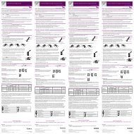
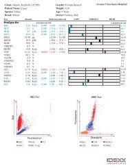
Interpreting <strong>IDEXX</strong> <strong>LaserCyte</strong>* Dx/<strong>IDEXX</strong> <strong>LaserCyte</strong>* Hematology Analyzer <strong>Dot</strong> <strong>Plot</strong>s<strong>Dot</strong> plots are a visual representation of the complete blood count. They are beneficial for quickly interpreting and verifying results. This poster will help you identify various diseases on feline or canine dot plots generated from the <strong>LaserCyte</strong> Dx/<strong>LaserCyte</strong> analyzer.Normal RBC <strong>Dot</strong> <strong>Plot</strong> (Canine)Abnormal RBC <strong>Dot</strong> <strong>Plot</strong> (Canine)Normal WBC <strong>Dot</strong> <strong>Plot</strong> (Canine)Abnormal WBC <strong>Dot</strong> <strong>Plot</strong> (Canine)Regenerative AnemiaLight AbsorptionMature red blood cellsPlateletsGranularityLight AbsorptionReticulocytosisGranularityRed Blood Cells PlateletsReticulocytes DoubletsRBC FragmentsLeukopenia/NeutropeniaLight AbsorptionEosinophilsNeutrophilsMonocytesLymphocytesqualiBeadsGranularityLight AbsorptionGranularityNeutrophils EosinophilsLymphocytes qualiBeadsBasophils uRBCMonocytesNormal RBC <strong>Dot</strong> <strong>Plot</strong> (Canine)Abnormal RBC <strong>Dot</strong> <strong>Plot</strong> (Canine)Normal WBC <strong>Dot</strong> <strong>Plot</strong> (Canine)Abnormal WBC <strong>Dot</strong> <strong>Plot</strong> (Canine)ThrombocytopeniaLight AbsorptionMature red blood cellsNormal platelet densityLight AbsorptionDecreased platelet densityRed Blood Cells PlateletsReticulocytes DoubletsRBC FragmentsLymphoid LeukemiaLight AbsorptionEosinophilsNeutrophilsMonocytesLymphocytesqualiBeadsLight AbsorptionNeutrophils EosinophilsLymphocytes qualiBeadsBasophils uRBCMonocytesGranularityGranularityGranularityGranularityNormal WBC <strong>Dot</strong> <strong>Plot</strong> (Feline)Abnormal WBC <strong>Dot</strong> <strong>Plot</strong> (Feline)Normal WBC <strong>Dot</strong> <strong>Plot</strong> (Feline)Abnormal WBC <strong>Dot</strong> <strong>Plot</strong> (Feline)EosinophiliaLight AbsorptionLight AbsorptionNeutrophilsEosinophilsMonocytesLymphocytesqualiBeadsGranularityNormal WBC <strong>Dot</strong> <strong>Plot</strong> (Canine)EosinophilsNeutrophilsMonocytesLymphocytesqualiBeadsLight AbsorptionLight AbsorptionGranularityEosinophilsAbnormal WBC <strong>Dot</strong> <strong>Plot</strong> (Canine)Neutrophils EosinophilsLymphocytes qualiBeadsBasophils uRBCMonocytesNeutrophils EosinophilsLymphocytes qualiBeadsBasophils uRBCMonocytesPlatelet ClumpingLight AbsorptionNeutrophilsMonocytesLymphocytesGranularityFor more information about <strong>LaserCyte</strong> dot plots,contact <strong>IDEXX</strong> Technical Support.<strong>IDEXX</strong> Technical SupportU.S./Canada/Latin America 1-800-248-2483Europe 00800 1234 3399EosinophilsqualiBeadsLight AbsorptionAustralia 1300 44 33 99New Zealand 0800-102-084Asia 0800-291-018GranularityNeutrophils EosinophilsLymphocytes qualiBeadsBasophils uRBCMonocytesGranularityGranularity
Regenerative AnemiaReticulocytosis (an increased number of reticulocytes) is the hallmark and most objective indicator of a bone marrow response.It is most commonly used to accurately identify a regenerative anemia but may also be seen with a variety of other conditions,including partial and complete compensated hemolytic disease. Reticulocytes are easily identified as the magenta dots to theright of the red blood cell population (red dots). The new methylene blue stain in the CBC5R tubes precipitates the residualreticulum in these immature red blood cells, which gives these cells increased granularity and therefore the shift to the right.In the normal dot plot, there are dramatically reduced numbers of the reticulocytes, noted as a decreased density of magentadots, when compared to the patients with significant increases in reticulocyte numbers. Rapid review of the dot plot allows quickvalidation of the reticulocyte count and therefore confidence in results generated.ThrombocytopeniaThrombocytopenia can be a critical finding in a CBC, and rapid validation of results from the hematology analyzer is essential.In the red blood cell and platelet dot plot, severe thrombocytopenia is easily validated. In the normal patient dot plot, there aredense accumulations of blue dots that represent individual platelet optical profiles. During severe thrombocytopenia, the densityof the blue dots is dramatically reduced compared to normal. Because platelet clumping or partial sample clotting may interferewith the recognition of platelets by the analyzer and they may not appear on the dot plot, rapid microscopic evaluation of a bloodfilm in any case of reported decreased platelet counts is warranted. This is true for all hematology analyzers used for in-housetesting as well as commercial and academic reference laboratories.EosinophiliaRecognition of increases in eosinophils (eosinophilia) is an important observation that directs diagnostic investigations towardsspecific diseases, such as allergies, parasites, and many others. Because they are of such value, rapid validation of reportedeosinophilia is quite important. In the dot plots, eosinophils (green) are located to the left of the neutrophils in the dog and to theright of the neutrophils in the cat. Different patterns are seen for different species because of their somewhat unique morphologicfeatures. In the cases where a significant eosinophilia is reported, the increased density of the eosinophil dot clouds make therapid confirmation of increased numbers of these cells simple.Leukopenia/NeutropeniaLeukopenia or a decrease in total leukocyte numbers and, in particular, neutropenia or decreased in neutrophil numbers oftenhave high clinical significance related to overwhelming inflammatory disease and possible effects of chemotherapy—immediateknowledge of these situations is critical to the veterinarian. Marked decreases in leukocytes can be rapidly validated by examiningthe WBC dot plot. When an isolated cell type such as the neutrophil is significantly decreased, it is easily recognized becauseof the obvious lack of or dramatic decrease in density of the dot plot cloud associated with that particular leukocyte. In thesecases, there is a leukopenia characterized by a marked neutropenia; note the absence of the cloud of purple dots that representindividual neutrophils in the sample.Lymphoid LeukemiaLeukemia has multiple presentations; one of the most common is lymphoid leukemia either as a result of progression ofmalignant lymphoma or primary lymphoid leukemia originating in the bone marrow. Most advanced hematology analyzers cannotaccurately characterize these circulating malignant cells. In many cases, the analyzers attempt cellular characterization, butbecause of difficulty in differentiating the different types of leukocytes, an Abnormal WBC Distribution message is reported toassure that there is follow-up evaluation of a blood film or submission to a reference laboratory for validation of the analyzersattempts. In the normal WBC dot plots, there are distinctly identified clouds of different colored dots that represent the differentpopulations of leukocytes typically seen in the peripheral blood. However, in the dot plots from the lymphoid leukemia patients,clear distinction between the different leukocyte clouds is not present; there is a continuum between different colored clouds. Inthese cases, including the appropriate message code indicating that the analyzer had difficulty in making an accurate leukocytecharacterization along with a blood film or EDTA sample submission to the reference laboratory is recommended.Platelet ClumpingPlatelet clumping is a common problem in veterinary medicine, especially with feline samples. Any time there is difficultyin sample collection resulting in a delay in placing sample into the EDTA tube or delay in proper mixing, there is a potentialfor platelet clumping. There are different degrees of platelet clumping, and most advanced analyzers have the capability ofrecognizing large platelet clumps. When identified, an appropriate message is relayed to the operator along with qualificationof selected results that could be impacted by the platelet clumping. The analyzer may still provide values; however, if there arequalifiers relayed or message codes reported, further evaluation and confirmation of the reported values is essential. A rapidreview of the dot plots can also provide the operator with very quick validation that large platelet clumps are present. Plateletclumps will be recognized as a linear cloud of dots almost parallel to the lymphocyte/monocyte populations. In the dog, plateletclumping could impact eosinophil and neutrophil counts, and especially in the cat, basophil and eosinophil counts may beimpacted. A rapid blood film review can allow timely recognition of large platelet clumps and verification of results reported. Ifplatelet clumps are reported or observed on the blood film, collection of a new sample for analysis is recommended.© 2012 <strong>IDEXX</strong> <strong>Laboratories</strong>, Inc. All rights reserved. •06-28904-00*<strong>LaserCyte</strong> is a trademark or registered trademark of <strong>IDEXX</strong> <strong>Laboratories</strong>, Inc. or its affiliates in the United States and/or other countries.