Brock Linden - Sheridan Memorial Hospital
Brock Linden - Sheridan Memorial Hospital
Brock Linden - Sheridan Memorial Hospital

Create successful ePaper yourself
Turn your PDF publications into a flip-book with our unique Google optimized e-Paper software.
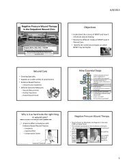
An Algorithm for the ManagementofLower Extremity Diabetic WoundsBased on UT Wound ClassificationUsing a Human Acellular DermalRegenerative Tissue Matrix<strong>Brock</strong> A. Liden, DPM,
Introduction• Most Common Reason of <strong>Hospital</strong>ization for Diabetics =Infected Foot Ulcers• Diabetic Foot Complications =Main Cause of Nontraumatic Lower Extremity Amputations• Most Common Pathway to Amputation =Diabetic Neuropathic Foot UlcerArmstrong DG et al., Clin Podiatr Med Surg 1998
Introduction• Up to 85% of diabetic footand leg amputations maybe prevented with:– Appropriate knowledgeof risk factors– Application ofevidence-basedmultidisciplinarytreatmentArmstrong DG et al., Clin Podiatr Med Surg 1998
Evidenced-Based Algorithmfor Management ofLower Extremity Diabetic WoundsBased on UT Classification
Algorithm Purpose• To develop an evidence-based algorithm for managingdiabetic lower extremity wounds based on theUniversity of Texas Diabetic Wound Classification System– User-friendly– Streamline management of wounds– Employ use of hADM grafts and calcium sulfateantibiotic delivery systemLavery LA et al, J Foot Ankle Surg 1996
Algorithm PurposeGradeWound Description0 Completely epithelialized pre- or post-ulcerative lesion1 Superficial wounds penetrating through epidermis only orboth epidermis & dermis2 Penetrating into tendon or capsule, but not bone3 Penetrating to bone or into jointStageA No infection or ischemiaB Infection onlyC Ischemia onlyD Infection & ischemiaLavery LA et al, J Foot Ankle Surg 1996
Algorithm Overview• Stratifies management according to:– Infection– Ischemia– Wound depth– Involvement of underlying structures• Adds additional factor for consideration:– Wound bed status
Algorithm Overview• Preliminary Work-Up for Diabetic Patientswith Lower Extremity Wounds• Step 1: UT Classification &Wound Bed Determination- Grade 0 Wounds- Grade I Wounds- Grade II Wounds- Grade III Wounds• Step 2: Treatment by UT Classification Grade• Dressing and advanced modality guidence
Wound ContinuumStalledHealingHealedInfectedSenescent CellsPVDNecroticEdemaWet or DryElevated MMPPressureWound edge statusColonizedImproving wound environmentImproved blood supplyMixed GranularControlled edemaMoist Wound bedBalanced MMP’sOffloadedReduced edge effectCleanEpithelizingBlood SupplyClosingSustainedmanagementMWHAccelerating WHLong term solutionCellular migration
Algorithm Goals• Revert Wound Progression to Maximize Healing Potential– First address infection and ischemia to convert:Stage B, C, & D wounds → Stage A wounds– Manage wound bed status to progress:Fibrinous & necrotic wounds → Granular wounds– Employ aggressive debridement to restore:Chronic wounds → Acute wounds
Preliminary Work-UpDiabetic Patient with Lower Extremity WoundPatient History/Clinical PresentationPhysical Examination• Complete Medical/Surgical History– Arterial or Vascular Disease– Level of Diabetic Control• Social History• Nutritional Status• Medication History• Wound History– Cause and Duration– Treatment History– Diagnostic Imaging History• Compliance History• Vital Signs• Physical Examination• Lower Leg Assessment– Vascular Status– Pain– Edema– Infection• Wound Assessment– Location– Measurement– Description
Preliminary Work-Up of DiabeticPatients with Lower ExtremityWoundsDiabetic Patient with Lower Extremity WoundLaboratory TestingDiagnostic Imaging• Laboratory Tests– A1C– CBC– Chem 8– Nutritional Labs• ABI• Wound Culture(if necessary)• RadiographsIf Osteomyelitis Suspected• CT• MRI• Bone Scan
Tests and Lab Day OneBlood Work• CBC• Chem 8• A1c• PrealbuminStudies• ABI with segmental andtoes• Reflux (if venous)• X-rays• Suspect osteo CAT
Step 1A: UT ClassificationDeterminationDetermine UT Classification: Stage & GradeGrade 0 =Completely epithelializedpre- or postulcerative lesionStage A =No infection or ischemiaGrade I =Superficial wound penetrating toepidermis and possibly dermisGrade II =Wound penetrating totendon or capsuleUTClassification(Grade/Stage)Stage B =Infection onlyStage C =Ischemia onlyGrade III =Wound penetrating tobone or jointGrade D =Infection and ischemia
Step 1B: Wound StatusDeterminationDetermine Wound Bed StatusG =GranularF =FibrinousN =NecroticWound Bed Status
Step 1:UT Classification & Wound Bed Status DeterminationUT Classification(Grade/Stage)+Wound Bed Status(G, F, or N)Treatment Priority1. Grade2. Stage3. Wound Bed
Step 2:Treatment by UT Classification Grade• For each wound grade, treatment steps areidentical:– Address infection &/or ischemiaStage B, C, and D wounds → Stage A wounds– Manage wound bed statusFibrinous and necrotic wounds → Granular wounds
Step 2: Treatment Algorithm by UT GradeWound GradeStage A =Non compromisedStage B =Infection onlyStage C =Ischemia onlyStage D =Infection and ischemiaGTreatFTreatNTreatTreat Infection•Mild•MildTreat IschemiaTreatInfection•MildTreatIschemia•Mild•Moderate•ModerateEdema•Severe•Severe•Severe•SevereTreat Edema
Step 2: Treatment Algorithm by Stage(Wound Stages B and D)InfectionMild InfectionModerate Infection•More Aggressive Imaging•Tissue CultureConservative Treatment•Oral Antibiotics•Antimicrobial TopicalsTreatment•Oral Antibiotics•IV Antibiotics•Advanced Topical TherapiesApplies to UT Stage B and D Wounds
Step 2: Treatment Algorithm by Stage(Wound Stages B and D)Severe Infection•More Aggressive Imaging•Tissue Culture•Abscess•Gangrene•OsteomyelitisTreatment•Oral Antibiotics•IV Antibiotics•I&D/Resection•Advanced Topical TherapiesTreatment•Oral Antibiotics•IV Antibiotics•I&D/Resection•Advanced Topical TherapiesAdditional Treatments•Wound VAC•hADM Application
Infection
Infected ?
Infection ?PallorRuborDolorTumorLoss of function
Infected ?
Step 2: Treatment Algorithm by Stage(Wound Stages C and D)IschemiaConservative Treatment•Oral TherapiesReferral for Possible Surgery•Vascular Surgeon•Endovascular Interventionalist
Negative Pressure TherapyProperties• Controls Heavy Exudate• Promotes Granulation/Fill Depth• Increases Blood Supply→ Oxygenate Region• Reduces InfectionUsage• Combine with Silver Dressing for InfectionControl HOCL• Combine with Enzymatic Debriding Agents• Use as Dressing Over Matrix– Different protocols depending on product
Advanced Topical TherapiesW/WO Negative Pressure• Calcium Sulfate– Beads– injectable• HOCl• Trivalent Silver• Doxycycline paste
Step 2: Treatment Algorithm by Stage(Stage D Wounds)Ischemia TreatmentFollowing aforementioned algorithm• Conservative• SurgicalInfection TreatmentFollowing aforementioned algorithm• Mild• Moderate• SevereWhen both issues are concurrent,management of dominant issue takes precedence.
Step 2: Management of Wound Bed AlgorithmStage ANo Infection or IschemiaG =GranularF =FibrinousN =NecroticTreatment•Exudate-Sensitive Dressings•Collagen•Advanced ModalitiesTreatment•Exudate-Sensitive Dressings•Enzymatic Debridement•Sharp Debridement•Negative Pressure•Select Advanced Modalities
Step 2: Management of Wound Bed AlgorithmAfter Treating Wound Bed…EdemaTreatment•Compression Therapy(if adequate blood flow)
Management of Granular WoundsWound Ointments & Dressings• Minimal Exudate– Collagen– Non-Adhering Dressing– Living substitutes / Growth Factors• Minimal to Moderate Exudate– Collagen– Foams– Alginates– Advanced Modalities• Moderate to Heavy Exudate– Collagen– PVA foams– Alginate– Hydrofibre• Heavy Exudate– Collagen– Cavity– Negative PressrureOverlapBetweenGroups
Management of EdemaCompression Therapy• Minimal to Moderate Exudate– Unna Boot w/ Coban– Self-Adherent Wrap or Ace-StyleWrapMultilayer providesbetter compression for– Multi-Layeredema management• Heavy Exudate
Management of GranularWoundsAdvanced Modalities• Living Grafts• Collagen Matrix• Growth Factors• Skin Grafts
Management of Fibrous and Necrotic WoundsWound Dressings• Minimal to Moderate Exudate– Hydrogel– Enzymes– Foams• Moderate to Heavy Exudate– Calcium Alginate– Foams– Cavity• Heavy Exudate– PVA foam– Cavity– Hydrofibre– Negative PressureOverlapBetweenGroups
Management of Fibrinous and Necrotic WoundsCompression Therapy• Minimal to Moderate Exudate– Unna Boot w/ Coban– Self-Adherent Wrap or Ace-Style Wrap• Heavy Exudate– Multi-LayerMultilayer providesbetter compressionfor edemamanagement
Management of Fibrinous & Necrotic WoundsEnzymatic Debriding Agents• Collagenase Santyl® OintmentHydrolytic Debriding Agents• Hydrogel• Hydrocolloids• Honey• Negative Pressure– Lower Pressure / Intermittent
Management of Fibrinous & Necrotic WoundsNegative Pressure Therapy• V.A.C.® with White or Black Foam, asAppropriate
Management of Fibrinous & Necrotic WoundsBioengineered Grafts• Collagen Matrix with abx beads• Stem Cell pucksDeep Wounds
Conclusions
ConclusionsSimilar Successful Results Found Between:• UT Wound Stages• UT Wounds Grades• Chronic and Acute WoundsLending Support to Algorithm Goals…
Algorithm Goals• Revert Wound Progression to Maximize Healing Potential– First address infection and ischemia to convert:Stage B, C, & D wounds → Stage A wounds– Manage wound bed status to progress:Fibrinous & necrotic wounds → Granular wounds– Employ aggressive debridement to restore:Chronic wounds → Acute wounds
Conclusions• This Evidence-Based Algorithm– Streamlines management oflower extremity wounds indiabetic patients– Provides predictable outcomes– May reduce lower extremityamputation incidence
Thank You!
![Adult - Medical History Form [PDF] - Sheridan Memorial Hospital](https://img.yumpu.com/40577874/1/190x245/adult-medical-history-form-pdf-sheridan-memorial-hospital.jpg?quality=85)