Dr. Gheorghe Jurj, MD, DSc Course: Visual Homeopathic Semiology ...
Dr. Gheorghe Jurj, MD, DSc Course: Visual Homeopathic Semiology ...
Dr. Gheorghe Jurj, MD, DSc Course: Visual Homeopathic Semiology ...

Create successful ePaper yourself
Turn your PDF publications into a flip-book with our unique Google optimized e-Paper software.
<strong>Dr</strong>. <strong>Gheorghe</strong> <strong>Jurj</strong>, <strong>MD</strong>, <strong>DSc</strong><strong>Course</strong>: <strong>Visual</strong> <strong>Homeopathic</strong> <strong>Semiology</strong>XXIX Brazilian Congress of HomeopathySão Paulo, September 2008
• That for a same disease, different patient willpresent different traits, which allow forindividualization. Consequently, the chosenremedies are equally different.• However, how does one remedy express itselfin different diseases and patients?
We cannot “identify” remedies, only indicatorsare available: signs which point out to certainremedies.1) Repertory rubrics – help to restrict the field ofpossible remedies2) However, what truly helps to restrict the field ofpossible remedies are not isolated signs, butconfigurations
Groups of symptoms that appear together To find configurations, it is useful to know thepicture of intoxications. For instance, when we know how atropinintoxication is manifested, it easier to find aconfiguration of Belladona. In intoxications, symptoms don’t appear isolated,but in complexes, due to the physio-pathologicalprocesses elicited by the toxic substance.
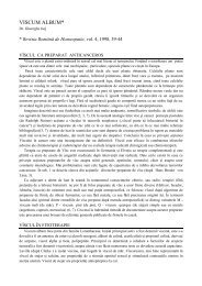
1. Large bullae2. Swelling3. Ecchymosis4. Cyanosis5. Lymphaticdisturb
• The action of a TOXIC substance• Producing:1. Microthrombosis with2. Dilaceration of the superficial layer of the skin,which is detached, forming bullae,3. Disturb in coagulation,4. Disturb in microcirculation, affecting particularlythe veins (cyanosis)5. Edema
• It could be some disease, like e.g. bullouspemphigoid• Or an intoxicationPhotograph Brent Lindon www.venomous.com
• When a patient presents a similarconfiguration, it points out to a snake remedy,as Lachesis• However, it must be kept in mind that we willnot find in the patient the full picture of anintoxication, but only its main characteristics
• Patient consults in poor general state;dyspneic; very irritable.• Signs of chronic pulmonary swelling onauscultation.• Hospitalized for the 3 previous weeks, withoutimprovement.• Ulcers in the legs.
• Peripheral edema• Cyanosis• Dark blue hue on the distalthird of the legs• Congestion on the proximalthird• Ulcers
As it is an unusual localization, it is apeculiar sign
Bleeding in the place of venopuncture
1. Bifid2. <strong>Dr</strong>awn to the left3. <strong>Dr</strong>amatic cyanosis4. Toxic white coating
• Alternation of red(congestion) andblue (cyanosis) smallareas• Wet secretion
Lack of granulation tissue= atrophic ulcer =necrosis Only a few remediespresent atrophic ulcers:Lach; Merc; Carb-an;Carb-v; Kali-bi; someacids (Ph-ac; Sul-ac;Fl-ac)
• In every place and lesion, the same phenomena:– Alternation of congestion and cyanosis on small areas– Toxic aspect– Infiltration edema– Ecchymosis (disturb in coagulation)– Microthrombosis, venous stasis, venectasis– Necrosis
• Improvement in circulation• Improvement of dyspnea• Improvement of mental symptoms• Improvement of weakness• Improvement of pulmonary edema onauscultation• Could decrease diuretic agents
Severe dyspnea:must fixate thechest
with Hippocratic nails
1. Cyanosis2. Toxic white coating3. Deviated to the left
• Severe cyanosis• Dyspnea• Characteristic aspect of thetongue• Does not need as muchventilation as Carb-v• Epitelioma on the nose isdid not ulcerate as fast as inCarb-an
1. Deep ulcer2. Blue margins3. Black background4. Alternation of red andblue small areas5. Lack of granulation6. Grayish areas ofnecrosis7. Edema around
1. Necrosis2. Black hue3. Edema4. Congestionaround
• But the hue isblue, instead ofred• Wet, instead ofdry
1. Cyanosis2. Toxic whitecoating3. Cyanosis on lips
At the first sight: typicalaspect of psoriasis There are no 2 skindiseases exactly alike:we must find whatbelongs “to thepatient”, instead of “thedisease” It is not an abstractpsoriasis, but thepsoriasis of THIS patient
• Alternation of blueand red areas• The background ofthe psoriasis lesionis cyanotic
1. Bifid2. <strong>Dr</strong>awn to the left3. BlueLips: alternation of redand blueFace: congestion