3P-Seq protocol - Bartel Lab - MIT
3P-Seq protocol - Bartel Lab - MIT
3P-Seq protocol - Bartel Lab - MIT

Create successful ePaper yourself
Turn your PDF publications into a flip-book with our unique Google optimized e-Paper software.
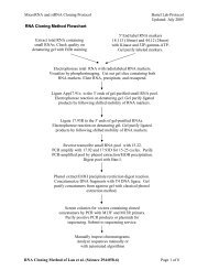
Poly(A)-position profiling by sequencing (<strong>3P</strong>-<strong>Seq</strong>)Jan, C.H., R.C. Friedman, J.G. Ruby, and D.P. <strong>Bartel</strong>. Formation, regulation and evolution of Caenorhabditis elegans3´UTRs. Nature, 10.1038/nature09616, published online Nov 17, 2010.Overview1Oligo(dT) select RNABTTTT 2AAAAAAAAAAAAAnneal oligosSplint ligateAAAAAAAAAAAATTTTPartial digest3with RNase T1BEnrich for 3´ ends ofpoly(A) RNATTTT 4HOHOAAAAAAAAAAAATTTTAAAAAAAAAAAA4AAAAAAAAAAAATTTTTTTTTTTT4BBBBind to StreptavidinWashAnneal primerReverse transcribeDigest with RNase HHOAAAAAAAAAAAA BTTTTTTTTTTTT4HOAAACollect supernatantA ppC3C 3C 35a Ligate 3´ adaptorHOAAA5b Phosphorylate 5´ endsPrepare 3´ end fragmentsfor sequence analysisHOHOPAAAAAAC 35c Ligate 5´ adaptorC 35d RT-PCRAAA6 <strong>Seq</strong>uence & Analysis1
<strong>3P</strong>-<strong>Seq</strong> <strong>Bartel</strong> <strong>Lab</strong>November 2010Step 1: Enrich for polyA + RNAMaterials required (per sample):160 µl Dynal oligo dT 25 magnetic beads (Invitrogen)60 µg* Total RNA (From trizol extraction)300 µl Binding buffer [20 mM Tris-Cl pH 7.5, 1 M LiCl, 2 mM EDTA]400 µl Buffer B [10 mM Tris-Cl pH 7.5, .15 M LiCl, 1 mM EDTA]100 µl 10 mM Tris-Cl pH 7.5 (pre-warm to 75 o C)1 µl Glycoblue (Ambion)6.4 µl 5M NaCl270 µl 100% EthanolProcedure:- Magnetize beads for 30–60 seconds and discard supernatant.- Wash beads by resuspending in 100 µl binding buffer by pipetting or vortexing, magnetize and discardsupernatant as before.- Resuspend in 100 µl binding buffer; the beads are now ready for binding.- Dilute 60 µg of total RNA to a final volume of 100 µl in water.- Add 100 µl binding buffer to diluted RNA and heat at 65 o C for 2 minutes, then place on ice for 2 minutesto denature.- Add denatured RNA to beads, and rotate tubes at room temperature for 5 minutes to anneal.- Magnetize beads for 30–60 seconds and discard the supernatant. Resuspend beads in 200 µl buffer Band wash 1 minute rotating at room temperature.- Repeat wash in buffer B.- Resuspend beads in 100 µl pre-warmed 10 mM Tris-Cl pH 7.5.- Incubate at 75 o C for 2 minutes, then magnetize 30 seconds. Collect the supernatant, which contains thepolyA + RNA.- Add 1 µl glycoblue, 6.4 µl 5M NaCl and 270 µl ethanol to the supernatant and precipitate at –20 o C for atleast 2 hours.* n.b. – We have had success starting with 20–60 µg total RNA.3
<strong>3P</strong>-<strong>Seq</strong> <strong>Bartel</strong> <strong>Lab</strong>November 2010Step 2: Biotin attachment to polyadenylate 3´ endsMaterials required (per sample):5 µl PolyA+ RNA from step 1, resuspended in 5 µl water1 µl 25 µM 3´ biotin adaptor1 µl 25 µM 3´ biotin bridge1 µl 10x annealing buffer [100 mM Tris-Cl pH 7.5, 500 mM NaCl, 10 mM EDTA]1 µl 10 mM MgCl 22 µl 10x ligase buffer (–ATP) [500 mM Tris-Cl pH 7.8, 100 mM MgCl 2 , 100 mM DTT]1 µl 4 mM ATP1 µl T4 RNL2 (wt, NEB).5 µl T4 RNL2 (truncated K227Q, NEB) – optionalProcedure:- Mix the following in a pcr tube:5 µl RNA1 µl 25 µM 3´ biotin adaptor1 µl 25 µM 3´ biotin bridge1 µl 10x annealing buffer2 µl water- Heat at 75 o C for 5 minutes, then slowly ramp down to 22 o C (In a pcr machine, 0.1 o C/sec).- Once the tube has reached room temperature, combine the remaining reagents:1 µl 10 mM MgCl 22 µl 10x ligase buffer (–ATP)1 µl 4 mM ATP5 µl water (4.5 µl if also using RNL2)1 µl T4 RNL2optional .5 µl T4 RNL2 mutant- Incubate at 22 o C overnight, ~16 hours.- Freeze at –20 o C or proceed directly to step 3 below,4
<strong>3P</strong>-<strong>Seq</strong> <strong>Bartel</strong> <strong>Lab</strong>November 2010Step 3: RNase T1 DigestionThe RNase T1 digestions may require optimization. To ensure a partial digestion, we recommend cordecypinlabeling of an in vitro transcribed RNA ≥ 200 nt in length. This RNA can be spiked into the reaction to monitor theextent of digestion by T1. Optimize such that the majority of the RNA has been cut and a ladder of products isvisible. We have had good results with the biochemistry grade T1 nuclease from Ambion using the volume andtime described below.Materials required:Ambion RNase T1 (biochemsitry grade)0.1 M EDTAGlycoblueTracer rna – optional, for monitoring the T1 digestProcedure:- Add the following directly to the ligation reaction:4 µl 0.1 M EDTA80 µl <strong>Seq</strong>uence buffer (ambion).75 µl glycoblueoptional - 0.75 µl radiolabeled rna- Heat to 50 o C in pcr machine with heated lid for 5 minutes.- Ramp down to 22 o C and hold.- Add 3 µl RNase T1.- Incubate at 22 o C for 20 minutes.- Add 220 µl stop solution, precipitate at –20 o C for at least 2 hours.5
<strong>3P</strong>-<strong>Seq</strong> <strong>Bartel</strong> <strong>Lab</strong>November 2010Step 4: Biotin capture and 3´ end tag releaseMaterials required (per sample):100 µl Dynabeads M-280 streptavidin200 µl Bead prep buffer [0.1M NaOH, 50mM NaCl]200 µl 0.1 M NaCl600 µl 2x B&W buffer [10mM Tris-Cl pH 7.5, 1mM EDTA, 2M NaCl]800 µl Wash buffer [10mM Tris-Cl pH 7.5, 1mM EDTA, 50 mM NaCl], pre-warmed to 50 o C200 µl 1x superscript III buffer [40 µl 5x buffer + 12 µl 0.1 M DTT + 148 µl water]1 µl 100 µM 3´ biotin rt primer1 µl 10 mM dTTP5 µl 5x SSIII buffer1.5 µl 0.1 M DTT1 µl Superscript III (invitrogen)1 µl RNase H (invitrogen)Proceedure:- Prep beadso Aliquot 100 µl beads and magnetize for 30~60 seconds.o Discard supernatant and resuspend in 100 µl bead prep buffer to wash (rotate for 2’ at roomtemp).o Repeat wash with 100 µl bead prep buffer.o Wash with 100 µl 0.1 M NaCl.o Repeat wash with 100 µl 0.1 M NaCl.o Wash beads in 200 µl B&W buffer.o Resuspend beads in 200 µl B&W buffer.- Bind and wash RNAo Resuspend RNA from step 3 above in 200 µl water.o Add to prepared beads, rotate at room temp for 15 minutes.o Very briefly spin down tube and magnetize for 1 minute.o Collect and precipitate supernatant if evaluating T1 with tracer RNA.o Wash once in 400 µl 1x B&W buffer.o Wash in 400 µl wash buffer at 50 o C for 2 minutes, occasionally inverting or gently vortexing thetube to mix.o Repeat wash in 400 µl wash buffer at 50 o C.- Release tags into supernatanto Wash beads in 200 µl 1x superscript III buffer at room temp for 2 minutes.o Make RT Mix:1 µl 100 µM 3´ biotin rt primer1 µl 10 mM dTTP5 µl 5x buffer1.5 µl 0.1 M DTT15.5 µl Water1 µl SSIIIo Magnetize beads and remove supernatant.o Add RT Mix to beads and transfer to a PCR tube.o Incubate at 48 o C for 20 minutes in PCR machine.o Ramp down to 37 o C, then add 1 µl RNase H.o Incubate for 25 minutes, gently vortex occasionally to mix beads.o Add 25 µl water to beads and transfer to a 1.5 ml tube.o Magnetize for 1 minute and recover the supernatant (contains end tags).o Ethanol precipitate supernatant with glycoblue at –20 o C for at least 2 hours.6
<strong>3P</strong>-<strong>Seq</strong> <strong>Bartel</strong> <strong>Lab</strong>November 2010Step 5a: Library preparation - 3´ ligationMaterials required (per sample):7.5 µl RNA (tags from step 4, resuspended in 7.5 µl water)1 µl 100 µM adenylated 3´ solexa adaptor (3´ ligation adaptor)1 µl 10x ligase buffer (–ATP) [500 mM Tris-Cl pH 7.8, 100 mM MgCl 2 , 100 mM DTT]0.5 µl T4 RNL1 (neb)Procedure:- Combine the above reagents and incubate at 22 o C (room temp) for 2 hours.- Add 0.75 µl glycoblue, .63 µl 5M NaCl, 30 µl Ethanol. Precipitate at –20 o C for at least 2 hours.- Gel purify RNA ranging from 75 nt ~ 300 nt* on a denaturing 6% acrylamide gel. The gel is run short,such that the bromophenol blue dye migrates ~5 cm. This will limit the size of the gel slab excised.o Elute overnight in 800 µl 0.3 M NaCl.o Split eluate in half and precipitate each half with 1 µl Glycoblue and 2.5 volumes ethanol at least2 hours.o I use radiolabeled RNA markers run in separate lanes to estimate sizes. Decade and centurymarkers from Ambion cover the desired range.o I run thin gels to minimize acrylamide and maximize elution.* n.b. – a larger range is more inclusive from a tag diversity perspective, but will make estimating the library molarconcentration less accurate.Step 5b: Library preparation – phosphorylate 5´ endsMaterials required (per sample):21 µl Ligated RNAs (from step 5a resuspended in 21 µl water)2.5 µl 10x T4 DNA ligase buffer (NEB).5 µl Rnase inhibitor1 µl T4 PNKProcedure:- Combine the above reagents and incubate at 37 o C for 1 hour.- Phenol/chloroform extract and precipitate for at least 2 hours at –20 o C.Step 5c: Library preparation - 5´ ligationMaterials required (per sample):pellet Kinased RNA (from step 5b, pelleted)1 µl 100 µM 26.71 (5´ ligation adaptor)0.5 µl 10x T4 RNL1 buffer (includes ATP, from NEB)0.5 µl T4 RNL1 (NEB)0.1 µl RNasin2.9 µl WaterProcedure:- Combine the water, buffer, RNase inhibitor and 5´ adaptors in a PCR tube.- Use this mix to resuspend the RNA pellet from step 5b.- Add 0.5 µl T4 RNL1, and incubate overnight (~16 hrs) at 22 o C.- Gel purify RNA ranging from ~100 nt ~ 325 nt on a denaturing 6% acrylamide gel as in step 5a.o The reaction volume is low so you can load the entire reaction without precipitation.ooElute overnight in 800 µl 0.3 M NaCl.Split eluate in half and precipitate each half with 1 µl Glycoblue and 2.5 volumes ethanol at least2 hours.7
<strong>3P</strong>-<strong>Seq</strong> <strong>Bartel</strong> <strong>Lab</strong>November 2010Step 5d: Library preparation – RT-PCRMaterials required (per sample):14.6 µl RNA from step 5c resuspended in 14.6 µl water1 µl 100 µM solexa rt primer6.4 µl 5x RT buffer (superscript)3 µl 0.1 M DTT17 µl 10x dNTPs1 µl Superscript III5 µl 1M NaOH25 µl 1M HEPES pH 71 µl 20 µM solexa pcr fwd primer1 µl 20 µM solexa pcr rev primer20 µl 5x pcr buffer (phusion)1 µl Phusion hot start DNA polymerase (NEB/Finnzymes)1 G-25 desalting columnsybr gold stain (Invitrogen)minelute gel purification kit (Qiagen)Procedure:- Mix the following in a PCR tube:14.6 µl RNA1 µl 100 µM solexa rt primer- Heat at 65 o C for 5 minutes, then place on ice for 2 minutes.- Add the following:6.4 µl 5x RT buffer3 µl 0.1 M DTT7 µl 10x dNTPs- Incubate at 48 o C for 3 minutes.- Remove 3 µl and freeze for a no RT control (for PCR).- Add 1 µl Superscript III and incubate at 48 o C for 1 hour.- Add 5 µl 1 M NaOH and incubate at 90 o C for 10 minutes.- Place on ice for 1 minute to cool, then add 25 µl 1M HEPES pH 7.- Desalt 30 µl of the RT reaction over G-25, freeze the rest.- Setup the PCR by mixing the following in a pcr tube:10 µl desalted RT reaction (or 3 µl of no RT control)20 µl 5x pcr buffer10 µl 10x dNTPs57 µl water (64 µl for no RT control)1 µl 20 µM solexa pcr fwd primer1 µl 20 µM solexa pcr rev primer1 µl phusion- Amplify for 16–20 cycles with the following program:1 - 98 o C x 1 min2 - 98 o C x 15 sec |3 - 60 o C x 30 sec | x 15–19 extra cycles4 - 72 o C x 30 sec |5 - 72 o C x 10 minutes6 - hold at 4 o C- Ethanol precipitate for at least 1 hour.- Gel purify 150 ~ 375 bp range.o Run a 2% agarose gel and stain with sybr gold.o Purify using Qiagen’s minelute gel extraction kit.o Elute in EB.8
<strong>3P</strong>-<strong>Seq</strong> <strong>Bartel</strong> <strong>Lab</strong>November 2010Step 6: Library sequencing and analysis considerationsLibraries should be sequenced with Illumina’s gDNA primer. The resulting sequences are antisense to thesample RNA. <strong>3P</strong>-<strong>Seq</strong> libraries will be, by design, heavily biased towards T bases for the first several cycles ofsequencing. This homogeneity can lead to difficulties in cluster definition and base-calling, as neighboringclusters are less likely to have contrasting bases in the first several cycles. When possible, we recommend thatusers cyclically permute the flow cell images such that base calling is initiated with images starting at cycle 6 andimages from cycles 1–5 are appended to the end of the run. The data are subsequently de-convoluted back tothe actual sequence.There are a number of tools available to align reads to a reference genome or transcriptome. For simplicity, wereverse complement reads before alignment. It is imperative to allow mismatches for the oligo(A) tails at the endof reads. This is accomplished by either relaxing alignment stringency at the 3´ end of the read, or stripping the 3´A’s and checking that one or more of these A’s was non-genomic after the read has been aligned. We thenconsider only those reads that terminated in 2 or more consecutive adenylates, at least one of which did notoriginate from the genomic template. The remaining reads may be treated as strand-specific RNA-<strong>Seq</strong> if desired.We cluster reads based on the coordinates of the alignment of their 3´-most non-A nucleotide. Reads areiteratively clustered by identifying the coordinate at which the greatest number of reads terminate then groupingall reads which terminate within 10 bases. Reads falling in this cluster are removed and the process repeateduntil all reads have been accounted for.Because of the high depth of library sampling attainable by high-throughput sequencing, we require that clustersbe definitively derived from multiple independent RNA molecules. This is achieved by requiring that clusters meetat least one of the following criteria:1) Contain reads with different 3´ termini after removing consecutive 3´ adenylates.2) Contain reads with different numbers of consecutive 3´ adenylates (remnants of the poly(A) tail)3) Contain reads sequenced from independent librariesPairing <strong>3P</strong>-<strong>Seq</strong> clusters to transcript models depends greatly on the quality of the genome assembly and geneannotations. Due to the limited read lengths, parsimony is invoked to relate <strong>3P</strong>-clusters to neighboring transcriptmodels (ie, for 3´UTRs, clusters most likely represent the 3´ ends of transcripts from the nearest annotated gene).If available, RNA-<strong>Seq</strong> data can improve the robustness of the pairing between transcript models and 3´ ends, orcan be used for ab initio construction of transcript models. The pertinent issues for correct 3´ end assignmentdepend on the organism being studied, but can relate to gene density (are genes sparse or are they close to eachother? do they overlap?), gene structure (operons, trans-splicing, genome rearrangements?), uniqueness (arethere many recent gene duplications?), etc.9