

FGF-signalling in the differentiation of mouse ES cells towards ...
FGF-signalling in the differentiation of mouse ES cells towards ...
FGF-signalling in the differentiation of mouse ES cells towards ...

You also want an ePaper? Increase the reach of your titles
YUMPU automatically turns print PDFs into web optimized ePapers that Google loves.
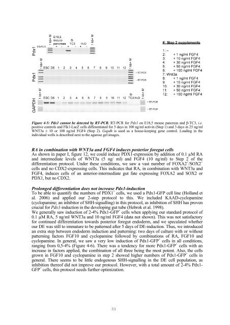
Figure 4-5: Pdx1 cannot be detected by RT-PCR. RT-PCR for Pdx1 on E18,5 <strong>mouse</strong> pancreas and β-TC3, i.e.positive controls and Flk1-LacZ <strong>cells</strong> differentiated for 5 days <strong>in</strong> 100 ng/ml activ<strong>in</strong> (Step 1) and 3 days <strong>in</strong> 25 ng/mlWNT3a ± 10 or 100 ng/ml <strong>FGF</strong>4 (Step 2). Gapdh is used as a house-keep<strong>in</strong>g gene control. Load<strong>in</strong>g <strong>in</strong> <strong>the</strong><strong>in</strong>dividual wells is described next to <strong>the</strong> agarose gel images.RA <strong>in</strong> comb<strong>in</strong>ation with WNT3a and <strong>FGF</strong>4 <strong>in</strong>duces posterior foregut <strong>cells</strong>As shown <strong>in</strong> paper I, figure 12, we could <strong>in</strong>duce PDX1-expression by addition <strong>of</strong> 0.1 µM RAand <strong>in</strong>termediate levels <strong>of</strong> WNT3a (5 ng/ ml) and <strong>FGF</strong>4 (10 ng/ml) to Step 2 <strong>of</strong> <strong>the</strong><strong>differentiation</strong> protocol. Under <strong>the</strong>se conditions, we saw a vast number <strong>of</strong> FOXA2 + /SOX2 +<strong>cells</strong> and no CDX2-express<strong>in</strong>g <strong>cells</strong>. This <strong>in</strong>dicates that RA, <strong>in</strong> comb<strong>in</strong>ation with WNT3a and<strong>FGF</strong>4, <strong>in</strong>duces <strong>cells</strong> <strong>of</strong> an anterior-<strong>in</strong>termediate gut fate express<strong>in</strong>g FOXA2 and SOX2 orPDX1, but no CDX2.Prolonged <strong>differentiation</strong> does not <strong>in</strong>crease Pdx1-<strong>in</strong>ductionTo be able to quantify <strong>the</strong> numbers <strong>of</strong> PDX1 + <strong>cells</strong>, we used a Pdx1-GFP cell l<strong>in</strong>e (Holland etal. 2006) and applied our 2-step protocol to this. We <strong>in</strong>cluded KAAD-cyclopam<strong>in</strong>e(cyclopam<strong>in</strong>e; an <strong>in</strong>hibitor <strong>of</strong> SHH-<strong>signall<strong>in</strong>g</strong>) <strong>in</strong> this protocol, as <strong>in</strong>hibition <strong>of</strong> SHH has provencrucial for Pdx1-<strong>in</strong>duction <strong>in</strong> <strong>the</strong> develop<strong>in</strong>g gut tube (Hebrok et al. 1998).We generally saw <strong>in</strong>duction <strong>of</strong> 2-4% Pdx1-GFP + <strong>cells</strong> when apply<strong>in</strong>g our standard protocol <strong>of</strong>0.1 µM RA, 5 ng/ml WNT3a and 10 ng/ml <strong>FGF</strong>4 (data not shown). This was not satisfactoryfor cont<strong>in</strong>ued <strong>differentiation</strong> <strong>towards</strong> posterior foregut endoderm, and we speculated whe<strong>the</strong>rour DE was still to immature to be patterned after 5 days <strong>of</strong> DE-<strong>in</strong>duction. Thus, we <strong>in</strong>troducedan extra step between endoderm <strong>in</strong>duction and pattern<strong>in</strong>g: two days <strong>of</strong> culture with or withoutpattern<strong>in</strong>g factors <strong>FGF</strong>10 and cyclopam<strong>in</strong>e followed by comb<strong>in</strong>ations <strong>of</strong> RA, <strong>FGF</strong>10 andcyclopam<strong>in</strong>e. In general, we saw a very low <strong>in</strong>duction <strong>of</strong> Pdx1-GFP + <strong>cells</strong> <strong>in</strong> all conditions,rang<strong>in</strong>g from 0,5-4% (Figure 4-6). There was a tendency for more Pdx1-GFP + <strong>cells</strong> with an<strong>in</strong>crease <strong>in</strong> factors applied, <strong>the</strong> comb<strong>in</strong>ation <strong>of</strong> all three be<strong>in</strong>g <strong>the</strong> most potent. Also, <strong>the</strong> <strong>cells</strong>grown <strong>in</strong> <strong>FGF</strong>10 and cyclopam<strong>in</strong>e <strong>in</strong> step 2 showed higher numbers <strong>of</strong> Pdx1-GFP + <strong>cells</strong> <strong>in</strong>general. There seems to be little endogenous SHH-<strong>signall<strong>in</strong>g</strong> <strong>in</strong> <strong>the</strong> DE cell population, as<strong>in</strong>hibition <strong>the</strong>re<strong>of</strong> did not improve our protocol. However, with a total amount <strong>of</strong> 2-4% Pdx1-GFP + <strong>cells</strong>, this protocol needs fur<strong>the</strong>r optimization.53


