RGS2 Determines Short-Term Synaptic Plasticity in Hippocampal ...
RGS2 Determines Short-Term Synaptic Plasticity in Hippocampal ...
RGS2 Determines Short-Term Synaptic Plasticity in Hippocampal ...

You also want an ePaper? Increase the reach of your titles
YUMPU automatically turns print PDFs into web optimized ePapers that Google loves.
Neuron 51, 575–586, September 7, 2006 ª2006 Elsevier Inc. DOI 10.1016/j.neuron.2006.07.012<br />
<strong>RGS2</strong> <strong>Determ<strong>in</strong>es</strong> <strong>Short</strong>-<strong>Term</strong> <strong>Synaptic</strong> <strong>Plasticity</strong><br />
<strong>in</strong> <strong>Hippocampal</strong> Neurons by Regulat<strong>in</strong>g Gi/o-<br />
Mediated Inhibition of Presynaptic Ca 2+ Channels<br />
J<strong>in</strong>g Han, 1 Melanie D. Mark, 1 Xiang Li, 1 Mian Xie, 1<br />
Sayumi Waka, 1 Jens Rettig, 2 and Stefan Herlitze 1, *<br />
1<br />
Department of Neurosciences<br />
Case Western Reserve University<br />
School of Medic<strong>in</strong>e<br />
Room E 604<br />
10900 Euclid Avenue<br />
Cleveland, Ohio 44106<br />
2<br />
Department of Physiology<br />
Saarland University<br />
66421 Homburg<br />
Germany<br />
Summary<br />
<strong>RGS2</strong>, one of the small members of the regulator of G<br />
prote<strong>in</strong> signal<strong>in</strong>g (RGS) family, is highly expressed <strong>in</strong><br />
bra<strong>in</strong> and regulates G i/o as well as G q-coupled receptor<br />
pathways. <strong>RGS2</strong> modulates anxiety, aggression, and<br />
blood pressure <strong>in</strong> mice, suggest<strong>in</strong>g that <strong>RGS2</strong> regulates<br />
synaptic circuits underly<strong>in</strong>g animal physiology<br />
and behavior. How <strong>RGS2</strong> <strong>in</strong> bra<strong>in</strong> <strong>in</strong>fluences synaptic<br />
activity is unknown. We therefore analyzed the synaptic<br />
function of <strong>RGS2</strong> <strong>in</strong> hippocampal neurons by<br />
compar<strong>in</strong>g electrophysiological record<strong>in</strong>gs from <strong>RGS2</strong><br />
knockout and wild-type mice. Our study provides<br />
a general mechanism of the action of the RGS family<br />
conta<strong>in</strong><strong>in</strong>g <strong>RGS2</strong> by demonstrat<strong>in</strong>g that <strong>RGS2</strong> <strong>in</strong>creases<br />
synaptic vesicle release by downregulat<strong>in</strong>g<br />
the G i/o-mediated presynaptic Ca 2+ channel <strong>in</strong>hibition<br />
and therefore provides an explanation of how regulation<br />
of <strong>RGS2</strong> expression can modulate the function<br />
of neuronal circuits underly<strong>in</strong>g behavior.<br />
Introduction<br />
G prote<strong>in</strong> signal<strong>in</strong>g couples extracellular signals with <strong>in</strong>tracellular<br />
effectors. G prote<strong>in</strong> coupled receptor (GPCR)<br />
activation via neurotransmitters <strong>in</strong>itiates the exchange<br />
of GDP for GTP on the Ga subunit, allow<strong>in</strong>g the dissociation<br />
of the Gbg subunit and enabl<strong>in</strong>g it to <strong>in</strong>teract with<br />
different effectors, such as ion channels. The hydrolysis<br />
of GTP to GDP on the Ga subunit leads to the reassociation<br />
of the Gbg dimer with the Ga subunit and term<strong>in</strong>ation<br />
of the signal (Hamm, 1998). The term<strong>in</strong>ation of the G<br />
prote<strong>in</strong> signal is accelerated by a superfamily of GTPase<br />
accelerat<strong>in</strong>g prote<strong>in</strong>s (GAPs) known as RGS prote<strong>in</strong>s.<br />
Besides their function as GAPs for the term<strong>in</strong>ation of<br />
the G prote<strong>in</strong> signals, RGS prote<strong>in</strong>s can act as effector<br />
antagonists by block<strong>in</strong>g the G q pathway (De Vries<br />
et al., 2000).<br />
GPCRs are found at presynaptic and postsynaptic term<strong>in</strong>als<br />
and are <strong>in</strong>volved <strong>in</strong> the regulation of neuronal excitability.<br />
RGS prote<strong>in</strong>s accelerate both the onset and<br />
decay of G prote<strong>in</strong>-mediated signals (Herlitze et al.,<br />
1999; Zerangue and Jan, 1998). This implies that RGS<br />
*Correspondence: sxh106@cwru.edu<br />
is essential for precise physiological signal<strong>in</strong>g events<br />
such as synaptic transmission <strong>in</strong> the central nervous<br />
system, which <strong>in</strong>volves G prote<strong>in</strong>-coupled receptor cascades<br />
and ion channels. In addition, several studies<br />
demonstrate the modulation of presynaptic Ca 2+ channels<br />
of the N-, P/Q- and R-type (Jarvis and Zamponi,<br />
2001) <strong>in</strong> heterologous expression systems. These studies<br />
reveal that RGS accelerates the onset and offset of<br />
transmitter-mediated <strong>in</strong>hibition of presynaptic Ca 2+<br />
channels and also po<strong>in</strong>t to a role of RGS <strong>in</strong> alter<strong>in</strong>g the<br />
amount of <strong>in</strong>hibition for the presynaptic Ca 2+ channels.<br />
Among the RGS family, <strong>RGS2</strong> plays a prom<strong>in</strong>ent role<br />
<strong>in</strong> the bra<strong>in</strong>. A regulatory role of <strong>RGS2</strong> <strong>in</strong> synaptic transmission<br />
and plasticity was suggested when <strong>RGS2</strong><br />
knockout mice revealed a decrease <strong>in</strong> synaptic activity<br />
<strong>in</strong> hippocampal CA1 neurons probably correlated with<br />
<strong>in</strong>creased anxiety <strong>in</strong> the mice (Oliveira-Dos-Santos<br />
et al., 2000). Genetic dissection of a behavioral quantitative<br />
trait locus also revealed that <strong>RGS2</strong> is a modulator of<br />
anxiety <strong>in</strong> mice (Yalc<strong>in</strong> et al., 2004). <strong>RGS2</strong> expression<br />
has been shown to be rapidly upregulated <strong>in</strong> various<br />
bra<strong>in</strong> regions, such as the hippocampus, by excitatory<br />
stimuli (Burchett et al., 1998; Ingi et al., 1998), and it is<br />
therefore likely that regulation of <strong>RGS2</strong> prote<strong>in</strong> levels<br />
<strong>in</strong> neurons regulates synaptic output and behavior.<br />
Thus, the abundance of GPCRs at presynaptic term<strong>in</strong>als<br />
and their <strong>in</strong>volvement <strong>in</strong> presynaptic Ca 2+ channel<br />
<strong>in</strong>hibition, the functional <strong>in</strong>teraction between <strong>RGS2</strong><br />
and presynaptic Ca 2+ channels <strong>in</strong> heterologous expression<br />
systems as well as neurons, and the fact that<br />
<strong>RGS2</strong> 2/2 mice reveal reduced synaptic activity accompanied<br />
with behavioral changes suggest that <strong>RGS2</strong><br />
acts at the presynaptic term<strong>in</strong>al. However, direct evidence<br />
for this hypothesis is still lack<strong>in</strong>g. We therefore<br />
exam<strong>in</strong>ed the effect of <strong>RGS2</strong> on synaptic transmission<br />
by exogenously express<strong>in</strong>g <strong>RGS2</strong> <strong>in</strong> cultured hippocampal<br />
neurons and compar<strong>in</strong>g the results with record<strong>in</strong>gs<br />
from <strong>RGS2</strong> 2/2 neurons. Our data suggest that <strong>RGS2</strong><br />
regulates synaptic output via modulation of basal G prote<strong>in</strong><br />
activity of the G i/o, but not the G q, pathway at the<br />
presynaptic term<strong>in</strong>al. This result is surpris<strong>in</strong>g, given<br />
the fact that <strong>RGS2</strong> has been described for its high aff<strong>in</strong>ity<br />
for the Gq, but not the Gi/o, pathway.<br />
Results<br />
<strong>RGS2</strong> Is Endogenously Expressed <strong>in</strong> Cultured<br />
<strong>Hippocampal</strong> Neurons and Colocalizes<br />
with the <strong>Synaptic</strong> Marker Synaptobrev<strong>in</strong>-2<br />
The goal of this study was to understand if and how<br />
<strong>RGS2</strong> regulates synaptic transmission. A well-established<br />
model system for perform<strong>in</strong>g such experiments<br />
is to compare def<strong>in</strong>ed synaptic parameters between<br />
knockout and wild-type autaptic hippocampal neurons<br />
and to rescue the effects observed <strong>in</strong> knockout cultures<br />
by exogenously express<strong>in</strong>g the wild-type prote<strong>in</strong> (see<br />
for example Bekkers and Stevens, 1991; Calakos et al.,<br />
2004; Rhee et al., 2002). S<strong>in</strong>ce no specific antibodies<br />
are commercially available to detect <strong>RGS2</strong> prote<strong>in</strong> (see<br />
Figure S1 <strong>in</strong> the Supplemental Data), we performed
Neuron<br />
576<br />
<strong>in</strong> situ hybridization and found that <strong>RGS2</strong> mRNA is<br />
detected <strong>in</strong> hippocampal neurons from wild-type, but<br />
not <strong>RGS2</strong> 2/2 , mice (Figure 1A). Exogenous expression<br />
of <strong>RGS2</strong> tagged with YFP at its N and/or C term<strong>in</strong>i<br />
revealed a punctate sta<strong>in</strong><strong>in</strong>g pattern (Figure 1B). These<br />
puncta are partially colocalized with the synaptic marker<br />
synaptobrev<strong>in</strong>-2 (Figure 1B), suggest<strong>in</strong>g that <strong>RGS2</strong> is<br />
transported to synaptic sites.<br />
<strong>RGS2</strong> Regulates <strong>Short</strong>-<strong>Term</strong> <strong>Synaptic</strong> <strong>Plasticity</strong><br />
To determ<strong>in</strong>e if <strong>RGS2</strong> is able to modulate synaptic transmission,<br />
we characterized short-term synaptic plasticity<br />
of autaptic synapses, which hippocampal neurons make<br />
onto themselves when cultured alone on microislands.<br />
<strong>Synaptic</strong> transmitter release via two depolariz<strong>in</strong>g pulses<br />
separated by 50 ms (20 Hz stimulation) and the ratio be-<br />
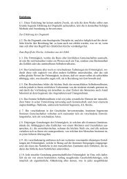
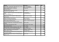
Figure 1. <strong>RGS2</strong> Expressed <strong>in</strong> <strong>Hippocampal</strong><br />
Neurons Targets to <strong>Synaptic</strong> Sites and Regulates<br />
<strong>Short</strong>-<strong>Term</strong> <strong>Synaptic</strong> <strong>Plasticity</strong><br />
(A) <strong>RGS2</strong> mRNA is expressed <strong>in</strong> neurons from<br />
wild-type hippocampal cultures, but not <strong>in</strong><br />
cultures from <strong>RGS2</strong> 2/2 mice. In situ hybridization<br />
of neurons from low-density hippocampal<br />
cultures of wild-type mice (upper<br />
panel) and <strong>RGS2</strong> 2/2 mice (lower panel).<br />
(Left) Nuclei of neurons were visualized with<br />
DAPI sta<strong>in</strong><strong>in</strong>g. <strong>RGS2</strong> mRNA was detected<br />
with a DIG-labeled probe and visualized<br />
with an Alexa546-coupled anti-DIG antibody.<br />
Scale bar, 10 mm.<br />
(B) Exogenously expressed <strong>RGS2</strong>-YFP<br />
reveals somato-dendritic sta<strong>in</strong><strong>in</strong>g and colocalization<br />
with the presynaptic marker<br />
synaptobrev<strong>in</strong>-2 at synaptic sites. (Left) <strong>Hippocampal</strong><br />
neurons were sta<strong>in</strong>ed with an<br />
anti-synaptobrev<strong>in</strong>-2 antibody and visualized<br />
with an Alexa 546-coupled secondary antibody.<br />
(Middle) Fluorescence patterns of neurons<br />
from low-density hippocampal cultures<br />
<strong>in</strong>fected with <strong>RGS2</strong>-YFP reveal a punctuate<br />
sta<strong>in</strong><strong>in</strong>g. A punctuate sta<strong>in</strong><strong>in</strong>g similar to that<br />
seen with <strong>RGS2</strong>-YFP was observed. (Right)<br />
Overlay of the left and middle picture demonstrates<br />
that <strong>RGS2</strong>-YFP is partially colocalized<br />
with the presynaptic marker synaptobrev<strong>in</strong>-<br />
2, as <strong>in</strong>dicated <strong>in</strong> the yellow sta<strong>in</strong><strong>in</strong>g. Scale<br />
bar, 25 mm.<br />
(C–E) Comparison of the paired-pulse ratio<br />
(PPR) from autaptic cultures of rat (D), mice,<br />
and <strong>RGS2</strong> 2/2 mice (E) <strong>in</strong> the absence and<br />
presence of exogenously expressed <strong>RGS2</strong>.<br />
S<strong>in</strong>gle neuron autapses were voltage-clamped<br />
at a hold<strong>in</strong>g potential of –60 mV. EPSCs were<br />
evoked by pairs of 2 ms depolariz<strong>in</strong>g pulses<br />
to 10 mV (50 ms <strong>in</strong>terpulse <strong>in</strong>terval [20 Hz])<br />
every 2 s. Representative sample traces of<br />
the summary data <strong>in</strong> (D) and (E) are shown<br />
<strong>in</strong> panel (C). PPR was calculated as the ratio<br />
of the second peak EPSC amplitude to the<br />
first one. (D) Exogenously expressed <strong>RGS2</strong><br />
significantly reduced the 20 Hz PPR (depression)<br />
compared with wild-type controls<br />
(**p < 0.005). (E) <strong>RGS2</strong> 2/2 cultures show<br />
significantly <strong>in</strong>creased PPR (facilitation) compared<br />
with wild-type controls (**p < 0.005),<br />
and PPR is reduced <strong>in</strong> <strong>RGS2</strong> 2/2 and wildtype<br />
neurons when <strong>RGS2</strong> is exogenously<br />
expressed (*p < 0.05). Error bars = SEM.<br />
tween the first and the second elicited excitatory postsynaptic<br />
current (EPSC) were compared (paired-pulse<br />
ratio [PPR]). To <strong>in</strong>vestigate the possibility that <strong>RGS2</strong><br />
may function dur<strong>in</strong>g synaptic transmitter release, our <strong>in</strong>itial<br />
experiments were performed on rat hippocampal<br />
neurons, while later studies were performed on mouse<br />
cultures. Exogenous expression of <strong>RGS2</strong> <strong>in</strong> wild-type<br />
autaptic hippocampal cultures from rat as well as from<br />
mice reduced the PPR and <strong>in</strong>duced paired-pulse depression<br />
(PPD) (Figures 1C–1E). In contrast, record<strong>in</strong>gs<br />
from autaptic hippocampal neurons from <strong>RGS2</strong> 2/2<br />
mice revealed paired-pulse facilitation (PPF) and thus<br />
an <strong>in</strong>crease <strong>in</strong> the PPR (Figures 1C and 1E). Furthermore,<br />
the PPF was depressed by exogenous expression of<br />
<strong>RGS2</strong> to PPRs found <strong>in</strong> the wild-type cultures (Figure<br />
1E). These results suggest that <strong>RGS2</strong> regulates
<strong>RGS2</strong> Regulates <strong>Short</strong>-<strong>Term</strong> <strong>Synaptic</strong> <strong>Plasticity</strong><br />
577<br />
short-term synaptic plasticity, lead<strong>in</strong>g to PPF when<br />
absent or at low concentrations (Figure 1E), and lead<strong>in</strong>g<br />
to PPD when available at high concentrations <strong>in</strong> hippocampal<br />
neurons (Figure 1D). To determ<strong>in</strong>e if the decrease<br />
<strong>in</strong> PPR is due to changes <strong>in</strong> expression of various<br />
molecules <strong>in</strong>volved <strong>in</strong> RGS signal<strong>in</strong>g, we compared expression<br />
levels of several G prote<strong>in</strong>s and RGS prote<strong>in</strong>s<br />
<strong>in</strong> wild-type and <strong>RGS2</strong> 2/2 neurons. No significant differences<br />
<strong>in</strong> prote<strong>in</strong> levels of Ga i1, Ga i3, Ga o,Ga q/11, Gb 2,<br />
and RGS4 were observed by Western blot analysis <strong>in</strong><br />
wild-type neurons versus <strong>RGS2</strong> 2/2 neurons (Figure 2A).<br />
In addition, we compared the relative mRNA expression<br />
levels of RGS5, 7, and 8 between wild-type and <strong>RGS2</strong> 2/2<br />
cultures and also observed no significant changes<br />
(Figure 2B). Increases <strong>in</strong> PPR may also reflect changes<br />
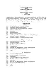
Figure 2. Cultured <strong>Hippocampal</strong> Neurons<br />
from <strong>RGS2</strong> 2/2 and Wild-Type Mice Have<br />
Comparable G Prote<strong>in</strong> Expression and<br />
Make Comparable Amounts of <strong>Synaptic</strong> Contacts<br />
(A) Western blot from 14-day-old wild-type<br />
(left) and <strong>RGS2</strong> 2/2 (middle) cultured hippocampal<br />
neurons. The expression of the follow<strong>in</strong>g<br />
endogenous prote<strong>in</strong>s was evaluated<br />
with antibodies: Ga i1, Ga i3, Ga o,Ga q/11, Gb 2,<br />
and RGS4. Load<strong>in</strong>g control was a-tubul<strong>in</strong>.<br />
As a positive control for the antibody, we<br />
transfected HEK293 cells with cDNAs for<br />
the <strong>in</strong>dicated prote<strong>in</strong>s (right).<br />
(B) Real time quantitative PCR reveals that<br />
neither the absence nor the overexpression<br />
of <strong>RGS2</strong> (lower panel) changes the relative<br />
gene expression of RGS5, 7, and 8. Data are<br />
represented relative to the expression of<br />
<strong>RGS2</strong> <strong>in</strong> the wild-type neurons. The experiments<br />
were performed with three <strong>in</strong>dependent<br />
neuronal cultures <strong>in</strong> duplicates (n = 6).<br />
A 1:100 dilution of sample was used for 18S<br />
as an <strong>in</strong>ternal control.<br />
(C) Examples of low-density hippocampal<br />
neurons (14 days <strong>in</strong> culture) from wild-type<br />
(upper) and <strong>RGS2</strong> 2/2 (lower) mice sta<strong>in</strong>ed<br />
with MAP2-antibody (green) and synaps<strong>in</strong><br />
I-antibody (red). (Middle) Magnification of<br />
the <strong>in</strong>dicated region with<strong>in</strong> the neuron (left)<br />
and (right). Three-dimensional reconstruction<br />
of dendritic arbors from 0.36 mm z-stacks<br />
reveal synaptic contacts between the postsynaptic<br />
MAP2-labeled neuron and the presynaptic<br />
site labeled by synaps<strong>in</strong> I.<br />
(D and E) The number of synaptic contacts (D)<br />
(i.e., the number of synaps<strong>in</strong> I punctuates per<br />
10 mm MAP2-sta<strong>in</strong>ed dendrite) and the synaps<strong>in</strong><br />
I/MAP2 sta<strong>in</strong><strong>in</strong>g ratio (E) (i.e., pixel<br />
area sta<strong>in</strong>ed by synaps<strong>in</strong> I/MAP2) is comparable<br />
between neurons from wild-type and<br />
<strong>RGS2</strong> 2/2 mice.<br />
(F) Comparison of the EPSC amplitudes between<br />
autaptic hippocampal neurons from<br />
wild-type and <strong>RGS2</strong> 2/2 mice from the first<br />
and second EPSC elicited by two 2 ms long<br />
test pulses to +10 mV separated by 50 ms.<br />
The EPSCs elicited by the first pulse are significantly<br />
different, while the second EPSCs<br />
of wild-type and <strong>RGS2</strong> 2/2 are not, <strong>in</strong>dicat<strong>in</strong>g<br />
that functional synaptic contacts are<br />
sufficiently and comparably formed <strong>in</strong> both<br />
cultures.<br />
Error bars = SEM.<br />
<strong>in</strong> neuronal circuit formation due to a reduced number<br />
of sp<strong>in</strong>es and synaptic contacts formed, as orig<strong>in</strong>ally<br />
suggested for CA1 hippocampal neurons from <strong>RGS2</strong> 2/2<br />
mice (Oliveira-Dos-Santos et al., 2000). We did not detect<br />
a reduction <strong>in</strong> the number of synapses formed <strong>in</strong><br />
<strong>RGS2</strong> 2/2 neurons when they were compared with<br />
wild-type neurons (Figures 2C–2E). This is well-supported<br />
by the comparison of the mean EPSC amplitudes.<br />
While the EPSC amplitudes are significantly different<br />
between the <strong>RGS2</strong> 2/2 and wild-type neurons for<br />
the first EPSC dur<strong>in</strong>g a 20 Hz stimulation, comparison<br />
of the EPSC amplitudes elicited by the second pulse<br />
dur<strong>in</strong>g the 20 Hz stimulation revealed no differences,<br />
s<strong>in</strong>ce the <strong>RGS2</strong> 2/2 neurons have a larger facilitation<br />
than wild-type neurons (Figure 2F).
Neuron<br />
578<br />
The Probability of <strong>Synaptic</strong> Vesicle Release Is<br />
Reduced <strong>in</strong> <strong>RGS2</strong> 2/2 Mice Due to a Shift <strong>in</strong><br />
the Ca 2+ Dependence of Transmitter Release<br />
To ga<strong>in</strong> a deeper understand<strong>in</strong>g of the precise action of<br />
<strong>RGS2</strong> on synaptic transmitter release, we analyzed several<br />
parameters of synaptic transmission <strong>in</strong> more detail.<br />
A change <strong>in</strong> the PPR may be caused by an alteration <strong>in</strong><br />
the vesicle release probability (Thomson, 2000). A low<br />
release probability may underlie PPF, while a high release<br />
probability may cause PPD. We therefore first<br />
compared the probability of synaptic vesicle release between<br />
<strong>RGS2</strong> knockout and wild-type neurons, which<br />
can be exam<strong>in</strong>ed by compar<strong>in</strong>g the size of the readily<br />
releasable vesicle pool (RRP) to the number of vesicles<br />
released by a s<strong>in</strong>gle action potential. The RRP is def<strong>in</strong>ed<br />
as the number of vesicles released dur<strong>in</strong>g application of<br />
a hypertonic solution (Rosenmund and Stevens, 1996).<br />
We found that the vesicle release probability is reduced<br />
from the wild-type value of 2.2% to 1.7% <strong>in</strong> <strong>RGS2</strong> 2/2<br />
mice when the RRP was compared to the EPSC <strong>in</strong><br />
each experiment (Figures 3A and 3B), while no differences<br />
<strong>in</strong> the mean RRP size nor the mean EPSC size<br />
could be detected due to the small number of neurons<br />
analyzed (wild-type: EPSC, 15.4 6 2.38 [pC], n = 21;<br />
RRP, 763 6 127 [pC], n = 21; <strong>RGS2</strong> 2/2 : EPSC, 14.1 6<br />
2.18 [pC], n = 24; RRP, 873 6 108 [pC], n = 24).<br />
This reduction <strong>in</strong> the probability of release could be<br />
caused by a decrease <strong>in</strong> the amount of available vesicles,<br />
an alteration <strong>in</strong> the recruitment and recycl<strong>in</strong>g of<br />
the vesicles, a change <strong>in</strong> the coupl<strong>in</strong>g of the vesicles<br />
to the release mach<strong>in</strong>ery, or a reduction <strong>in</strong> Ca 2+ <strong>in</strong>flux<br />
through presynaptic Ca 2+ channels. We first analyzed<br />
if the vesicle recycl<strong>in</strong>g process with<strong>in</strong> the synaptic term<strong>in</strong>al<br />
was altered. It has previously been observed that<br />
GABA B receptors <strong>in</strong> the calyx of Held reduce the refill<strong>in</strong>g<br />
of synaptic vesicles via cAMP-dependent signal<strong>in</strong>g (Sakaba<br />
and Neher, 2003) and that <strong>RGS2</strong> negatively regulates<br />
several forms of adenylyl cylcase, which could<br />
lead to a change <strong>in</strong> the presynaptic cAMP levels (Kehrl<br />
and S<strong>in</strong>narajah, 2002). We therefore depleted the vesi-<br />
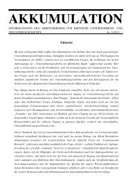
Figure 3. <strong>RGS2</strong> Regulates the Probability of<br />
<strong>Synaptic</strong> Vesicle Release and Ca 2+ Dependence<br />
of Transmitter Release<br />
(A) (Left) Examples of EPSCs evoked by 2 ms<br />
depolariz<strong>in</strong>g pulses from 260 mV to 10 mV<br />
are shown for wild-type neurons (upper) and<br />
<strong>RGS2</strong> 2/2 neurons (lower). (Right) Examples of<br />
the hypertonically mediated release of quanta<br />
from the same neuron shown on the left upon<br />
application of 500 mM sucrose for 4 s.<br />
(B) Probability of synaptic vesicle release was<br />
evaluated by calculat<strong>in</strong>g the ratio of release<br />
evoked by the action potential to that evoked<br />
by hypertonic sucrose. In autaptic neurons<br />
from <strong>RGS2</strong> 2/2 mice, the vesicular release<br />
probability is significantly reduced compared<br />
with wild-type controls (*p < 0.05). Error bars =<br />
SEM.<br />
(C and D) Vary<strong>in</strong>g external Ca 2+ and Mg 2+ concentrations<br />
from 1 to 8–10 mM and 8–10 to<br />
1 mM, respectively, were applied us<strong>in</strong>g an amplifier-controlled perfusion system. (C) Example EPSC traces of wild-type (with extracellular<br />
[Ca 2+ ] o 1 mM, 4 mM, and 8 mM) and <strong>RGS2</strong> 2/2 neurons (with extracellular [Ca 2+ ] o 1 mM, 6 mM, and 10 mM). (D) EPSC amplitudes were normalized<br />
to the maximal response determ<strong>in</strong>ed by the free dose response fit for the s<strong>in</strong>gle experiment. In the absence of <strong>RGS2</strong>, the midpo<strong>in</strong>t of the curve is<br />
shifted to the right, <strong>in</strong>dicat<strong>in</strong>g that <strong>RGS2</strong> 2/2 autapses need higher external Ca 2+ concentrations to obta<strong>in</strong> the same synaptic response as the wildtype<br />
ones. Error bars = SEM.<br />
cles from the synapses us<strong>in</strong>g 30 depolariz<strong>in</strong>g stimuli at<br />
a frequency of 20 Hz and analyzed how long it took for<br />
the synapse to recover the EPSC amplitude to the level<br />
measured before vesicle depletion (Figure 4A). Neither<br />
the time constant of EPSC depletion (Figure 4A) or recovery<br />
(Figure 4B) nor the amount of recovery of the<br />
EPSC (Figure 4B) or the size of the RRP (Figures 4C<br />
and 4D) was altered when <strong>RGS2</strong> was absent. This suggests<br />
that vesicle recycl<strong>in</strong>g or prim<strong>in</strong>g was not altered<br />
<strong>in</strong> the <strong>RGS2</strong> 2/2 mice. To exclude postsynaptic and<br />
structural/morphological changes of the synapse<br />
caused by the absence of <strong>RGS2</strong>, we also analyzed the<br />
m<strong>in</strong>iature EPSCs (mEPSCs) (Figure 4E). mEPSC amplitude<br />
(Figure 4F) and mEPSC frequency (Figures 4G<br />
and 4H) were not altered <strong>in</strong> <strong>RGS2</strong> 2/2 neurons <strong>in</strong> comparison<br />
to wild-type neurons, suggest<strong>in</strong>g that <strong>RGS2</strong> does<br />
not alter vesicle size or the postsynaptic response to<br />
transmitter release.<br />
Differences <strong>in</strong> PPF and PPD can be caused by<br />
changes <strong>in</strong> the Ca 2+ entry dur<strong>in</strong>g repetitive stimulations,<br />
result<strong>in</strong>g <strong>in</strong> an alteration of the number of transmitter<br />
quanta (vesicles) released (Fisher et al., 1997; Zucker,<br />
1999). Therefore, we analyzed the Ca 2+ dependence of<br />
the transmitter release by <strong>in</strong>creas<strong>in</strong>g the external Ca 2+<br />
concentration between 1 to 10 mM Ca 2+ and measured<br />
the change <strong>in</strong> EPSC size dur<strong>in</strong>g 0.2 Hz depolariz<strong>in</strong>g<br />
pulses. In the absence of <strong>RGS2</strong> prote<strong>in</strong>, the midpo<strong>in</strong>t<br />
of the Ca 2+ dependence of transmitter release curve<br />
was shifted to a higher Ca 2+ concentration (Figures 3C<br />
and 3D). This suggests alterations <strong>in</strong> the Ca 2+ <strong>in</strong>flux<br />
through voltage-dependent Ca 2+ channels, an alteration<br />
<strong>in</strong> the coupl<strong>in</strong>g between presynaptic Ca 2+ channels and<br />
the release mach<strong>in</strong>ery, and/or coupl<strong>in</strong>g of Ca 2+ to the<br />
vesicle release.<br />
<strong>RGS2</strong> Acts on <strong>Synaptic</strong> Transmission via Modulat<strong>in</strong>g<br />
Gi/o, but Not Gq, Pathways<br />
<strong>RGS2</strong> accelerates and/or <strong>in</strong>hibits G i/o pathways and<br />
<strong>in</strong>hibits Gq-coupled receptor pathways <strong>in</strong> cells (Kehrl<br />
and S<strong>in</strong>narajah, 2002). G i/o, as well as downstream
<strong>RGS2</strong> Regulates <strong>Short</strong>-<strong>Term</strong> <strong>Synaptic</strong> <strong>Plasticity</strong><br />
579<br />
Figure 4. <strong>Synaptic</strong> Vesicle Recycl<strong>in</strong>g and<br />
Spontaneous Release Properties Are Not<br />
Altered <strong>in</strong> <strong>Hippocampal</strong> Neurons from<br />
<strong>RGS2</strong> 2/2 Mice<br />
To evaluate whether <strong>RGS2</strong> affects synaptic<br />
vesicle recycl<strong>in</strong>g, two approaches were applied.<br />
(A and B) The amount and time course<br />
of depletion of the RRP as well as the recovery<br />
of the RRP follow<strong>in</strong>g activity is not different<br />
between <strong>RGS2</strong> 2/2 cultures and wild-type<br />
controls. The depletion was determ<strong>in</strong>ed by<br />
measur<strong>in</strong>g the recovery of the EPSC amplitude<br />
at vary<strong>in</strong>g time po<strong>in</strong>ts follow<strong>in</strong>g depletion<br />
<strong>in</strong>duced by 30 2 ms long voltage pulses<br />
to 10 mV at 20 Hz. Example traces are shown<br />
on top. In (A) (lower), the EPSCs were normalized<br />
to the largest EPSC dur<strong>in</strong>g the 20 Hz<br />
stimuli tra<strong>in</strong>. In (B) (lower), the recovered<br />
EPSCs were normalized to the first EPSC <strong>in</strong><br />
the 20 Hz stimuli tra<strong>in</strong>.<br />
(C and D) The refill<strong>in</strong>g of the RRP was also<br />
measured by apply<strong>in</strong>g paired pulses of hypertonic<br />
solution (500 mM sucrose, each for<br />
4 s) with vary<strong>in</strong>g <strong>in</strong>terpulse <strong>in</strong>tervals (1 s, 4 s,<br />
7 s, 10 s, 13 s, 30 s, and 60 s). Example traces<br />
are shown <strong>in</strong> (C). The second response was<br />
normalized to the first response, and the<br />
quantified data from wild-type and knockout<br />
cultures are shown <strong>in</strong> (D). Aga<strong>in</strong>, there was<br />
no significant difference between the recovery<br />
rates of neurons from wild-type and<br />
<strong>RGS2</strong> 2/2 mice.<br />
(E) Examples of mEPSCs recorded <strong>in</strong> the<br />
presence of 200 nM TTX from wild-type and<br />
<strong>RGS2</strong> 2/2 autaptic hippocampal cultures.<br />
The analysis <strong>in</strong>dicated that the amplitude distribution<br />
(F) and the frequency (shown as the<br />
mean frequency [G] and the <strong>in</strong>terevent <strong>in</strong>terval<br />
cumulative fraction [H]) of the spontaneous<br />
vesicle release were not significantly<br />
altered <strong>in</strong> the absence of <strong>RGS2</strong>. This suggests<br />
that postsynaptic properties did not change <strong>in</strong> neuronal cultures from <strong>RGS2</strong> 2/2 mice. The mean values for the amplitude distribution<br />
were: wild-type 11.5 6 0.1 pA (4510 events, n = 32), <strong>RGS2</strong> 2/2 11.4 6 0.1 pA (6441 events, n = 37). Statistical significance of the <strong>in</strong>terevent <strong>in</strong>terval<br />
cumulative fraction plot and the mEPSC amplitude distribution was evaluated with a Kolmogorov-Smirnov 2 sample test (p > 0.1).<br />
Error bars = SEM.<br />
effectors of the G q pathways, modulates synaptic transmission.<br />
For example, classical presynaptic <strong>in</strong>hibition is<br />
caused by the <strong>in</strong>hibition of presynaptic Ca 2+ channels<br />
via G i/o-pertussis tox<strong>in</strong> (PTX) sensitive GPCR activation<br />
(Stevens, 2004). Downstream components of the G q<br />
pathway, such as diacylglycerol (DAG), <strong>in</strong>crease vesicle<br />
prim<strong>in</strong>g (Brose et al., 2000), and PKC has been suggested<br />
to regulate the size and the refill<strong>in</strong>g rate of the<br />
vesicle pool (Morgan et al., 2005). Therefore, alterations<br />
<strong>in</strong> the G q as well as G i/o pathway <strong>in</strong> the <strong>RGS2</strong> 2/2 neurons<br />
might underlie the observed changes <strong>in</strong> PPR. In order to<br />
<strong>in</strong>vestigate whether the <strong>in</strong>creased PPF <strong>in</strong> <strong>RGS2</strong> 2/2 neurons<br />
is mediated by G i/o or G q-coupled pathways, we<br />
analyzed transmission <strong>in</strong> the presence of PTX, which<br />
blocks the G i/o pathway, and YM-254890, a specific<br />
blocker of the G q/11 pathway (Takasaki et al., 2004).<br />
We found that <strong>in</strong>cubation of the autaptic neurons with<br />
PTX 24 hr prior to the experiments abolished PPF and<br />
reduced the PPR of <strong>RGS2</strong> 2/2 neurons from 1.16 to<br />
0.98, i.e., to levels observed <strong>in</strong> the wild-type neurons<br />
(1.03 6 0.02 [n = 25], Figure 5). In wild-type neurons<br />
PTX did not alter the PPR (Figure 5B). In contrast, block<strong>in</strong>g<br />
the Gq pathway by application of 10 ng/ml<br />
YM-254890 did not change PPF <strong>in</strong> neurons from knock-<br />
out or wild-type mice (Figures 5A and 5B). To demonstrate<br />
that YM-254890 can effectively <strong>in</strong>hibit G q pathways<br />
at 10 ng/ml, we monitored the G q-<strong>in</strong>duced PIP 2<br />
hydrolysis with the described PIP 2 sensor PH-EGFP <strong>in</strong><br />
HEK293 cells (Stauffer et al., 1998). HEK293 cells were<br />
transfected with mAChR-M1 receptor and PH-EGFP <strong>in</strong><br />
a 5:1 molar ratio to guarantee that cells conta<strong>in</strong><strong>in</strong>g the<br />
PIP 2 sensor also conta<strong>in</strong> the G q-coupled receptor<br />
mAChR-M1. As shown <strong>in</strong> Figures 5C and 5D, YM-<br />
254890 blocks mAChR-M1-mediated PIP 2 hydrolysis<br />
at the plasma membrane. These results taken together<br />
suggest that the G i/o, but not the G q, pathway is <strong>in</strong>volved<br />
<strong>in</strong> the <strong>in</strong>creased PPF observed <strong>in</strong> <strong>RGS2</strong> 2/2 neurons.<br />
To obta<strong>in</strong> further evidence that <strong>RGS2</strong> mediates its effect<br />
via the G i/o, but not the G q, pathway, we analyzed<br />
the effect of the <strong>RGS2</strong> mutant (N149A) on the G i/o and<br />
G q pathway <strong>in</strong> Xenopus oocytes and on synaptic transmission<br />
<strong>in</strong> neurons. The correspond<strong>in</strong>g am<strong>in</strong>o acid <strong>in</strong><br />
RGS4 (N128A), which belongs to the same RGS subfamily,<br />
has been shown to determ<strong>in</strong>e RGS aff<strong>in</strong>ity toward<br />
the G prote<strong>in</strong> a subunits (Posner et al., 1999). S<strong>in</strong>ce<br />
<strong>RGS2</strong> has a low potency as a GAP <strong>in</strong> the G i/o pathway,<br />
but a high potency as an <strong>in</strong>hibitor of the Gq pathway<br />
(Heximer et al., 1997, 1999), we <strong>in</strong>troduced the po<strong>in</strong>t
Neuron<br />
580<br />
Figure 5. <strong>RGS2</strong> Regulates <strong>Synaptic</strong> <strong>Plasticity</strong> through PTX-Sensitive<br />
Pathways<br />
S<strong>in</strong>gle neuron autapses were voltage-clamped at a hold<strong>in</strong>g potential<br />
of –60 mV. EPSCs were evoked by pairs of 2 ms depolariz<strong>in</strong>g pulses<br />
(10 mV) at 50 ms <strong>in</strong>terpulse <strong>in</strong>tervals (20 Hz) every 2 s. (A) Example<br />
traces of wild-type and <strong>RGS2</strong> 2/2 neurons <strong>in</strong> the presence and absence<br />
(left) of PTX (middle) and YM-254890 (right) elicited by<br />
a two-pulse 20 Hz stimulation. (B) PTX (Gi/o blocker) pretreatment<br />
(100 ng/mL, 24 hr) abolished the <strong>in</strong>creased 20 Hz PPR <strong>in</strong> <strong>RGS2</strong> 2/2<br />
autapses (**p < 0.005), while YM-254890 (G q blocker) pretreatment<br />
(10 ng/mL, 18 hr) had no significant effect. (C and D) Monitor<strong>in</strong>g of<br />
<strong>in</strong>tracellular Gq pathways activation by PH-EGFP. (C) Confocal images<br />
of HEK293 cells transfected with mAChR-M1 and PH-EGFP before<br />
(left) and after (right) application of 100 mM Mch. Images are<br />
shown as negative contrast images. Black circles show the cytoplasmic<br />
area which was used for compar<strong>in</strong>g the fluorescence <strong>in</strong>tensity.<br />
(D) Time course of fluorescence ratio changes (F/F 0) with<strong>in</strong> the<br />
cytoplasm dur<strong>in</strong>g 100 mM Mch application <strong>in</strong> the presence (lower)<br />
and absence (upper) of YM-254890 (10 m<strong>in</strong> and 18 hr pretreatment<br />
10 ng/mL). Error bars = SEM.<br />
mutation N149A <strong>in</strong>to <strong>RGS2</strong> under the assumption that<br />
the reduced aff<strong>in</strong>ity for both pathways would elim<strong>in</strong>ate<br />
the effect of <strong>RGS2</strong> on the Gi/o pathway, but would still<br />
be able to block the G q pathway. To characterize the effect<br />
of the mutant <strong>RGS2</strong> on the two signal<strong>in</strong>g pathways,<br />
we made use of the Xenopus oocyte expression system.<br />
We and others have demonstrated that <strong>RGS2</strong> accelerates<br />
the deactivation k<strong>in</strong>etics of the G prote<strong>in</strong> <strong>in</strong>wardrectify<strong>in</strong>g<br />
potassium (GIRK) channel when the channels<br />
were activated by the M 2 muscar<strong>in</strong>ic acetychol<strong>in</strong>e receptor<br />
(mAChR-M 2), a G i/o-coupled receptor (Doupnik<br />
et al., 1997; Herlitze et al., 1999; Mark et al., 2000a). In<br />
addition, it was shown that activat<strong>in</strong>g the G q-coupled<br />
pathways <strong>in</strong> Xenopus oocytes via GPCRs activates an<br />
endogenous Ca 2+ -activated Cl 2 current. The activation<br />
of this Cl 2 current is blocked by <strong>RGS2</strong> and other RGS<br />
prote<strong>in</strong>s, and it has been suggested that the RGS effect<br />
is due to the <strong>in</strong>hibitory effect of the RGS on the G q<br />
pathway (Mark et al., 2000a; Saugstad et al., 1996). To<br />
evaluate if the <strong>RGS2</strong> mutant was still capable of <strong>in</strong>hibit<strong>in</strong>g<br />
the G q pathway, we coexpressed <strong>RGS2</strong> wild-type or<br />
<strong>RGS2</strong>(N149A) together with GIRK1/4 channels and P 2Y 2<br />
receptors. P 2Y 2 receptors, when expressed <strong>in</strong> Xenopus<br />
oocytes, activate both the G i/o and G q pathway (Mosbacher<br />
et al., 1998). Activation of the G i/o pathway leads<br />
to GIRK channel activation, while stimulation of the G q<br />
pathway activates the endogenous Ca 2+ -activated Cl 2<br />
channels. This current can be measured as an outward<br />
current (see arrow <strong>in</strong> Figure 6A, second trace). S<strong>in</strong>ce<br />
very little outward K + current is detected due to the<br />
<strong>in</strong>ward-rectify<strong>in</strong>g properties of the GIRK channel, most<br />
of the outward current is mediated by the Cl 2 channel.<br />
Coexpression of <strong>RGS2</strong> (1:2 dilution) or <strong>RGS2</strong>(N149A)<br />
(1:2 dilution) completely suppressed the Ca 2+ -activated<br />
outward current, suggest<strong>in</strong>g that both the wild-type<br />
and the <strong>RGS2</strong> mutant <strong>in</strong>hibit the G q pathway (Figures<br />
6A and 6B). Even upon further dilution, the <strong>RGS2</strong>(N149A)<br />
mutant (1:20) was still able to reduce the G q-activated<br />
Cl 2 current significantly, but not completely (Figure 6B).<br />
The same effects were observed <strong>in</strong> our previous study<br />
when <strong>RGS2</strong> was diluted to 1:40 (see Mark et al.,<br />
2000a). We next analyzed if the <strong>RGS2</strong> mutant is able to<br />
accelerate the deactivation k<strong>in</strong>etics of GIRK channels<br />
once they are activated via the G i/o pathway. We<br />
coexpressed the <strong>RGS2</strong> prote<strong>in</strong>s together with GIRK1/4<br />
subunits and mAChR-M 2. Activation of mAChR-M 2<br />
via application of 10 mM ACh leads to an <strong>in</strong>crease <strong>in</strong><br />
GIRK current. Once ACh is washed out the GIRK channel<br />
deactivates. The deactivation time depends on the<br />
term<strong>in</strong>ation of the G prote<strong>in</strong> cycle and is accelerated<br />
by RGS prote<strong>in</strong>s <strong>in</strong>clud<strong>in</strong>g <strong>RGS2</strong> (Herlitze et al., 1999;<br />
Mark et al., 2000a). As shown <strong>in</strong> Figures 6C and 6D,<br />
the GIRK channel deactivation is faster <strong>in</strong> the presence<br />
of <strong>RGS2</strong> but is slowed <strong>in</strong> the presence of <strong>RGS2</strong>(N149A).<br />
The slow<strong>in</strong>g of the GIRK channel deactivation can be expla<strong>in</strong>ed<br />
by a dom<strong>in</strong>ant-negative effect of <strong>RGS2</strong>(N149A)<br />
on the G i/o pathway, s<strong>in</strong>ce <strong>RGS2</strong>(N149A) may compete<br />
with the endogenous RGS prote<strong>in</strong>s for b<strong>in</strong>d<strong>in</strong>g to the<br />
Ga i/o prote<strong>in</strong>s <strong>in</strong> Xenopus oocytes.<br />
The experiments described above suggest that<br />
<strong>RGS2</strong>(N149A) is still capable of block<strong>in</strong>g the G q pathway<br />
but has lost its GAP activity on the G i/o pathway, where it<br />
now acts as a dom<strong>in</strong>ant-negative mutant. Thus, if <strong>RGS2</strong><br />
affects short-term synaptic plasticity via the G i/o pathway,<br />
we should not be able to depress PPF with this<br />
mutant <strong>in</strong> <strong>RGS2</strong> 2/2 neurons, but we should be able to <strong>in</strong>duce<br />
PPF <strong>in</strong> the wild-type neurons due to the dom<strong>in</strong>antnegative<br />
effect of the mutant. In contrast, if <strong>RGS2</strong> affects<br />
short-term plasticity via block of the G q pathway, we<br />
should be able to reduce PPF <strong>in</strong> the knockout neurons,<br />
but should see no effect on the wild-type neurons. Exogenous<br />
expression of <strong>RGS2</strong>(N149A) <strong>in</strong> neurons from<br />
<strong>RGS2</strong> 2/2 mice revealed no effect on PPF, but PPF was<br />
<strong>in</strong>creased <strong>in</strong> wild-type neurons, provid<strong>in</strong>g additional<br />
evidence that the <strong>RGS2</strong> mutant acts on the G i/o pathway
<strong>RGS2</strong> Regulates <strong>Short</strong>-<strong>Term</strong> <strong>Synaptic</strong> <strong>Plasticity</strong><br />
581<br />
Figure 6. The <strong>RGS2</strong> Mutant N149A, which Is<br />
Able to Affect the Gq Pathway, But Not the<br />
Gi/o Pathway, Is Unable to Rescue the Increased<br />
PPR <strong>in</strong> <strong>RGS2</strong> 2/2 Autapses But Acts<br />
as a Dom<strong>in</strong>ant-Negative Mutant <strong>in</strong> Wild-<br />
Type Neurons<br />
(A and B) Both <strong>RGS2</strong> and <strong>RGS2</strong>(N149A) <strong>in</strong>hibit<br />
the Gq pathway. (A) Example voltage<br />
ramp traces of GIRK1/4 currents elicited<br />
from Xenopus oocytes coexpress<strong>in</strong>g<br />
mAChR-M2, P2Y2-R, P2Y2-R and <strong>RGS2</strong>, and<br />
P2Y2-R and <strong>RGS2</strong>(N149A) by application of<br />
10 mM ACh to activate mAChR or 10 mM<br />
ATP to activate P2Y2-R. Note that the P2Y2- R activates not only the Gi/o (as mAChR-R<br />
does) but also the Gq pathway. This becomes<br />
evident <strong>in</strong> the large Ca 2+ -activated Cl 2<br />
current (arrow, outward current). This outward<br />
current is absent when <strong>RGS2</strong> or<br />
<strong>RGS2</strong>(N149A) are coexpressed with the<br />
P2Y2-R, s<strong>in</strong>ce the Gq pathway is blocked by<br />
the <strong>RGS2</strong> prote<strong>in</strong>s. (B) In the presence of<br />
<strong>RGS2</strong> or <strong>RGS2</strong>(N149A) mutant, the P2Y2- R-mediated outward current is drastically<br />
reduced or absent, suggest<strong>in</strong>g that the Gq<br />
pathway is <strong>in</strong>hibited. Values from outward<br />
currents are the largest outward current detected<br />
after application of ATP and were<br />
measured at +40 mV.<br />
(C and D) <strong>RGS2</strong>, but not <strong>RGS2</strong>(N149A), is able<br />
to accelerate the Gi/o pathway. (C) Example<br />
traces measured at 260 mV of GIRK1/4 currents<br />
recorded from Xenopus oocytes by activation<br />
of mAChR-M2 coexpressed with or<br />
without <strong>RGS2</strong> or <strong>RGS2</strong>(N149A). GIRK deactivation<br />
time is accelerated <strong>in</strong> the presence<br />
of <strong>RGS2</strong> and slowed <strong>in</strong> the presence of<br />
<strong>RGS2</strong>(N149A), <strong>in</strong>dicat<strong>in</strong>g that <strong>RGS2</strong> but not<br />
<strong>RGS2</strong>(N149A) accelerates the Gi/o pathway.<br />
(D) Comparison of deactivation time constants, derived from a s<strong>in</strong>gle exponential fit of the deactivation curve as shown <strong>in</strong> (C).<br />
(E and F) Exogenous expression of <strong>RGS2</strong>(N149A) <strong>in</strong> <strong>RGS2</strong> 2/2 autaptic cultures did not rescue PPR, but <strong>in</strong>creased the PPR <strong>in</strong> wild-type neurons,<br />
suggest<strong>in</strong>g that <strong>RGS2</strong>(N149A) can act as a dom<strong>in</strong>ant-negative mutant on the Gi/o pathway. (E) Example EPSC traces of <strong>RGS2</strong>(N149A) exogenously<br />
expressed <strong>in</strong> wild-type and <strong>RGS2</strong> 2/2 neurons elicited by a two-pulse 20 Hz stimulation. (F) Comparison between the PPRs of wildtype<br />
and <strong>RGS2</strong> 2/2 neurons exogenously express<strong>in</strong>g <strong>RGS2</strong> or <strong>RGS2</strong>(N149A).<br />
Error bars = SEM.<br />
<strong>in</strong> a dom<strong>in</strong>ant-negative way (Figures 6E and 6F) and that<br />
<strong>RGS2</strong> <strong>in</strong>fluences synaptic transmitter release at the<br />
presynaptic term<strong>in</strong>al.<br />
Non-L-type Ca 2+ Channels Exhibit Increased<br />
G Prote<strong>in</strong> Modulation <strong>in</strong> <strong>Hippocampal</strong> Neurons<br />
from <strong>RGS2</strong> 2/2 Mice<br />
The results suggest that the basal activity of G prote<strong>in</strong>s<br />
of the G i/o family is higher <strong>in</strong> <strong>RGS2</strong> 2/2 mice s<strong>in</strong>ce PTX <strong>in</strong>hibits<br />
the activation of this G prote<strong>in</strong> family via ADP-ribosylation<br />
of the G prote<strong>in</strong> a subunit. The <strong>in</strong>creased activity<br />
of the G prote<strong>in</strong> is probably due to the reduced level of<br />
RGS prote<strong>in</strong>s, which <strong>in</strong> turn prolong the activation of<br />
the G i/o prote<strong>in</strong> cycle. This predicts a higher concentration<br />
of active G prote<strong>in</strong> subunits (i.e., Ga i/o or Gbg subunits)<br />
with<strong>in</strong> the cell and the presynaptic term<strong>in</strong>al. We<br />
have shown that presynaptic Ca 2+ channels are modulated<br />
directly via G prote<strong>in</strong> bg subunits, lead<strong>in</strong>g to the <strong>in</strong>hibition<br />
of the channels (Herlitze et al., 1996; Ikeda,<br />
1996). In addition, Gbg subunits mediate presynaptic<br />
Ca 2+ channel <strong>in</strong>hibition at the calyx of Held (Kajikawa<br />
et al., 2001). The voltage-dependent <strong>in</strong>hibition of the<br />
Ca 2+ channels can be released by high positive prepulses,<br />
a process def<strong>in</strong>ed as prepulse facilitation<br />
(Elmslie et al., 1990). The amount of prepulse facilitation<br />
provides an <strong>in</strong>dication of the degree to which the channels<br />
are modulated by G prote<strong>in</strong> bg subunits (Zamponi<br />
and Snutch, 1998). We therefore tested if the prepulse<br />
facilitation of somatic non-L-type channels (ma<strong>in</strong>ly P/<br />
Q- and N-type channels) would be <strong>in</strong>creased <strong>in</strong> the<br />
<strong>RGS2</strong> 2/2 neurons, suggest<strong>in</strong>g <strong>in</strong>creased levels of active<br />
Gi/o prote<strong>in</strong>s. In the presence of TTX to block Na + channels<br />
and DHPs to block L-type channels, somatic Ca 2+<br />
currents were elicited by two test pulses to the same<br />
test potentials. Before the second test pulse, a high<br />
positive prepulse was elicited to release G prote<strong>in</strong> modulation<br />
(Figure 7B). The peak current ratios of the currents<br />
before and after the prepulse were compared to<br />
determ<strong>in</strong>e the amount of facilitation. Indeed, prepulse<br />
facilitation was <strong>in</strong>creased from 1.0 to 1.8 <strong>in</strong> <strong>RGS2</strong> 2/2<br />
neurons (Figures 7B and 7C). To further verify this result,<br />
we compared the IV relationship of the non-L-type Ca 2+<br />
currents <strong>in</strong> the neuronal cultures. As predicted for G<br />
prote<strong>in</strong>-modulated channels, the peak amplitude of the<br />
Ca 2+ current was shifted by 4 mV to more depolarized<br />
potentials with no change <strong>in</strong> the reversal potential (Figures<br />
7D and 7F). This most likely reflects the slower<br />
open<strong>in</strong>g of the channel and/or a shift <strong>in</strong> the voltage
Neuron<br />
582<br />
dependence of activation to more depolarized potentials.<br />
Both effects have been attributed to the modulation<br />
of voltage-gated Ca 2+ channels by G prote<strong>in</strong> bg<br />
subunits (Herlitze et al., 1996; Ikeda, 1996). In addition,<br />
the average amplitudes of the peak Ca 2+ currents were<br />
reduced <strong>in</strong> the <strong>RGS2</strong> 2/2 neurons (Figure 6E), thereby<br />
support<strong>in</strong>g the <strong>in</strong>creased <strong>in</strong>hibition of the Ca 2+ channels<br />
by Gbg subunits.<br />
The results suggest that <strong>RGS2</strong> regulates Ca 2+ <strong>in</strong>flux<br />
through voltage-gated Ca 2+ channels <strong>in</strong>to presynaptic<br />
term<strong>in</strong>als by reduc<strong>in</strong>g the basal activity of the Gi/o prote<strong>in</strong><br />
family.<br />
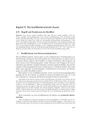
Figure 7. G Prote<strong>in</strong>-Mediated Ca 2+ Channel<br />
Inhibition Is Increased <strong>in</strong> <strong>Hippocampal</strong> Neurons<br />
from <strong>RGS2</strong> 2/2 Mice, while BoNT-A Has<br />
Similar Effects on the Release Probability<br />
and PPR of Neurons from Wild-Type and<br />
<strong>RGS2</strong> Knockout Mice<br />
(A) A diagram to show that <strong>in</strong>creased Gbg<br />
levels <strong>in</strong> the presynaptic term<strong>in</strong>al may reduce<br />
transmitter release by <strong>in</strong>hibition of presynaptic<br />
Ca 2+ channels or by b<strong>in</strong>d<strong>in</strong>g to SNAP-25 to<br />
<strong>in</strong>terfere with vesicle fusion. BoNT-A partially<br />
cleaves SNAP-25, which causes PPF <strong>in</strong> hippocampal<br />
neurons.<br />
(B and C) Ca 2+ currents were elicited from<br />
a hold<strong>in</strong>g potential of 260 mV by a 10 ms<br />
test pulse to +5 mV. After 2 s, a 10 ms prepulse<br />
to +100 mV was applied, and a second<br />
10 ms test pulse to +5 mV was elicited after<br />
stepp<strong>in</strong>g back for 10 ms to 260 mV. Facilitation<br />
ratios were determ<strong>in</strong>ed by divid<strong>in</strong>g the<br />
peak current of test pulse 2 by the peak current<br />
of test pulse 1. (B) Examples of traces recorded<br />
from wild-type and <strong>RGS2</strong> 2/2 neurons<br />
<strong>in</strong>dicat<strong>in</strong>g that, <strong>in</strong> the absence of <strong>RGS2</strong>, G<br />
prote<strong>in</strong> modulation of the Ca 2+ channels is <strong>in</strong>creased.<br />
(C) A diagram of the quantified Ca 2+<br />
current facilitation ratios <strong>in</strong>dicates aga<strong>in</strong> that<br />
<strong>in</strong> the absence of <strong>RGS2</strong>, G prote<strong>in</strong> modulation<br />
of Ca 2+ currents is <strong>in</strong>creased (***p < 0.0005).<br />
(D) Example current traces (IV curve) of non-<br />
L-type currents from wild-type and <strong>RGS2</strong> 2/2<br />
neurons elicited by a 500 ms voltage ramp<br />
from 260 to +90 mV. The comparison of the<br />
traces reveals a positive shift <strong>in</strong> the peak <strong>in</strong>ward<br />
current with no change <strong>in</strong> the reversal<br />
potential.<br />
(E) Diagram of the averaged peak currents of<br />
currents elicited by the voltage ramp demonstrates<br />
that the non-L-type Ca 2+ currents are<br />
reduced <strong>in</strong> the absence of <strong>RGS2</strong> (*p < 0.04).<br />
(F) Diagram of the voltage at which the peak<br />
current appears dur<strong>in</strong>g the voltage ramp.<br />
The diagram shows that the peak current<br />
is shifted to more positive potentials <strong>in</strong><br />
<strong>RGS2</strong> 2/2 neurons (**p < 0.007).<br />
(G and H) Application of BoNT-A <strong>in</strong>creases PPR of both wild-type and <strong>RGS2</strong> 2/2 neurons, suggest<strong>in</strong>g that the ma<strong>in</strong> action of Gbg <strong>in</strong> reduc<strong>in</strong>g<br />
transmitter release is via <strong>in</strong>hibition of presynaptic Ca 2+ channels. (G) Examples of EPSC traces of wild-type and <strong>RGS2</strong> 2/2 neurons <strong>in</strong> the presence<br />
and absence of BoNT-A elicited by a two-pulse 20 Hz stimulation. (H) Comparison of the PPR from autaptic cultures of wild-type mice and<br />
<strong>RGS2</strong> 2/2 mice <strong>in</strong> the presence or absence of BoNT-A. S<strong>in</strong>gle-neuron autapses were voltage-clamped at a hold<strong>in</strong>g potential of –60 mV. EPSCs<br />
were evoked by pairs of 2 ms depolariz<strong>in</strong>g pulses (10 mV) (50 ms <strong>in</strong>terpulse <strong>in</strong>terval [20 Hz]) every 2 s. PPR was calculated as the ratio of the<br />
second peak EPSC amplitude to the first one. In the presence of BoNT-A <strong>in</strong> <strong>RGS2</strong> 2/2 as well as wild-type cultures, PPR (facilitation) is significantly<br />
<strong>in</strong>creased (*p < 0.05; **p < 0.01).<br />
(I) Western blot analysis of endogenous SNAP-25 from wild-type and <strong>RGS2</strong> 2/2 hippocampal cultures before and after BoNT-A (0.2 nM) treatment<br />
for 1 and 3 hr. a-tubul<strong>in</strong> was used as a load<strong>in</strong>g control. As a positive control for the antibody, we transfected HEK293 cells with cDNAs for SNAP-<br />
25 A and B.<br />
(J) Quantification of the relative amount of SNAP-25 after 3 hr of BoNT-A treatment. After 3 hr of BoNT-A treatment, SNAP-25 prote<strong>in</strong> is reduced<br />
by 50%–60% when compared with control prote<strong>in</strong> levels <strong>in</strong> wild-type and <strong>RGS2</strong> 2/2 neurons.<br />
Error bars = SEM.<br />
Botul<strong>in</strong>um Tox<strong>in</strong> A Decreases the Release<br />
Probability and Induces PPF of both <strong>RGS2</strong> Knockout<br />
and Wild-Type <strong>Hippocampal</strong> Neurons<br />
It has been shown that Gbg <strong>in</strong>hibits synaptic transmission<br />
<strong>in</strong> the lamprey neurons downstream of Ca 2+ entry<br />
(Blackmer et al., 2001) (Figure 7A). Increased Gbg prote<strong>in</strong><br />
levels may therefore, <strong>in</strong> addition to modulat<strong>in</strong>g<br />
Ca 2+ <strong>in</strong>flux, also act directly at the presynaptic vesicle<br />
release mach<strong>in</strong>ery. More detailed studies show that<br />
Gbg most likely <strong>in</strong>terferes with vesicle fusion by b<strong>in</strong>d<strong>in</strong>g<br />
to SNAP-25 (Blackmer et al., 2005), thereby caus<strong>in</strong>g a reduction<br />
<strong>in</strong> transmitter release. In those experiments the
<strong>RGS2</strong> Regulates <strong>Short</strong>-<strong>Term</strong> <strong>Synaptic</strong> <strong>Plasticity</strong><br />
583<br />
specific cleavage of SNAP-25 by BoNT-A prevented<br />
additional transmitter- and Gbg-mediated synaptic <strong>in</strong>hibition.<br />
Therefore, <strong>in</strong> our system, if Gbg is act<strong>in</strong>g on<br />
SNAP-25 to prevent transmitter release, partial cleavage<br />
of SNAP-25 <strong>in</strong> neurons from <strong>RGS2</strong> knockout mice<br />
should not significantly <strong>in</strong>crease PPF as would be expected<br />
<strong>in</strong> the wild-type mice (Young, 2005). We therefore<br />
<strong>in</strong>cubated the wild-type and <strong>RGS2</strong> 2/2 autaptic hippocampal<br />
neurons with BoNT-A for 3 hr prior to the<br />
experiment, which leads to a 50%–60% reduction <strong>in</strong><br />
the SNAP-25 prote<strong>in</strong> levels (Figures 7I and 7J), and compared<br />
the PPR of these with those of neurons <strong>in</strong>cubated<br />
without BoNT-A. As expected, application of BoNT-A <strong>in</strong>creased<br />
the PPR from 1.02 to 1.23 <strong>in</strong> wild-type mice (Figures<br />
7G and 7H). Interest<strong>in</strong>gly, an <strong>in</strong>crease <strong>in</strong> PPR from<br />
1.17 to 1.34 was also observed <strong>in</strong> the <strong>RGS2</strong> 2/2 neurons<br />
(Figures 7G and 7H). The PPR <strong>in</strong> <strong>RGS2</strong> 2/2 neurons<br />
treated with BoNT-A was significantly larger than the<br />
PPR <strong>in</strong> wild-type neurons, reveal<strong>in</strong>g that cleavage of<br />
SNAP-25 is affect<strong>in</strong>g synaptic transmission of <strong>RGS2</strong> 2/2<br />
and wild-type neurons <strong>in</strong> a similar fashion. This suggests<br />
that <strong>in</strong>hibition of synaptic transmission by Gbg is mediated<br />
via <strong>in</strong>hibition of presynaptic Ca 2+ channels and not<br />
mediated via b<strong>in</strong>d<strong>in</strong>g to the SNARE complex. However,<br />
s<strong>in</strong>ce it is not possible to completely cleave SNAP-25<br />
without block<strong>in</strong>g transmitter release, the experiments<br />
have to be <strong>in</strong>terpreted carefully <strong>in</strong> respect to variations<br />
<strong>in</strong> SNAP-25 cleavage and to the possibility that the<br />
rema<strong>in</strong><strong>in</strong>g SNAP-25 is the target of Gbg action.<br />
Discussion<br />
We demonstrate here that one of the small RGS family<br />
members, <strong>RGS2</strong>, regulates synaptic strength. In the<br />
presence of <strong>RGS2</strong>, transmitter release probability is<br />
high, while <strong>in</strong> the absence of <strong>RGS2</strong>, synaptic transmitter<br />
release probability is reduced. Our data help to expla<strong>in</strong><br />
the effects previously observed <strong>in</strong> <strong>RGS2</strong> knockout<br />
mice (i.e., reduced synaptic activity [Oliveira-Dos-Santos<br />
et al., 2000]), give mechanistic <strong>in</strong>sight <strong>in</strong>to the action<br />
of <strong>RGS2</strong> on synaptic function, and po<strong>in</strong>t to an importance<br />
<strong>in</strong> regulat<strong>in</strong>g <strong>RGS2</strong> expression dur<strong>in</strong>g neuronal<br />
activity for regulat<strong>in</strong>g neuronal circuits and behavior.<br />
The physiological consequences and the molecular<br />
mechanisms of <strong>RGS2</strong> controll<strong>in</strong>g synaptic transmitter<br />
release are discussed below.<br />
Physiological Consequences of <strong>RGS2</strong> Expression<br />
<strong>RGS2</strong> knockout mice reveal <strong>in</strong>creased anxiety and reduced<br />
male aggression, which is correlated with a reduction<br />
<strong>in</strong> synaptic transmission <strong>in</strong> CA1 hippocampal<br />
neurons (Oliveira-Dos-Santos et al., 2000). Further studies<br />
of the <strong>RGS2</strong> knockout mice reveal an important function<br />
for <strong>RGS2</strong> <strong>in</strong> blood pressure control (Tang et al.,<br />
2003). Genetic dissection of trait loci also suggests<br />
that <strong>RGS2</strong> plays an important role <strong>in</strong> anxiety (Yalc<strong>in</strong><br />
et al., 2004). These studies suggest that <strong>RGS2</strong> is <strong>in</strong>volved<br />
<strong>in</strong> regulat<strong>in</strong>g neuronal circuits underly<strong>in</strong>g autonomic<br />
nervous system regulation and animal behavior.<br />
Our study provides an explanation of how <strong>RGS2</strong> can<br />
modulate the synaptic output <strong>in</strong> the bra<strong>in</strong>: high expression<br />
levels of <strong>RGS2</strong> <strong>in</strong>crease synaptic strength, while<br />
low expression levels decrease synaptic strength. S<strong>in</strong>ce<br />
<strong>RGS2</strong> is an immediate early gene, which is upregulated<br />
very efficiently dur<strong>in</strong>g activity-dependent processes, at<br />
least <strong>in</strong> certa<strong>in</strong> bra<strong>in</strong> areas (Burchett, 2005; Burchett<br />
et al., 1998; Ingi et al., 1998), our results suggest that<br />
the level of <strong>RGS2</strong> prote<strong>in</strong>s with<strong>in</strong> a certa<strong>in</strong> neuronal population<br />
will def<strong>in</strong>e their synaptic efficacy via regulat<strong>in</strong>g<br />
the basal activity of G prote<strong>in</strong>s of the G i/o family.<br />
Mechanistic Insight <strong>in</strong>to the Function of <strong>RGS2</strong><br />
<strong>in</strong> Neurons<br />
Our <strong>in</strong>itial f<strong>in</strong>d<strong>in</strong>g, i.e., that PPF is <strong>in</strong>creased <strong>in</strong> the absence<br />
of <strong>RGS2</strong>, suggested a reduction <strong>in</strong> the synaptic<br />
release probability. A reduction <strong>in</strong> the release probability<br />
can be caused by alter<strong>in</strong>g several processes dur<strong>in</strong>g<br />
transmitter release which <strong>in</strong>volve G i/o and/or G q-coupled<br />
receptor pathways. Classical presynaptic <strong>in</strong>hibition<br />
is mediated by GPCRs, which couple to the PTX-sensitive<br />
G i/o prote<strong>in</strong>s, where the Gbg subunits <strong>in</strong>hibit presynaptic<br />
Ca 2+ channels and reduce Ca 2+ <strong>in</strong>flux dur<strong>in</strong>g<br />
potential changes at the synaptic term<strong>in</strong>al (Herlitze<br />
et al., 1996; Ikeda, 1996; Kajikawa et al., 2001). We found<br />
<strong>in</strong> the <strong>RGS2</strong> knockout mice that non-L-type channels<br />
(i.e., mostly the presynaptic channel types N and P/Q)<br />
<strong>in</strong> hippocampal neurons reveal a stronger voltagedependent<br />
G prote<strong>in</strong> <strong>in</strong>hibition <strong>in</strong> comparison with<br />
wild-type neurons, suggest<strong>in</strong>g that the level of G prote<strong>in</strong><br />
bg subunits <strong>in</strong> the <strong>RGS2</strong> 2/2 neurons is higher. An <strong>in</strong>creased<br />
basal activity level of the G i/o pathway <strong>in</strong> neurons<br />
would expla<strong>in</strong> the elevated Gbg prote<strong>in</strong> levels.<br />
This basal activity is normally very low, as has been<br />
estimated by the tonic <strong>in</strong>hibition of Ca 2+ currents at the<br />
presynaptic term<strong>in</strong>al of the calyx of Held (Cuttle et al.,<br />
1998). In fact, <strong>in</strong>creased constitutive activity of GPCRs<br />
has been shown to play a role <strong>in</strong> caus<strong>in</strong>g diseases<br />
such as cancer, cardiac hypertrophy, and hypertension<br />
(Seifert and Wenzel-Seifert, 2002). Our data also correlate<br />
well with the recent f<strong>in</strong>d<strong>in</strong>g that <strong>in</strong>creased expression<br />
of RGS4 <strong>in</strong> striatal chol<strong>in</strong>ergic <strong>in</strong>terneurons<br />
decreases mAChR-M4-mediated Ca 2+ channel <strong>in</strong>hibition<br />
(D<strong>in</strong>g et al., 2006).<br />
G prote<strong>in</strong> bg subunits act on several synaptic effector<br />
prote<strong>in</strong>s besides the presynaptic Ca 2+ channels. It has<br />
been shown that Gbg <strong>in</strong>teracts with several components<br />
of the synaptic release mach<strong>in</strong>ery (Jarvis and Zamponi,<br />
2001) and that Gbg can lead to decreased transmitter release<br />
downstream of Ca 2+ entry (Blackmer et al., 2001),<br />
most likely via direct b<strong>in</strong>d<strong>in</strong>g to the SNARE prote<strong>in</strong><br />
SNAP-25 (Blackmer et al., 2001, 2005; Gerachshenko<br />
et al., 2005). This conclusion was drawn based on the<br />
fact that BoNT-A, which cleaves SNAP-25, prevented<br />
the seroton<strong>in</strong>-<strong>in</strong>duced, Gbg-mediated synaptic <strong>in</strong>hibition.<br />
In contrast, BoNT-B, which specifically cleaves<br />
synaptobrev<strong>in</strong>, did not prevent seroton<strong>in</strong>’s effect on<br />
transmission. We therefore tested the possibility that<br />
the <strong>in</strong>creased concentration of free Gbg <strong>in</strong> the presynaptic<br />
term<strong>in</strong>al would occlude effects of BoNT-A, as would<br />
be expected if Gbg <strong>in</strong>hibits transmitter release via b<strong>in</strong>d<strong>in</strong>g<br />
to SNAP-25. S<strong>in</strong>ce partial cleavage of SNAP-25 further<br />
reduced transmitter release <strong>in</strong> the <strong>RGS2</strong> 2/2 neurons<br />
<strong>in</strong> comparison with wild-type neurons, our results suggest<br />
that the reduction of transmitter release by <strong>in</strong>creased<br />
Gbg levels is mediated primarily by reduc<strong>in</strong>g<br />
Ca 2+ <strong>in</strong>flux through presynaptic Ca 2+ channels. Another<br />
possibility for <strong>RGS2</strong> function at the presynaptic term<strong>in</strong>al<br />
is that the G i/o pathway may <strong>in</strong>hibit adenylate cyclase
Neuron<br />
584<br />
(for example, via GABAB receptors [Sakaba and Neher,<br />
2003]), lead<strong>in</strong>g to a decrease <strong>in</strong> cAMP levels. Indeed,<br />
<strong>RGS2</strong> has been described to <strong>in</strong>hibit the activity of certa<strong>in</strong><br />
adenylyl cyclase isoforms and thus reduce cAMP levels<br />
(Kehrl and S<strong>in</strong>narajah, 2002). Decreased cAMP levels<br />
and <strong>in</strong>hibition of adenylate cyclase <strong>in</strong> the presynaptic<br />
term<strong>in</strong>al results <strong>in</strong> attenuation of vesicle recruitment to<br />
the RRP, an effect which is mediated by the cAMP-dependent<br />
guanos<strong>in</strong>e exchange factor (cAMP-GEF) and<br />
not by PKA. PKA by itself seems to play a role <strong>in</strong> synaptic<br />
transmission by <strong>in</strong>creas<strong>in</strong>g or ma<strong>in</strong>ta<strong>in</strong><strong>in</strong>g the vesicle<br />
pool size (RRP and slowly releasable pool [SRP]), an effect<br />
which is counteracted by the Ca 2+ /calmodul<strong>in</strong>dependent<br />
prote<strong>in</strong> phosphatase calc<strong>in</strong>eur<strong>in</strong> (Nagy<br />
et al., 2004). However, we did not observe any alterations<br />
<strong>in</strong> synaptic vesicle cycl<strong>in</strong>g, suggest<strong>in</strong>g that<br />
<strong>RGS2</strong> <strong>in</strong>hibits synaptic transmitter release by modulat<strong>in</strong>g<br />
Ca 2+ entry <strong>in</strong>to the presynaptic term<strong>in</strong>al.<br />
<strong>RGS2</strong> has the capacity to accelerate G i/o and to antagonize<br />
G q pathways (Kehrl and S<strong>in</strong>narajah, 2002), but it<br />
has been suggested to have a greater aff<strong>in</strong>ity for Ga q<br />
than for Ga i/o prote<strong>in</strong>s (Heximer et al., 1999). Therefore,<br />
recent studies have concentrated on the effects of<br />
<strong>RGS2</strong> on modulat<strong>in</strong>g G q-coupled pathways <strong>in</strong> heterologous<br />
expression systems and <strong>in</strong>tact tissues, where<br />
<strong>RGS2</strong>, for example, regulates the Ca 2+ oscillation, adaptation,<br />
and excitability of pancreatic ac<strong>in</strong>i via regulat<strong>in</strong>g<br />
<strong>in</strong>tracellular IP 3 levels (Wang et al., 2004). It was therefore<br />
surpris<strong>in</strong>g to observe that the loss of <strong>RGS2</strong> prote<strong>in</strong> and<br />
overexpression of <strong>RGS2</strong> <strong>in</strong> neuronal hippocampal cultures<br />
seems to act via the G i/o pathway rather than the<br />
Gq pathway, at least <strong>in</strong> the presynaptic term<strong>in</strong>al. Neither<br />
blockers of the G q pathway nor the po<strong>in</strong>t mutation of<br />
<strong>RGS2</strong>, which abolishes its effects on the Gi/o pathway<br />
while ma<strong>in</strong>ta<strong>in</strong><strong>in</strong>g modulation of the G q pathway, were<br />
effective <strong>in</strong> rescu<strong>in</strong>g <strong>RGS2</strong> deficiencies. On the other<br />
hand, PTX was sufficient to block the PPF effect <strong>in</strong><br />
<strong>RGS2</strong> 2/2 mice, and the <strong>RGS2</strong>(N149A) mutant acted<br />
as a dom<strong>in</strong>ant-negative mutant for <strong>RGS2</strong> function by<br />
<strong>in</strong>creas<strong>in</strong>g the PPF. These results suggest that the major<br />
targets of <strong>RGS2</strong> with<strong>in</strong> the presynaptic term<strong>in</strong>al are<br />
GPCRs, which couple to the G i/o pathway. This conclusion<br />
is also supported by the fact that vesicle recycl<strong>in</strong>g<br />
<strong>in</strong> <strong>RGS2</strong> 2/2 mice is normal, s<strong>in</strong>ce several second messengers<br />
with<strong>in</strong> the G q pathway, <strong>in</strong>clud<strong>in</strong>g DAG and<br />
PKC, have been described to modulate synaptic transmitter<br />
release at the level of vesicle prim<strong>in</strong>g and recycl<strong>in</strong>g<br />
(Morgan et al., 2005; Nagy et al., 2004; Rhee et al., 2002).<br />
Therefore, <strong>in</strong>creased G q signal<strong>in</strong>g <strong>in</strong> the absence of<br />
<strong>RGS2</strong> would have been expected to cause <strong>in</strong>creased<br />
PLC activation, <strong>in</strong>creased DAG synthesis, and <strong>in</strong>creased<br />
Munc13 activation, result<strong>in</strong>g <strong>in</strong> faster vesicle prim<strong>in</strong>g and<br />
less depression dur<strong>in</strong>g high-frequency tra<strong>in</strong>s. In contrast,<br />
<strong>in</strong>hibition of G q pathways by <strong>RGS2</strong> would have<br />
been expected to <strong>in</strong>crease depression. In both cases<br />
the time course of vesicle recycl<strong>in</strong>g should be altered.<br />
This was, however, not the case, po<strong>in</strong>t<strong>in</strong>g aga<strong>in</strong> to<br />
<strong>RGS2</strong> act<strong>in</strong>g primarily on the G i/o pathway, rather than<br />
the G q pathway, dur<strong>in</strong>g short-term synaptic plasticity.<br />
In summary, we show that <strong>RGS2</strong> regulates synaptic<br />
output. The importance of this f<strong>in</strong>d<strong>in</strong>g lies <strong>in</strong> the underestimated<br />
effects of <strong>RGS2</strong> on modulat<strong>in</strong>g the G i/o pathway,<br />
the important function of <strong>RGS2</strong> <strong>in</strong> regulat<strong>in</strong>g the basal<br />
activity of the G i/o pathway with<strong>in</strong> a neuron, and <strong>in</strong> the<br />
possibility that upregulation of <strong>RGS2</strong> <strong>in</strong> certa<strong>in</strong> neuronal<br />
circuits will most likely modulate synaptic strength.<br />
Experimental Procedures<br />
cDNA Constructs, Viral Production and Infection,<br />
and Cell Culture<br />
Mouse <strong>RGS2</strong>, 4, 5, 8 cDNA (Herlitze et al., 1999; Mark et al., 2000b),<br />
mouse RGS7 (Accession number BC051133), G prote<strong>in</strong> a i1,3,o and<br />
Gb 2 (Herlitze et al., 1996), mouse Ga q (Accession number M55412,<br />
gift from Dr. M. Simon, Pasadena, CA) and SNAP-25A and B (gift<br />
from Dr. M. Wilson, Albuquerque, NM) were cloned <strong>in</strong>to mammalian<br />
expression vectors (pcDNA variants) for expression <strong>in</strong> HEK293 cells.<br />
<strong>RGS2</strong> cDNA was also cloned <strong>in</strong>to the S<strong>in</strong>Rep(nsP2S726)dSP-EGFP<br />
virus vector and also cloned for the <strong>in</strong>itial record<strong>in</strong>gs <strong>in</strong> rat <strong>in</strong>to pSFV.<br />
For localization studies the <strong>RGS2</strong> was <strong>in</strong>serted <strong>in</strong>to pEYFP-N1 and<br />
pEYFP-C1 (Clontech). The po<strong>in</strong>t mutation <strong>RGS2</strong>(N149A) was <strong>in</strong>troduced<br />
<strong>in</strong>to <strong>RGS2</strong> us<strong>in</strong>g an overlap extension PCR method (Herlitze<br />
and Koenen, 1990; Ho et al., 1989). The po<strong>in</strong>t mutation was confirmed<br />
by DNA sequenc<strong>in</strong>g, and the cDNA carry<strong>in</strong>g the mutation<br />
was cloned <strong>in</strong>to S<strong>in</strong>Rep(nsP2S726)dSP-EGFP and pBF1 (oocyte expression).<br />
PH-EGFP was a gift from Dr. T. Meyer (Stauffer et al.,<br />
1998). S<strong>in</strong>dbis pseudovirions were prepared accord<strong>in</strong>g to Invitrogen’s<br />
<strong>in</strong>structions (S<strong>in</strong>dbis Expression System) and as published<br />
<strong>in</strong> our recent publication (Li et al., 2005). For <strong>RGS2</strong> overexpression<br />
and rescue experiments, cultured neurons were <strong>in</strong>fected with 50 ml<br />
of s<strong>in</strong>dbis pseudovirions conta<strong>in</strong><strong>in</strong>g the cDNAs of <strong>RGS2</strong> or<br />
<strong>RGS2</strong>(N149A) constructs. Record<strong>in</strong>gs were performed up to 24 hr<br />
post <strong>in</strong>fection. Microisland and cont<strong>in</strong>ental cultures of hippocampal<br />
neurons were prepared accord<strong>in</strong>g to a modified version of published<br />
procedures from mouse or rat pups (P0-3) (Bekkers and Stevens,<br />
1991; Wittemann et al., 2000).<br />
Immunocytochemistry, Imag<strong>in</strong>g, and Western Blot<br />
Cont<strong>in</strong>ental hippocampal cultures were prepared as described<br />
above and transfected with <strong>RGS2</strong>-YFP by us<strong>in</strong>g the Ca 2+ phosphate<br />
method (Park et al., 2004). 24 hr after transfection, neurons were<br />
fixed with 4% paraformaldehyde and permeabilized with 0.2% Triton<br />
X-100 <strong>in</strong> PBS. Anti-GFP (Molecular Probes) and anti-synaptobrev<strong>in</strong>-2<br />
(SYSY) antibodies were used to label <strong>RGS2</strong> and the synaptic<br />
marker synaptobrev<strong>in</strong>-2. Neurons were <strong>in</strong>cubated with the primary<br />
antibody overnight at 4 C, washed, then <strong>in</strong>cubated with Alexa 488and<br />
Alexa 568-conjugated secondary antibody (Molecular Probes)<br />
for 30 m<strong>in</strong> at room temperature. Cells were embedded <strong>in</strong> Prolong<br />
Gold antifade (Molecular Probes). For the calculation of the number<br />
of synapses and synaps<strong>in</strong>/MAP2 sta<strong>in</strong><strong>in</strong>g ratio, low-density hippocampal<br />
cultures were prepared, fixed, and sta<strong>in</strong>ed as described<br />
above. Anti-synaps<strong>in</strong> I (Invitrogen) and anti-MAP2 (Sigma) antibodies<br />
were visualized with Texas red and Alexa 488-conjugated<br />
secondary antibodies. Images were acquired with a Zeiss LSM<br />
410 or 510 confocal microscope and analyzed by us<strong>in</strong>g VOLOCITY<br />
software (Improvision). Western blots from 14-day-old hippocampal<br />
neurons from wild-type and <strong>RGS2</strong> 2/2 mice were performed accord<strong>in</strong>g<br />
to published procedures (Wittemann et al., 2000). Antibodies<br />
were from Santa Cruz (Ga i1, Ga i3, Ga o,Ga q/11, Gb 2) and RGS4 antibody<br />
was a gift from Dr. S. Mumby, Southwestern University, TX.<br />
For monitor<strong>in</strong>g the Gq-coupled receptor activation with the PH-<br />
EGFP constructs, HEK293 cells were transfected <strong>in</strong> a 5:1 molar ratio<br />
with mAChR-M1 and PH-EGFP. 24 hr after transfection cells were<br />
cont<strong>in</strong>uously perfused with extracellular record<strong>in</strong>g solution. GFP<br />
fluorescence was monitored on the Zeiss LSM 510 <strong>in</strong>verted confocal<br />
microscope at room temperature us<strong>in</strong>g a 633 air objective. Pictures<br />
were taken every 5 s and analyzed us<strong>in</strong>g VOLOCITY software (Improvision).<br />
100 mM Mch (acetyl-B-methylchol<strong>in</strong>e chloride, Sigma)<br />
was applied with<strong>in</strong> 2 m<strong>in</strong> and the average fluorescence <strong>in</strong>tensity <strong>in</strong><br />
a def<strong>in</strong>ed cytoplasmic region was normalized to the average <strong>in</strong>tensity<br />
before the application of the drug (F 0 3 (F/F 0)).<br />
In Situ Hybridization<br />
In situ hybridization was performed as described previously by<br />
Schaeren-Wiemers and Gerf<strong>in</strong>-Moser (1993) and as described <strong>in</strong><br />
the Supplemental Data.
<strong>RGS2</strong> Regulates <strong>Short</strong>-<strong>Term</strong> <strong>Synaptic</strong> <strong>Plasticity</strong><br />
585<br />
Real Time Quantitative PCR<br />
Total RNA was subtracted from 14 DIV cultured neurons with<br />
RNeasy M<strong>in</strong>i Kit (Qiagen Inc.) and purified with on-column DNase<br />
digestion us<strong>in</strong>g RNase-Free DNase Set (Qiagen Inc.). For RT-PCR,<br />
1 mg of RNA was used for reverse transcription with Advantage<br />
RT-for-PCR Kit (BD Biosciences) to generate 100 ml cDNA. 3 ml of<br />
the f<strong>in</strong>al RT product was used for real time PCR for <strong>RGS2</strong>, 5, 7, 8,<br />
and 18S RNA. Real time PCR quantification was performed on<br />
iCycler Iq Detection System (Bio-Rad) with CYBR Green assay<br />
(Bio-Rad), and DNA fragments of <strong>RGS2</strong>, 5, 7, 8, and 18S RNA were<br />
amplified with primer pairs given <strong>in</strong> the Supplemental Data. ThePCR<br />
reactions used a modified 2-step profile with an <strong>in</strong>itial denaturation<br />
step for 3 m<strong>in</strong> at 95 C, followed by 40 cycles of 15 s denaturation at<br />
95 C and 25 s polymerase reaction at 57 C. For <strong>in</strong>ternal control, 3 ml<br />
of 1:100 diluted RT product was used for the 18S reaction with every<br />
run so that the 18S cyber threshold (Ct) value was reported <strong>in</strong> the<br />
same range of those of the <strong>RGS2</strong> fragments to give more accurate<br />
comparison. Relative gene expression data was analyzed with<br />
2-DDCT method (Livak and Schmittgen, 2001).<br />
Electrophysiology and Data Analysis<br />
For EPSC measurements, currents were elicited by a 2 ms long test<br />
pulse to 10 mV and recorded and analyzed as published previously<br />
(Wittemann et al., 2000). Sucrose solution and various extracellular<br />
[Ca 2+ ]o solutions were applied directly onto the recorded neurons<br />
by us<strong>in</strong>g a fast-flow perfusion system (ALA Scientific Instruments).<br />
The EPSC and RRP charge was calculated by <strong>in</strong>tegrat<strong>in</strong>g the currents<br />
elicited by the s<strong>in</strong>gle action potential or the sucrose application.<br />
The Ca 2+ response curves were fitted accord<strong>in</strong>g to the Hill<br />
equation: EPSC = EPSCmaximal/(1 + (EC50/[Ca 2+ ]o) Hill coefficient ). The<br />
fit values from each curve were used to normalize the dose response<br />
curves. The mEPSCs amplitude histograms were plotted with 0.5 pA<br />
b<strong>in</strong>s and were fitted with a Gaussian function. mEPSC analysis was<br />
performed manually us<strong>in</strong>g M<strong>in</strong>i Analysis Program (Synaptosoft) with<br />
an amplitude threshold of 5 pA. 100 ng/ml PTX (Sigma), 10 ng/ml<br />
YM-254890 (gift from Dr. Brian Roth) and 0.2 nM BoNT-A (Sigma)<br />
were applied 24 hr, 18 hr, and 3 hr, respectively, prior to record<strong>in</strong>gs<br />
onto the neuronal cultures. Two microelectrode voltage-clamp record<strong>in</strong>gs<br />
were performed as extensively described <strong>in</strong> our recent paper<br />
(Mark et al., 2000a) and as given <strong>in</strong> the Supplemental Data.<br />
Briefly, Xenopus oocytes express<strong>in</strong>g GIRK1/4 and mAChR-M 2 or<br />
P 2Y 2-R together with or without <strong>RGS2</strong> or <strong>RGS2</strong>(N149A) (see Supplemental<br />
Data for mRNA synthesis, Xenopus oocyte preparation, and<br />
mRNA <strong>in</strong>jection) were voltage-clamped at 0 mV for 20 ms. Then a<br />
960 ms long voltage ramp from 2100 to +50 mV was applied, which<br />
then stepped back to 0 mV for 20 ms. This 1 s long protocol was<br />
repeated 100 times. Transmitters (10 mM ACh and 10 mM ATP)were<br />
typically applied after 10 s for 30 s and then washed out. Time constants<br />
for deactivation curves were calculated at 265 mV and were<br />
determ<strong>in</strong>ed with a s<strong>in</strong>gle exponential fit. The Ca 2+ -activated Cl 2 current<br />
(outward current) was measured at +40 mV. Here the largest outward<br />
current, which was elicited by 10 mM ATP, was used for analysis.<br />
The extracelluar record<strong>in</strong>g solution was a modified R<strong>in</strong>ger’s solution<br />
given <strong>in</strong> the Supplemental Data. Record<strong>in</strong>g solutions for EPSC<br />
measurements and non-L-type channel record<strong>in</strong>gs are described<br />
(Li et al., 2005) and given <strong>in</strong> the Supplemental Data.<br />
Statistical significance throughout the experiments was tested<br />
with ANOVA by us<strong>in</strong>g IGOR software unless otherwise <strong>in</strong>dicated.<br />
Standard errors are given as mean 6 SEM.<br />
All experiments were approved by the Institutional Animal<br />
Research Facility.<br />
Supplemental Data<br />
The Supplemental Data for this article can be found onl<strong>in</strong>e at http://<br />
www.neuron.org/cgi/content/full/51/5/575/DC1/.<br />
Acknowledgments<br />
We thank Drs. E.S. Deneris and L.T. Landmesser for read<strong>in</strong>g the<br />
manuscript, Dr. B. Roth for the YM-254890, Dr. S. Mumby for the<br />
RGS4 antibody, and Dr. M. Wilson for the SNAP-25 cDNAs. This<br />
work was supported by National Institutes of Health Grants<br />
NS0447752 and NS42623 to S.H.<br />
Received: February 27, 2006<br />
Revised: June 6, 2006<br />
Accepted: July 14, 2006<br />
Published: September 6, 2006<br />
References<br />
Bekkers, J.M., and Stevens, C.F. (1991). Excitatory and <strong>in</strong>hibitory autaptic<br />
currents <strong>in</strong> isolated hippocampal neurons ma<strong>in</strong>ta<strong>in</strong>ed <strong>in</strong> cell<br />
culture. Proc. Natl. Acad. Sci. USA 88, 7834–7838.<br />
Blackmer, T., Larsen, E.C., Takahashi, M., Mart<strong>in</strong>, T.F., Alford, S.,<br />
and Hamm, H.E. (2001). G prote<strong>in</strong> bg subunit-mediated presynaptic<br />
<strong>in</strong>hibition: regulation of exocytotic fusion downstream of Ca 2+ entry.<br />
Science 292, 293–297.<br />
Blackmer, T., Larsen, E.C., Bartleson, C., Kowalchyk, J.A., Yoon,<br />
E.J., Pre<strong>in</strong><strong>in</strong>ger, A.M., Alford, S., Hamm, H.E., and Mart<strong>in</strong>, T.F.<br />
(2005). G prote<strong>in</strong> bg directly regulates SNARE prote<strong>in</strong> fusion mach<strong>in</strong>ery<br />
for secretory granule exocytosis. Nat. Neurosci. 8, 421–425.<br />
Brose, N., Rosenmund, C., and Rettig, J. (2000). Regulation of transmitter<br />
release by Unc-13 and its homologues. Curr. Op<strong>in</strong>. Neurobiol.<br />
10, 303–311.<br />
Burchett, S.A. (2005). Psychostimulants, madness, memory. and<br />
RGS prote<strong>in</strong>s? Neuromolecular Med. 7, 101–127.<br />
Burchett, S.A., Volk, M.L., Bannon, M.J., and Granneman, J.G.<br />
(1998). Regulators of G prote<strong>in</strong> signal<strong>in</strong>g: rapid changes <strong>in</strong> mRNA<br />
abundance <strong>in</strong> response to amphetam<strong>in</strong>e. J. Neurochem. 70, 2216–<br />
2219.<br />
Calakos, N., Schoch, S., Sudhof, T.C., and Malenka, R.C. (2004).<br />
Multiple roles for the active zone prote<strong>in</strong> RIM1a <strong>in</strong> late stages of<br />
neurotransmitter release. Neuron 42, 889–896.<br />
Cuttle, M.F., Tsujimoto, T., Forsythe, I.D., and Takahashi, T. (1998).<br />
Facilitation of the presynaptic Ca 2+ current at an auditory synapse<br />
<strong>in</strong> rat bra<strong>in</strong>stem. J. Physiol. 512, 723–729.<br />
De Vries, L., Zheng, B., Fischer, T., Elenko, E., and Farquhar, M.G.<br />
(2000). The regulator of G prote<strong>in</strong> signal<strong>in</strong>g family. Annu. Rev. Pharmacol.<br />
Toxicol. 40, 235–271.<br />
D<strong>in</strong>g, J., Guzman, J.N., Tkatch, T., Chen, S., Goldberg, J.A., Ebert,<br />
P.J., Levitt, P., Wilson, C.J., Hamm, H.E., and Surmeier, D.J.<br />
(2006). RGS4-dependent attenuation of M(4) autoreceptor function<br />
<strong>in</strong> striatal chol<strong>in</strong>ergic <strong>in</strong>terneurons follow<strong>in</strong>g dopam<strong>in</strong>e depletion.<br />
Nat. Neurosci. 9, 832–842.<br />
Doupnik, C.A., Davidson, N., Lester, H.A., and Kofuji, P. (1997). RGS<br />
prote<strong>in</strong>s reconstitute the rapid gat<strong>in</strong>g k<strong>in</strong>etics of Gbg -activated <strong>in</strong>wardly<br />
rectify<strong>in</strong>g K + channels. Proc. Natl. Acad. Sci. USA 94,<br />
10461–10466.<br />
Elmslie, K.S., Zhou, W., and Jones, S.W. (1990). LHRH and GTPgS<br />
modify calcium current activation <strong>in</strong> bullfrog sympathetic neurons.<br />
Neuron 5, 75–80.<br />
Fisher, S.A., Fischer, T.M., and Carew, T.J. (1997). Multiple overlapp<strong>in</strong>g<br />
processes underly<strong>in</strong>g short-term synaptic enhancement.<br />
Trends Neurosci. 20, 170–177.<br />
Gerachshenko, T., Blackmer, T., Yoon, E.J., Bartleson, C., Hamm,<br />
H.E., and Alford, S. (2005). Gbg acts at the C term<strong>in</strong>us of SNAP-25<br />
to mediate presynaptic <strong>in</strong>hibition. Nat. Neurosci. 8, 597–605.<br />
Hamm, H.E. (1998). The many faces of G prote<strong>in</strong> signal<strong>in</strong>g. J. Biol.<br />
Chem. 273, 669–672.<br />
Herlitze, S., and Koenen, M. (1990). A general and rapid mutagenesis<br />
method us<strong>in</strong>g polymerase cha<strong>in</strong> reaction. Gene 91, 143–147.<br />
Herlitze, S., Garcia, D.E., Mackie, K., Hille, B., Scheuer, T., and<br />
Catterall, W.A. (1996). Modulation of Ca 2+ channels by G-prote<strong>in</strong><br />
bg subunits. Nature 380, 258–262.<br />
Herlitze, S., Ruppersberg, J.P., and Mark, M.D. (1999). New roles for<br />
<strong>RGS2</strong>, 5 and 8 on the ratio-dependent modulation of recomb<strong>in</strong>ant<br />
GIRK channels expressed <strong>in</strong> Xenopus oocytes. J. Physiol. 517,<br />
341–352.<br />
Heximer, S.P., Watson, N., L<strong>in</strong>der, M.E., Blumer, K.J., and Hepler,<br />
J.R. (1997). <strong>RGS2</strong>/G0S8 is a selective <strong>in</strong>hibitor of Gqa function.<br />
Proc. Natl. Acad. Sci. USA 94, 14389–14393.
Neuron<br />
586<br />
Heximer, S.P., Sr<strong>in</strong>ivasa, S.P., Bernste<strong>in</strong>, L.S., Bernard, J.L., L<strong>in</strong>der,<br />
M.E., Hepler, J.R., and Blumer, K.J. (1999). G prote<strong>in</strong> selectivity is<br />
a determ<strong>in</strong>ant of <strong>RGS2</strong> function. J. Biol. Chem. 274, 34253–34259.<br />
Ho, S.N., Hunt, H.D., Horton, R.M., Pullen, J.K., and Pease, L.R.<br />
(1989). Site-directed mutagenesis by overlap extension us<strong>in</strong>g the<br />
polymerase cha<strong>in</strong> reaction. Gene 77, 51–59.<br />
Ikeda, S.R. (1996). Voltage-dependent modulation of N-type calcium<br />
channels by G-prote<strong>in</strong> bg subunits. Nature 380, 255–258.<br />
Ingi, T., Krum<strong>in</strong>s, A.M., Chidiac, P., Brothers, G.M., Chung, S., Snow,<br />
B.E., Barnes, C.A., Lanahan, A.A., Siderovski, D.P., Ross, E.M., et al.<br />
(1998). Dynamic regulation of <strong>RGS2</strong> suggests a novel mechanism <strong>in</strong><br />
G-prote<strong>in</strong> signal<strong>in</strong>g and neuronal plasticity. J. Neurosci. 18, 7178–<br />
7188.<br />
Jarvis, S.E., and Zamponi, G.W. (2001). Interactions between presynaptic<br />
Ca 2+ channels, cytoplasmic messengers and prote<strong>in</strong>s of<br />
the synaptic vesicle release complex. Trends Pharmacol. Sci. 22,<br />
519–525.<br />
Kajikawa, Y., Saitoh, N., and Takahashi, T. (2001). GTP-b<strong>in</strong>d<strong>in</strong>g prote<strong>in</strong><br />
bg subunits mediate presynaptic calcium current <strong>in</strong>hibition by<br />
GABA(B) receptor. Proc. Natl. Acad. Sci. USA 98, 8054–8058.<br />
Kehrl, J.H., and S<strong>in</strong>narajah, S. (2002). <strong>RGS2</strong>: a multifunctional regulator<br />
of G-prote<strong>in</strong> signal<strong>in</strong>g. Int. J. Biochem. Cell Biol. 34, 432–438.<br />
Li, X., Gutierrez, D., Hanson, M.G., Han, J., Mark, M.D., Chiel, H.,<br />
Hegemann, P., Landmesser, L.T., and Herlitze, S. (2005). Fast non<strong>in</strong>vasive<br />
activation and <strong>in</strong>hibition of neural and network activity by<br />
vertebrate rhodops<strong>in</strong> and green algae channelrhodops<strong>in</strong>. Proc.<br />
Natl. Acad. Sci. USA 102, 17816–17821.<br />
Livak, K.J., and Schmittgen, T.D. (2001). Analysis of relative gene expression<br />
data us<strong>in</strong>g real-time quantitative PCR and the 2(-Delta<br />
Delta C(T)) Method. Methods. 25, 402–408.<br />
Mark, M.D., Ruppersberg, J.P., and Herlitze, S. (2000a). Regulation<br />
of GIRK channel deactivation by Galpha(q) and Galpha(i/o) pathways.<br />
Neuropharmacology 39, 2360–2373.<br />
Mark, M.D., Wittemann, S., and Herlitze, S. (2000b). G prote<strong>in</strong> modulation<br />
of recomb<strong>in</strong>ant P/Q-type Ca 2+ channels by regulators of G<br />
prote<strong>in</strong> signall<strong>in</strong>g prote<strong>in</strong>s. J. Physiol. 528, 65–77.<br />
Morgan, A., Burgoyne, R.D., Barclay, J.W., Craig, T.J., Prescott,<br />
G.R., Ciufo, L.F., Evans, G.J., and Graham, M.E. (2005). Regulation<br />
of exocytosis by prote<strong>in</strong> k<strong>in</strong>ase C. Biochem. Soc. Trans. 33, 1341–<br />
1344.<br />
Mosbacher, J., Maier, R., Fakler, B., Glatz, A., Crespo, J., and Bilbe,<br />
G. (1998). P2Y receptor subtypes differentially couple to <strong>in</strong>wardlyrectify<strong>in</strong>g<br />
potassium channels. FEBS Lett. 436, 104–110.<br />
Nagy, G., Reim, K., Matti, U., Brose, N., B<strong>in</strong>z, T., Rettig, J., Neher, E.,<br />
and Sorensen, J.B. (2004). Regulation of releasable vesicle pool<br />
sizes by prote<strong>in</strong> k<strong>in</strong>ase A-dependent phosphorylation of SNAP-25.<br />
Neuron 41, 417–429.<br />
Oliveira-Dos-Santos, A.J., Matsumoto, G., Snow, B.E., Bai, D.,<br />
Houston, F.P., Whishaw, I.Q., Mariathasan, S., Sasaki, T., Wakeham,<br />
A., Ohashi, P.S., et al. (2000). Regulation of T cell activation, anxiety,<br />
and male aggression by <strong>RGS2</strong>. Proc. Natl. Acad. Sci. USA 97, 12272–<br />
12277.<br />
Park, M., Penick, E.C., Edwards, J.G., Kauer, J.A., and Ehlers, M.D.<br />
(2004). Recycl<strong>in</strong>g endosomes supply AMPA receptors for LTP. Science<br />
305, 1972–1975.<br />
Posner, B.A., Mukhopadhyay, S., Tesmer, J.J., Gilman, A.G., and<br />
Ross, E.M. (1999). Modulation of the aff<strong>in</strong>ity and selectivity of RGS<br />
prote<strong>in</strong> <strong>in</strong>teraction with Ga subunits by a conserved asparag<strong>in</strong>e/ser<strong>in</strong>e<br />
residue. Biochemistry 38, 7773–7779.<br />
Rhee, J.S., Betz, A., Pyott, S., Reim, K., Varoqueaux, F., August<strong>in</strong>, I.,<br />
Hesse, D., Sudhof, T.C., Takahashi, M., Rosenmund, C., and Brose,<br />
N. (2002). b phorbol ester- and diacylglycerol-<strong>in</strong>duced augmentation<br />
of transmitter release is mediated by Munc13s and not by PKCs. Cell<br />
108, 121–133.<br />
Rosenmund, C., and Stevens, C.F. (1996). Def<strong>in</strong>ition of the readily releasable<br />
pool of vesicles at hippocampal synapses. Neuron 16,<br />
1197–1207.<br />
Sakaba, T., and Neher, E. (2003). Direct modulation of synaptic vesicle<br />
prim<strong>in</strong>g by GABA(B) receptor activation at a glutamatergic synapse.<br />
Nature 424, 775–778.<br />
Saugstad, J.A., Segerson, T.P., and Westbrook, G.L. (1996). Metabotropic<br />
glutamate receptors activate GIRK channels <strong>in</strong> Xenopus<br />
oocytes. J. Neurosci. 16, 5979–5985.<br />
Schaeren-Wiemers, N., and Gerf<strong>in</strong>-Moser, A. (1993). A s<strong>in</strong>gle protocol<br />
to detect transcripts of various types and expression levels <strong>in</strong><br />
neural tissue and cultured cells: <strong>in</strong> situ hybridization us<strong>in</strong>g digoxigen<strong>in</strong>-labelled<br />
cRNA probes. Histochemistry 100, 431–440.<br />
Seifert, R., and Wenzel-Seifert, K. (2002). Constitutive activity of<br />
G-prote<strong>in</strong>-coupled receptors: cause of disease and common property<br />
of wild-type receptors. Naunyn Schmiedebergs Arch. Pharmacol.<br />
366, 381–416.<br />
Stauffer, T.P., Ahn, S., and Meyer, T. (1998). Receptor-<strong>in</strong>duced transient<br />
reduction <strong>in</strong> plasma membrane PtdIns(4,5)P2 concentration<br />
monitored <strong>in</strong> liv<strong>in</strong>g cells. Curr. Biol. 8, 343–346.<br />
Stevens, C.F. (2004). Presynaptic function. Curr. Op<strong>in</strong>. Neurobiol. 14,<br />
341–345.<br />
Takasaki, J., Saito, T., Taniguchi, M., Kawasaki, T., Moritani, Y., Hayashi,<br />
K., and Kobori, M. (2004). A novel Gaq/11-selective <strong>in</strong>hibitor.<br />
J. Biol. Chem. 279, 47438–47445.<br />
Tang, K.M., Wang, G.R., Lu, P., Karas, R.H., Aronovitz, M., Heximer,<br />
S.P., Kaltenbronn, K.M., Blumer, K.J., Siderovski, D.P., Zhu, Y., and<br />
Mendelsohn, M.E. (2003). Regulator of G-prote<strong>in</strong> signal<strong>in</strong>g-2 mediates<br />
vascular smooth muscle relaxation and blood pressure. Nat.<br />
Med. 9, 1506–1512.<br />
Thomson, A.M. (2000). Facilitation, augmentation and potentiation<br />
at central synapses. Trends Neurosci. 23, 305–312.<br />
Wang, X., Huang, G., Luo, X., Penn<strong>in</strong>ger, J.M., and Muallem, S.<br />
(2004). Role of regulator of G prote<strong>in</strong> signal<strong>in</strong>g 2 (<strong>RGS2</strong>) <strong>in</strong> Ca 2+<br />
oscillations and adaptation of Ca 2+ signal<strong>in</strong>g to reduce excitability<br />
of <strong>RGS2</strong>2/2 cells. J. Biol. Chem. 279, 41642–41649.<br />
Wittemann, S., Mark, M.D., Rettig, J., and Herlitze, S. (2000). <strong>Synaptic</strong><br />
localization and presynaptic function of Ca 2+ channel b4-subunits<br />
<strong>in</strong> cultured hippocampal neurons. J. Biol. Chem. 275, 37807–37814.<br />
Yalc<strong>in</strong>, B., Willis-Owen, S.A., Fullerton, J., Meesaq, A., Deacon, R.M.,<br />
Rawl<strong>in</strong>s, J.N., Copley, R.R., Morris, A.P., Fl<strong>in</strong>t, J., and Mott, R. (2004).<br />
Genetic dissection of a behavioral quantitative trait locus shows that<br />
Rgs2 modulates anxiety <strong>in</strong> mice. Nat. Genet. 36, 1197–1202.<br />
Young, S.M., Jr. (2005). Proteolysis of SNARE prote<strong>in</strong>s alters facilitation<br />
and depression <strong>in</strong> a specific way. Proc. Natl. Acad. Sci. USA 102,<br />
2614–2619.<br />
Zamponi, G.W., and Snutch, T.P. (1998). Decay of prepulse facilitation<br />
of N type Ca 2+ channels dur<strong>in</strong>g G prote<strong>in</strong> <strong>in</strong>hibition is consistent<br />
with b<strong>in</strong>d<strong>in</strong>g of a s<strong>in</strong>gle Gb subunit. Proc. Natl. Acad. Sci. USA 95,<br />
4035–4039.<br />
Zerangue, N., and Jan, L.Y. (1998). G-prote<strong>in</strong> signal<strong>in</strong>g: f<strong>in</strong>e-tun<strong>in</strong>g<br />
signal<strong>in</strong>g k<strong>in</strong>etics. Curr. Biol. 8, R313–R316.<br />
Zucker, R.S. (1999). Ca 2+ - and activity-dependent synaptic plasticity.<br />
Curr. Op<strong>in</strong>. Neurobiol. 9, 305–313.