Case Reports - Indian Journal of Pharmacy Practice
Case Reports - Indian Journal of Pharmacy Practice
Case Reports - Indian Journal of Pharmacy Practice

You also want an ePaper? Increase the reach of your titles
YUMPU automatically turns print PDFs into web optimized ePapers that Google loves.
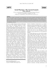
<strong>Indian</strong> <strong>Journal</strong> <strong>of</strong> <strong>Pharmacy</strong> <strong>Practice</strong>Association <strong>of</strong> Pharmaceutical Teachers <strong>of</strong> IndiaOccurrence <strong>of</strong> fixed Drug Eruptions in a Tertiary Care Hospital: <strong>Case</strong> <strong>Reports</strong>1 2Vijayakumar S *,RamChandra D, Narmadha R1Associate Pr<strong>of</strong>essor & Head, Department <strong>of</strong> <strong>Pharmacy</strong> <strong>Practice</strong>, MGM Hospital, Vaagdevi College <strong>of</strong> <strong>Pharmacy</strong>, Warangal, A.P2Pr<strong>of</strong>essor and Head, Department <strong>of</strong> Dermatology, Dr. NTR University <strong>of</strong> Health Sciences, KMC/MGM Hospital, Warangal, A.P.A B S T R A C TSubmitted: 10/1/2012Accepted: 3/2/2012Fixed drug eruption (FDE) is a unique pattern <strong>of</strong> cutaneous drug reaction, characterized by skin lesions that recur at the same site or sites eachtime the drug is administered. Acute lesions appear as round or oval, sharply marginated, erythmateous plaques that sometimes developcentral bullae. They are generally under reported with different rates in different health care systems. The present case studies were carried outin the department <strong>of</strong> dermatology in a multidisciplinary tertiary care government hospital for one month from June 12th to July 12th, 2011 by theDepartment <strong>of</strong> <strong>Pharmacy</strong> <strong>Practice</strong>. The cases were recorded and subjected to descriptive analysis. We observed four cases with FDE possiblyinduced by Isoniazid, Dicl<strong>of</strong>enac, Primaquine, Cipr<strong>of</strong>loxacin and Carbamazepine. Clinical patterns and drug causing cutaneous ADRs aresimilar to other researchers. The case studies highlighted that above medications belongs to the group <strong>of</strong> drugs that can induce fixed drugeruption.Keywords: Fixed Drug Eruptions, Skin rashes, Pruritus, and HypersensitivityINTRODUCTIONFixed drug eruption (FDE) is a unique pattern <strong>of</strong> cutaneousdrug reaction, characterized by skin lesions that recur at thesame site or sites each time the drug is administered. Acutelesions appear as round or oval, sharply marginatederythmateous plaques that sometimes develop central bullae.The lesions are usually found on the lips and genitalia,1,2although any skin or mucosal surface may be involved. Theeruption usually occurs within hours <strong>of</strong> administration <strong>of</strong> the<strong>of</strong>fending agent and resolves spontaneously without scarringafter few weeks <strong>of</strong> onset, usually with residual postinflammatory pigmentation. The most frequently implicateddrugs are sulphonamides, tetracycline, salicylates and3barbiturates. It is <strong>of</strong> utmost importance to recognize drugsthat induce these severe reactions and to identify the earlysymptoms signalling such a reaction, because prompt4withdrawal might decrease mortality. The present casestudies were carried out in the department <strong>of</strong> dermatology in amultidisciplinary tertiary care government hospital for onemonth from June 12th to July 12th, 2011 by the Department <strong>of</strong><strong>Pharmacy</strong> <strong>Practice</strong>.Address for Correspondence:Subash Vijaya Kumar,Associate Pr<strong>of</strong>essor & Head, Department <strong>of</strong> <strong>Pharmacy</strong><strong>Practice</strong>, MGM Hospital, Vaagdevi College <strong>of</strong> <strong>Pharmacy</strong>, Warangal, AndhraPradesh,India.CASE REPORT 1A 25-year-old woman was admitted to the hospitalcomplaining <strong>of</strong> skin rashes. The patient was a known case <strong>of</strong>epilepsy disorder for which medications (CBZ 200mg) werebeing used. Unfortunately, she noticed to develop skin rashesin lower and upper extremities as shown in the figure 1. Therash was associated with round lesions with blisters in thighand lumbar region. The patient was found to be suffering withlow grade fever. The laboratory investigations showed:Haemoglobin 9 g/dL, platelet count 120,000/cmm, and WBC4,000/cmm. Neutrophil 62%, lymphocyte 14%, and serumelectrolytes-normal, serum creatinine 165 μmol/L, serumalbumin 2 g/dL; serum calcium and serum magnesium werenormal. Urine culture showed no growth. Reticulocyte countwas 12%. After admission in the hospital CBZ 200 mg BDwas replaced by prescribing phenytoin 150mg OD, (cause forFDE) in addition to it, hydrocortisone 75mg/topical TID,chlorpheniramine maleate 5mg/OD/bed time, paracetamol500mg/TID and emollient cream were prescribed. I.V fluidswere given as a supportive therapy. After 3weeks <strong>of</strong> thetreatment, skin lesions started fading, patient's general wellbeingwas improved with no fever, relief <strong>of</strong> body pain. Shewas discharged on medication and asked to consult thephysician regularly till she becomes normal without anymanifestations <strong>of</strong> FDE.E-mail : vijayvijay66@yahoo.co.in<strong>Indian</strong> <strong>Journal</strong> <strong>of</strong> <strong>Pharmacy</strong> <strong>Practice</strong> Volume 5 Issue 1 Jan - Mar, 2012 67
Vijaykumar S - Occurrence <strong>of</strong> fixed Drug Eruptions in a Tertiary Care Hospital: <strong>Case</strong> <strong>Reports</strong>CASE REPORT 2A 33-year-old woman presented with malaria anderythematous rashes over the face as shown in figure 2. Shehad experienced sore throat, and skin lesions appeared oncheeks and nose. The preceding day, she had been diagnosedwith high fever and body pain and was administered with IVdicl<strong>of</strong>enac 50 mg BD and Primaquine 15mg/day. She did nottake any other medication, or nutritional supplement beforebeing treated with antimalarial drug. On repeated 3days <strong>of</strong>administration <strong>of</strong> primaquine, she developed erythematousmacules with darker purpuric centers over which flaccidblisters and a sheet-like epidermal detachment haddeveloped, especially on her face and nose regions. Because asevere form <strong>of</strong> drug eruption was strongly suspected, thepatient was hospitalized on the same day. After admission, theerythema and blisters further increased and extended rapidlyover the whole body, involving >20% <strong>of</strong> the body surfacearea, allowing definitive diagnosis <strong>of</strong> FDE. The center <strong>of</strong> eacherythema became dark red in color, followed by blisterformation. The patient's temperature was high, but other vitalsigns were stable. Laboratory findings were normal uponadmission, except for elevated erythrocyte sedimentation rate(ESR) (48 mm/h; reference range 0–15), Urine, blood, andthroat cultures were negative. Electrocardiogram, abdomenultrasonographic examination, and chest X-rays were normal.Oral prednisolone 60 mg daily, arsenuate 100 mg BD andsupportive topical therapy were given. Ringer's lactateinfusion <strong>of</strong> 2000 mL/day, parenteral nutrition were alsostarted. During the first week <strong>of</strong> the therapy, few new lesionscontinued to appear. The signs and symptoms <strong>of</strong> FDEprogressively resolved in 10 days, after which prednisolonedose was gradually tapered to discontinuation. Followingdischarge, there was no recurrence <strong>of</strong> the symptoms, and theskin lesions healed without any scars.CASE REPORT 3A 40 year old woman was admitted to the hospital with skinrashes all over the body ruptured leaving raw area like erosionand ulceration as depicted in figure 3. Patient was a knowncase <strong>of</strong> tuberculosis and had suffered with cough and fever for7 days and she was administered with anti-tuberculosis drugand paracetamol 500mg (OTC drugs) for 7 days. Uponinvestigation, the patient's condition was confirmed to beisoniazid induced fixed drug eruption. Laboratoryinvestigations revealed elevated WBC count and ESR. Thepatient's condition improved with antihistamine drugs,systemic steroids and supportive medications.CASE REPORT 4A severe form <strong>of</strong> eruptions over the trunk with pigmentedlesions and rashes was presented in a female patient <strong>of</strong> age 21years as shown in the figure 4, who was regularly on antituberculardrugs for one month, prescribed by apulmonologist. Her laboratory results showed: RBC3.4K/cmm, Hb 10g/dl, HCT 32%, erythrocyte sedimentationrate (ESR) 11 mm/hour. She was admitted to the hospital andtreated with hydrocortisone 75mg/topical TID,chlorpheniramine maleate 5mg/OD/bed time, and IV fluidswere given as supportive therapy. An extensive literaturesearch revealed few earlier reports <strong>of</strong> Isoniazid induced FDE.DISCUSSIONADRs are a threat to patient's health and quality <strong>of</strong> life, andthey can cause significant affect to the healthcare systems indeveloping and developed countries. Recently, the incidence<strong>of</strong> ADRs has increased significantly and the importance <strong>of</strong>this phenomenon will obviously vary in different healthcare6,7system. The present study examined ADRs in thedepartment <strong>of</strong> dermatology in a tertiary care governmenthospital which is attached to medical college. FDE ischaracterised by sudden onset <strong>of</strong> round and/or oval,oedematous dusty-red macules and plaques on the skin and/oritching and the re-appearance <strong>of</strong> the lesions over the8,9previously affected area when the <strong>of</strong>fending agent is reused.Histologically, there is basal hydropic degeneration,pigmentary incontinence, upper epidermal keratinocytenecrosis, dermal oedema, vasodilatation and perivascularinflammatory cells (lymphocytes, neutrophils, histocytes,10-14mast cells). The observations <strong>of</strong> the present studyimplicated Primaquine, Isoniazid, Carbamezepine andNSAIDS. As FDE are sometimes confused with multiplevenereal diseases, it is <strong>of</strong> utmost importance for all themedical specialists to identify FDE clinically by doing the15provocation test so that these cases are not missed. Earilerstudies reported that fixed drug eruptions seen, mostly due to1 6 1 7N S A I D S , C o - t r i m o x a z o l e , C e p h a l o s p o r i n ,18Carbamazepine and oral amoxicillin. In our present study,we found isoniazid induced fixed drug eruption. Our results19were similar with the study conducted by Noel M.V, et alwho reported that drugs such as anti-epileptics mainlyphenytoin and carbamazepine were responsible for themajority (44%) <strong>of</strong> the ADRs which strengthens our studyfindings in case <strong>of</strong> carbamazepine.<strong>Indian</strong> <strong>Journal</strong> <strong>of</strong> <strong>Pharmacy</strong> <strong>Practice</strong> Volume 5 Issue 1 Jan - Mar, 2012 68
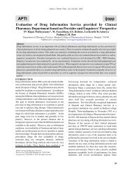
Vijaykumar S - Occurrence <strong>of</strong> fixed Drug Eruptions in a Tertiary Care Hospital: <strong>Case</strong> <strong>Reports</strong>Fig.1: Erythematous round lesions <strong>of</strong> reddish, featuring blisters on thigh and backFig.2: Round, black lesion on handFig.4: Pigmented lesions over the trunkFig.3: erosion and ulceration<strong>Indian</strong> <strong>Journal</strong> <strong>of</strong> <strong>Pharmacy</strong> <strong>Practice</strong> Volume 5 Issue 1 Jan - Mar, 2012 69
Vijaykumar S - Occurrence <strong>of</strong> fixed Drug Eruptions in a Tertiary Care Hospital: <strong>Case</strong> <strong>Reports</strong>CONCLUSIONIt can be concluded that the clinical patterns and the drugscausing fixed drug eruptions are remarkably similar to thoseobserved in other countries except with minor variations.ACKNOWLEDGEMENTSWe are grateful to Dr. E. Ashok kumar Ex. SuperintendentKMC/ MGM hospital, Warangal without whose inspirationand support this work would not have been possible.The authors are very much thankful to the patients for their cooperationto carry out this study and take the photographs <strong>of</strong>their manifestations. We also extend our thanks to Principal,Vaagdevi College <strong>of</strong> <strong>Pharmacy</strong>, Warangal, for their kindsupport.REFERENCES:1. Gaffoor PM, George WM. Fixed drug eruption occurring onthe male genitals cutis 1990;45:242-4.2. Jain VK,Dixit VB, Archana. Fixed drug eruption <strong>of</strong> the oralmucous membrane. Ann Dent 1991;50:9-11.3. Shukla SR. Drugs causing fixed drup eruptions.Dermatological 1981;163:160-163.4. Subash VK, Ramchandra D, Narmadha, Dheeraj Kumar G.Occurrence <strong>of</strong> Stevens Johnson Syndrome and ToxicEpidermal Necrolysis in a tertiary care hospital: case reports.J Hosp Clin Pharm 2011;2:9-15.5. Subash V K, Narmada R, Sasikala M, Ramchandra D.Cutaneous reactions due to Antibacterial drug(fluoroquinolone derivative). ijopp 2010;3:47-48.6. Mignot G, Biaggi J, Lethurgez P, Chichmanian RM, Analysefinanciere des hospitalisations pour effects indesirables.Therapie 1990; 45: 387-390.7. Dartnell J, Anderson RP, Chohan V et al, Hospitalisation foradverse events related to drug therapy : incidencebioavailability and costs, Med J Aust 1996;164:659-628. Sehgal VN, Srivastava G. Fixed drug eruption (FDE):changing scenario <strong>of</strong> incriminating drugs. Int J Dermatol 2006;45:897–908.9. Mahboob A, Haroon TS. Drugs causing fixed eruptions: astudy <strong>of</strong> 450 cases. Int J Dermatol 1998; 37:833–8.10. O zkaya-Bayazit E. Specific site involvement in fixed drugeruption. J Am Acad Dermatol 2003; 49:1003–7.11. Zanolli MD, McAlvany J, Krowchuk DP. Phenolphthaleininducedfixed drug eruption: a cutaneous complication <strong>of</strong>laxative use in a child. Paediatrics 1993; 91:1199–201.12. Alanko K, Kanerva L, Mohell-Talolahti M, et al. Nonpigmentedfixed drug eruption from pseudoephedrine. J AmAcad Dermatol 1996; 35:647–8.13. Young PC, Montemarano AD, Lee N, et al. Hypersensitivity topaclitaxel manifested as a bullous fixed drug eruption. J AmAcad Dermatol 1996; 34:313–4.14. Vilaplana J, Romaguera C. Fixed drug eruption from sodiumbenzoate. Contact Dermat 2004; 49:290–1.15. Kauppinen K. Cutaneous reactions to drugs, with specialreference to severe bullous muco-cutaneous eruptions andsulphonamides. Acta Derm Venereol Suppl (Stockh) 1972;52:68.16. Sanjay KK, AmoolyaKS, Shailjaratan S. A study on genitalfixed drug eruption in a tertiary care hospital. <strong>Journal</strong> <strong>of</strong> clinicaland diagnostic research 2011; 5:700-2.17. Patriarca G, Schiavino D,Buonoma A, Aruanno A et al.Desensitization to Co-trimoxazole in a patient with fixed drugeruption. J Investig Allergol Clin Immunol 2008; 18:309-311.18. Khoo BP, Glam YC. Drug eruptions in children: A review <strong>of</strong> 111cases seen in a tertiary skin referral centre.Singapore Med J2000; 525-529.19. Noel MV, Sushma M, Guido S. Cutaneous adverse drugreactions in hospitalised patients in a tertiary care center.<strong>Indian</strong> J Pharmacol 2004; 36:292-295.<strong>Indian</strong> <strong>Journal</strong> <strong>of</strong> <strong>Pharmacy</strong> <strong>Practice</strong> Volume 4 Issue 4 Oct - Dec, 2011 70