J. Crayton Pruitt Family Department Of BiomeDical Engineering
J. Crayton Pruitt Family Department Of BiomeDical Engineering
J. Crayton Pruitt Family Department Of BiomeDical Engineering

You also want an ePaper? Increase the reach of your titles
YUMPU automatically turns print PDFs into web optimized ePapers that Google loves.
J. <strong>Crayton</strong> <strong>Pruitt</strong> <strong>Family</strong><strong>Department</strong> of Biomedical <strong>Engineering</strong>
Wesley BOLCHProfessorPh.D. , 1988, University of FloridaRadiation DosimetryComputational PhantomsDosimetry Models of the SkeletonComputed TomographyInterventional FluoroscopyNuclear MedicineRadiotherapyHomeland SecurityRadiation DosimetryRadiation dosimetry encompasses the calculation andmeasurement of energy deposition within the humanbody resulting from exposure to radiation sources atthe workplace, in the environment, or from medical applicationsto imaging or cancer therapy. These exposures may result fromeither radiation sources external to the body (e.g., CT imaging)or internal to the body (e.g., radiopharmaceuticals). In mostinstances, radiation doses to the internal organs and tissues isnearly impossible to directly measure, and thus computationalsimulations of radiation transport and energy transfer arerequired using either patient or individual specific anatomicalmodels, or computerize replicas of patient anatomy.COMPUTATIONAL PHANTOMSThe Advanced Laboratory for Radiation Dosimetry Studies(ALRADS) at the University of Florida is a world leader in thedevelopment of hybrid computational phantoms of humananatomy – based upon the application of polygon mesh andNURBS surface modeling of internal organs and the outerbody contour. A series of reference (50th percentile) modelsof the newborn to 15-year adolescent were developed atUF and recently adopted as international standards by theInternational Commission on Radiological Protection (ICRP).Other work has resulted in a 400+ member phantom librarycovering pediatric and adult males and females representingthe current height/weight distribution of the US population.These phantoms are serving as the basis for pre-computed doselibraries for a broad range of medical imaging modalities – CT,fluoroscopy, and nuclear medicine. Other work has resultedin some of the most detailed anatomic models of developingembryo, fetus, and pregnant female for applications to bothenvironmental and medical dose assessment.DOSIMETRY MODELS OF THE SKELETAL TISSUESThe hematopoietically active tissues of bone marrow are themost radiosensitive tissues in the human body. At low radiationdoses, exposure can increase the risk of leukemia induction,while at high doses – such as during radionuclide therapy– marrow suppression and toxicity may result. Predictivemodels of radiation dose to bone marrow are exceedinglydifficult to develop owing to the complex microstructure oftrabecular spongiosa, variations in marrow cellularity, andvariations in bone mineral status of the individual or patient.UF has pioneered the use of CT and microCT imaging of humancadaveric bone to the application of radiation transportsimulation and predictive models of marrow dosimetry. MRtechniques for non-invasively assessing marrow cellularityhave been developed and validated in a canine model. Skeletalmodels have been developed for both adults and pediatricphantoms, as well as the developing fetus.APPLICATIONS TO MEDICAL IMAGING ANDTHERAPYThe ALRADS laboratory is actively engaged in thedevelopment of predictive models of patient radiation dosefor all major forms of diagnostic medical imaging. UF isworking with the National Cancer Institute to develop softwareto predict organ doses to patients undergoing computedtomography imaging, including those scanned undertube current modulation. UF is also developing real-timesoftware to predict and map skin dose to patients undergoingfluoroscopically-guided interventions, and to report organdoses using cloud-computing radiation transport simulation.Finally, UF is a key partner in national efforts to optimize thequantity of radiopharmaceuticals given to pediatric patientsthat will maximize image quality while minimizing risk ofsecond cancers. In radiation therapy, the ALRADS laboratoryhas partnered with Oraya Therapeutics, Inc. to develop a noninvasivex-ray treatment for age-related macular degeneration.APPLICATIONS TO EPIDEMIOLOGY & HOMELANDSECURITYThe computational models developed at UF have alsobeen applied to studies of the Southern Urals populationsexposed to radionuclides released during past USSRnuclear weapons development in the 1950s. These studieswill significantly enhance our understanding radiationrisks. Work at UF has also resulted in the development ofcomputer software to assist first-responders in performingradiological triage of victims following radiological terroristevents.Share identical anatomy except gender organsNewborn 1-year 5-year 10-year 15-year male 15-year female5
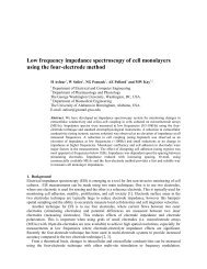
JON DOBSONProfessor & Director, Institute for Cell <strong>Engineering</strong> andRegenerative Medicine, ICERMPh.D. 1991, Swiss Federal Inst. of Technology, ETH-ZurichMagnetic BiomaterialsNanomagnetic Gene TransfectionNanomagnetic Cell ActuationBrain Iron and NerodegenerationThe main focus of our group falls into several interrelatedcategories as outlined below. The underlyingtheme of all this work is the novel use of magneticnanoparticles to develop technologies for bionanotechnology/nanomedicine applications in fields as diverse as gene therapy,stem cell therapy, tumour targeting and tissue engineering/regenerative medicine. In addition, we have been developingsynchrotron x-ray and MRI-based techniques to exploit naturallyoccurring magnetic iron oxides in the brain for diagnosticand mechanistic studies of neurodegenerative diseases, suchas Alzheimer’s and Parkinson’s.Nanomagnetic Actuation: tissue engineeringand stem cell therapyMagnetic nanoparticles are being used to target andmanipulate cellular processes and functions and to controlstem cell differentiation, primarily through the activationof ion channels. These nanoparticles are coated with afunctionalizable polymer to which surface antigens/targetingmolecules may be bound. These antigens may target eitherspecific ion channels, such as the mechanosensitive TREK-1potassium channel, or non-specific cell surface receptors suchas integrins. By applying a static or time-varying magneticfield, forces exerted on the particles activate either specific ionchannels or general membrane and cytoskeletal deformationactivates adjacent mechanosensitive ion channels, initiatingbiochemical processes within the cell. We have used thistechnology to speed up bone matrix production, to control thedifferentiation of stem cells without chemicals, and to enhanceproduction of cartilage and upregulate cartilage-related genesboth in vitro and in vivo. These devices, Magnetic Ion ChannelActivation (MICA) systems are being commercialized by a spinoffcompany.A B C DE F G HI J K LHBMSC HBMSC + TGFβ3 HBMSC + TREK-K+ HBMSC + RGDHistology and immunohisto-chemistry of HBMSCs only; HBMSCs and transforminggrowth factor b3; and HBMSCs labeled with TREK-1 or HBMSCs labeledwith RGD particles encapsulated into alginate/chitosan capsules, implantedsubcutaneously in MF-1 nu/nu mice, and exposed to a magnetic field for 21days, 1 h/day (Mon., Wed. & Fri.). Representative 6 mm tissue sections stainedfor Alcian blue/Sirius red (A–D), type-1 collagen (E–H), and type-2 collagen(I–L). Arrows indicate positively stained HBMSCs for type-1 collagen. Scalebars1/4100 mm.Nanomagnetic transfection of green fluorescentprotein (GFP) into PC12 neuronal cells.magnefect-LTNanomagnetic Gene transfection anddEliveryWith the sequencing of the human genome and the adventof gene therapy has come the need to develop effective deliveryand transfection agents. These agents must be able totarget therapeutic and reporter genes to the relevant cells andorgans both in vitro for basic investigations as well as in vivofor therapeutic applications. Recent safety concerns over theuse of viral vectors has begun to shift the emphasis toward thedevelopment of non-viral delivery agents, primarily cationiclipids. Our group has been working on the development of anovel magnetic nanoparticle-based gene transfection systemsbased on oscillating arrays of magnets. In these “magnefect”systems, DNA or siRNA is attached to magnetic nanoparticlesand oscillating arrays of magnets placed underneath a cell cultureplate are used to stimulate particle uptake and improvegene expression. In addition, we are developing novel, highgradientmagnet arrays and new motion control systems toimprove efficiency. These systems also have been commercialized.Our in vivo work in this area has focused not onlyon the delivery of nanoparticle/drug/gene complexes butalso cells. The aim of this work is to load cells with biocompatiblemagnetic nanoparticles and re-introduce them intothe body, using magnets to target them to repair sites or tumours.We have successfully enhanced the natural tumourhoming ability of human macrophages by loading themwith magnetic nanoparticle/reporter gene complexes. Theuptake of these “therapeutically armed” cells into the nonvascularized,hypoxic cores of solid tumours was enhancedby more than three-fold over non-magnetized cells. Onceinside the tumour, the cells can deliver a payload of “suicide”genes or cytotoxic compounds, after which AC electromagneticfields are used to heat the particles, destroyingthe macrophages before they build a new blood supply tothe tumour core. We have also used the technology to targethuman mesenchymal stem cells to tissue repair sites insmall animal models.magnetic nanoparticle synthesis andcharacterizationOur group is also active in the development of techniquesfor the synthesis of novel magnetic nanoparticles for biomedicalapplications. This work focuses on producing particleswith enhanced magnetic properties or surface chemistry,as well as investigating new methods for enhanced DNAloading. Techniques such as High- Resolution TransmissionElectron Microscopy and Superconducting Quantum InterferenceDevice (SQUID) magnetometry are used for characterizationof the particles.Nanoscale iron compounds in neurodeGenerativeDiseaseOver the past 15 years we have pioneered SQUID andsynchrotron-based detection techniques in order to quantify,characterize and map specific iron compounds in neurodegenerativetissue. This work aims to provide a betterunderstanding of the role of disrupted iron homeostasis inneuro-degenerative diseases and to guide the developmentof chelation therapies. More recently, data from these studieshas been used by us and other groups in the continuingdevelopment of MRI-based diagnostic techniques, whichaim to use iron compounds formed due to neurodegenera-7
David R. GillandAssociate ProfessorPh.D., 1989, University of North Carolina, Chapel HillMedical ImagingPositron Emission Tomography (PET)Single Photon Emission ComputedTomography (SPECT)Our lab focuses on advancements in medical imagingwith emphasis on emission tomography. We havedeveloped a new mobile PET/SPECT system for bedsideimaging. The system is capable of being moved within ahospital to image patients who cannot be easily transported toa conventional imaging facility, for example, patients in an intensivecare unit. This unique device, which is currently underclinical evaluation, promises to deliver PET and SPECT imagingtechnology to a critically-ill patient population. We are alsocurrently developing advanced motion compensation/imagereconstruction algorithms for cardiac imaging in PET, SPECT,and CT Angiography. These algorithms have the potential toimprove image quality by reducing motion blur due to cardiaccontraction. The algorithms also provide a means of spatiallyregistering images across imaging modalities.A B C(A) SPECT imaging of anthropomorphic phantoms, (B,C) Mobile PET/SPECT imaging system.We have initiated a project with the focus of developingan improved method for imaging prostate cancer (PCa). Theproject includes collaborators from Johns Hopkins University,who are involved in the design of radiotracers that targetprostate-specfic membrane antigens (PSMA), and fromGamma Medica-Ideas, Inc., an industry partner who are worldleaders in advanced SPECT imaging detector devices. We areinvestigating a unique detector design that can potentially deliverimproved spatial resolution and detection sensitivity for adedicated SPECT prostate cancer imager. The potential impactof this study is to improve the staging of PCa through effectiveimaging methods as well as to improve the detection ofrecurrent cancer following treatment. The higher spatial resolutionand detection sensitivity of the proposed imaging device,combined with more effective imaging agents that targetPSMA, have the potential to localize small lesions in the areaof the prostate gland and pelvic lymph nodes and determinethe extent of intra- and extra-glandular disease.Rigid motion estimation for cardiac CT and SPECTSchematic of a dedicated SPECT prostate imager.8
Aysegul GUNDUZAssistant ProfessorPh.D. , 2008, University of FloridaNeural <strong>Engineering</strong>Cognitive NeuroscienceNeurorehabilitationBrain-Computer InterfacesThe human brain consists of numerous networks distributedover space and connected over time to orchestratemeaningful interaction with the externalworld. Studying precursors to behavior and aftereffects ofsensory stimulation in these recruited networks enables directinterpretation and control of this interaction. Our researchaims to identify neural correlates of behavior and informationprocessing in electrocorticographic signals (ECoG) in humans,which are collected via subdural electrodes placed on the surfaceof the cortex. ECoG research has strong clinical ties, asthe signals are recorded from patients awaiting surgery for thetreatment of intractable epilepsy. This setting facilitates rareaccess to the human cortex and opens unparalleled avenuesfor human brain research. ECoG enables the investigation ofcortical networks with high spatial and temporal precision.ATTENTION AND MEMORYOur research program studies the neural bases and interactionof attention and memory in humans. These mechanismshave evolved in tandem because the human brain is limitedin its resources to process and store information. Attentionand memory are thought to be interdependent as attentionpromotes improved storage of information, and retrievedinformation from past experiences can guide what shouldbe attended in the current scenario. The contribution of thiswork would be significant as attention- and memory-deficitdisorders are highly associated with learning disabilities inchildren and aging adults.LANGUAGE AND MEMORYLanguage is a distinctly human trait. No other non-humancommunication system compares to human language in itscomplexity and expressive power, and no animal aptitude approachesthe universal human capacity for vocabulary. Ourresearch is aimed at identifying the functional neocortical organizationof semantic processing in humans, and to localizecortical areas of semantic memory that are distinctive fromarticulatory and comprehensive processing. Alterations inthe cortical mechanisms supporting semantic processing lieat the heart of many language-based learning disabilities. It isestimated that developmental disorders of language (whichinclude deficits in both oral and written language) occur in upto 20% of preschool and school-age children. Thus, revealingmechanisms by which the brain encodes comprehensionand semantic memory bears important implications for ourunderstanding of these disorders, and more importantly, willguide strategies for their amelioration.ACV2PMPPM1M1PPV2BV2PMPPM1CueBRAIN-COMPUTER INTERFACES & NEUROREHA-BILITATIONRehabilitation of chronically lost motor functions is currentlya challenge in the treatment of stroke survivors.Our goal is to determine whether surface-acquired brainsignals (EEG) can feasibly be trained for recovery of volitionalmotor control after stroke in humans. A potentialnovel approach for the restoration of function and improvingthe quality of life of these patients could be theuse of brain–computer interface (BCI) systems. So far,there is no information on whether training a patient toproduce more normal brain signal features will improvemotor function that involves the same areas that producethose signals. These unknown factors include the extentto which patients have detectable brain signals that cansupport training strategies; which brain signal featuresare best suited for use in restoring motor functions; andwhat the most effective formats are for the BCIs aimed atimproving motor functions (e.g., what guidance should beprovided to the user to maximize training that producesbeneficial changes in brain signals). The eventual value ofBCI technologies for improving motor function in individualswho have strokes or other neurological disorders dependson adequate answers to these questions.StimulusContrast ChangeButton Press200 Hz 100 Hz1 HzPMCue/StimulusContrast Change/Button Press-500 ms 0500 ms-0.5r0 0.5250 ms50 Hz9
david hintenlangAssociate ProfessorPh.D. , 1985, Brown UniversityDiplomate, American Board of RadiologyMedical PhysicsImaging and DosimetryComputed TomographyMammographyImage GuidancePATIENT RISK/BENEFIT FROM RADIOLOGICPROCEDURESThe benefits of ionizing radiation procedures for clinicalapplications have been long established but continueto find new applications through faster and newimaging technologies. Many procedures are incorporatedinto medical specialties that have not traditionally utilizedradiological techniques making it of paramount importance toaccurately characterize and balance patient risks and benefitsand subsequently optimize new procedures. Our laboratoryfocuses on the development of tools and techniques thatfacilitate the quantitative evaluations of dose assessment andimage quality.ANTHROPOMORPHIC PHANTOMSOur laboratory has a long history of designing and fabricatinganthropomorphic phantoms that accurately mimichuman anatomy. It is important that the materials used inthese phantoms represent the radiological properties of livinghuman tissues, and we have developed a variety of tissuesimulant materials that meet this goal. These “tissue-equivalent”materials provide the basis for fabricating whole bodyphantoms with accurate anatomical detail. The phantoms’anatomy are based on high resolution CT data sets and arefabricated with state-of-the-art computer controlled machiningand molding processes. Based on this methodology wehave developed a family of phantoms representing newborns,pediatrics at several ages, adult females, adult males,and obese adult males. Combined with our specializedplastic scintillation detector based array dosimetry system,we have developed unique abilities to quantify organ dosesFigure 1: Anatomical detail represented in the cross section of ananthropomorphic phantom of a newborn.and image quality for a wide variety of patients and clinicalprocedures.PSD DOSIMETRY SYSTEMIn order to measure radiation doses in organs distributedthroughout the anthropomorphic phantoms we have developeda dosimetry system to meet the specific requirementsof clinical based dosimetry. We have integrated small (a fewmm) plastic scintillation detectors with a coupled fiber opticarray, reader and laptop PC to provide a portable dosimetrysystem capable of accurately and rapidly measuring organdoses. The small physical size and near tissue-equivalence ofthe dosimeters and fibers are incorporated into the phantomswithout perturbing the radiological integrity of the phantomtissues. The system permits sampling from an array of organlocations with instantaneous and real-time monitoring capabilities.This high resolution data provides insight into thespatial and temporal dose delivery patterns associated withmodern imaging and radiation therapy systems. Current researchis advancing the dosimetry system development tolarger arrays and application specific detectors.CLINICAL APPLICATIONSOur laboratory leads the development of accurate organdose assessment from clinical procedures. The integratedFigure 2: An adult male phantom torso integrated with the PSD dosimetrysystem undergoing a CT procedurephantom/dosimetry system, along with image qualityevaluations, allows us to better characterize, understandand develop techniques that maximize patient benefitsand minimize the risks from radiological procedures.Some of the specific procedures that have been, or areunder investigation include, pediatric radiography andCT, mammography, multi-detector CT, and cone-beam CTimage guidance in radiation therapy. We are also activelyextending phantom and tissue simulants to develop usefulproducts for a variety of other clinical training simulatorsand tools.Figure 3: Image quality comparison from two cone beam CT image guidancesystems used in radiation therapy.10
Huabei JiangJ. <strong>Crayton</strong> <strong>Pruitt</strong> <strong>Family</strong> ProfessorPh.D., 1988, University of Electronic Scienceand Technology of ChinaPh.D., 1995, Dartmouth CollegeDiffuse Optical Tomographyof Breast CancerDiffuse Optical TomographyPhotoacoustic ImagingFluorescence Molecular TomographyMulti-modal ImagingPhotoacoustic Imaging of EpilepsyApproximately 2.5 million Americans live with epilepsy andepilepsy-related deficits today. However, 80 percent of individualswith medication resistant epilepsy might be cured through surgeryif one were able to precisely localize the seizure focus. Ourresearch aims to significantly advance the ability to localize the focus,and thereby offer curative epilepsy surgery for this devastatingdisease. Photoacoustic tomography (PAT) uniquely combinesthe high contrast advantage of optical imaging and the high resolutionadvantage of ultrasound imaging in a single modality. In additionto high resolution structural information, PAT is also ableto provide functional information that are strongly correlatedwith regional or focal seizure activity, including blood volume andblood oxygenation because of the high sensitivity of optical contrastto oxyhemoglobin and deoxyhemoglobin concentrations,and thus offers the possibility to non-invasively track dynamicalchanges during seizure occurrence.Multi-modal Imaging ofOsteoarthritisOsteoarthritis (OA) is the most common arthritic condition worldwideand is estimated to affect nearly 60 million Americans. Besides the kneesand hips, there is a subset of individuals with a predilection for developingOA of the hands and a more generalized form of OA. Due to the fact thatDOT can provide high-contrast joint tissue imaging with low resolution,while x-ray can offer high-resolution joint structure with low contrast insoft tissues, we present an optimized approach that combines x-ray andoptical imaging for early diagnosis of osteoarthritis in the finger joints.Breast cancer has been one of the leading causes ofdeath for women in the United States. Yet, the bestway of combating the increased incidence of breastcancer is early detection. Therefore, there is a critical need toinvestigate breast cancer detection methods that could serveeither a complementary or competitive role with respect toconventional x-ray mammography, which has unacceptablefalse negative rate for patients with radiodense breast tissues.The fundamental hypothesis of our research is that spatiallyand spectrally resolved NIR diffuse optical tomography (DOT)approaches offer unparallel opportunity to access the molecularand cellular signatures in breast tissue through endogenouscontrast mechanisms. To clinically evaluate optical tomographybased on absorption chromophores and scattering parameters,three clinical prototype imagers have been constructed.We have also developed a new contrast mechanism for NIRtomography based on refractive index/phase contrast: the initialclinical results show that the addition of refractive indexcan significantly improve our ability for distinguishing betweenmalignant and benign breast lesions. Further, the opportunityexists to explore the possibility of obtaining cellular densityand size from scattering spectra which together with functionalparameters and refractive index should form the foundationof next generation NIR tomography for more complete characterizationof breast abnormalities.Fluorescence MolecularTomography ofMargin Identificationof Breast CancerBreast-conserving surgery orlumpectomy is the most commonsurgical procedure for patientswith early invasive stages of breastcancer. However, there is no accuratemethod to identify tumormargins pre- or intra-operatively.To develop a sensitive approach forthe detection of residual tumors inbreast tissues, we have developedthe Cy5.5 ATF-IO tumor targetednanoparticles, and in combinationwith sensitive and high resolutionNIR fluorescence tomography system,should have great potentialfor determining tumor marginsduring surgery, preventing tumorreoccurrence and therefore, increasingsurvival of breast cancerpatients.PAT is able to image epileptic events as they are happening. The arrow in this image indicates the detected seizure.11
Benjamin G. KeselowskyAssistant ProfessorPh.D., 2004, Georgia Institute of TechnologyBiomaterialsCell AdhesionVaccinesType 1 DiabetesThe Biomaterial Immuno-<strong>Engineering</strong> Lab focuses onthe engineering of biomaterial-cell interactions, andtargeted controlled release of immune modulatingfactors in order to direct immune cell function. Biomaterialsundergo complex interactions with cells of the immune systemupon implantation. These interactions are incompletelyunderstood and poorly controlled, complicating the ability toachieve favorable outcomes in clinical applications. Our effortsfocus on both a basic understanding of interactions of immunecells with biomaterials as well as the engineering of biomaterialscapable of directing immunological processes. Thiswork has wide-ranging implications in diverse fields such asimplanted devices, therapeutic vaccines and tissue engineering.We are particularly interested in the biomaterials-basedmodulation of the phagocytic antigen present cell types ofdendritic cells and macrophages.Microparticle-based vaccines for type 1(autoimmune) diabetesWe are engineering polymeric biomaterials-basedmicroparticles as an injectable vaccine system to retrainthe immune system, correcting aberrant activation towardpancreatic self-antigens. Microparticles with encapsulatedimmunomodulatory factors and insulin antigen providetargeted, controlled delivery to both intracellular and cellsurface receptors of dendritic cells in vivo in order to promotetolerance in diabetes. While systemic administration ofimmune-modulating agents can often result in harmful offtargeteffects due to uncontrolled dosing of bystander tissues,encapsulation into biodegradable microparticles can reducethe total dose required and limiting off-target effects. (Middletwo panels)High-throughput screening of immunecell responses to immuno-modulatorymicroparticlesWe are developing high-throughput methods to screen invitro, microparticle-based vaccines targeting dendritic cells.The goal is to identify microparticle formulations able to shiftdendritic cell phenotype toward the ability to induce regulatoryT-cells and tolerance.Other research topics include:• Immune cell adhesion (to extracellular matrixproteins, to nanotopographies, and response tomechanical strain)• Receptor-mediated mechanisms of macrophagephagocytosis of orthopedic implant wear debrisfor the mitigation of peri-implant osteolysis in jointreplacement patientsFunding is gratefully acknowledged from the followingsources:• National Institutes of Health (R01 DK091658, R21AI094360)• National Science Foundation (CMMI 0927918)• Juvenile Diabetes Research Foundation• Arthritis Foundation12
Peter S. McFetridgeAssistant ProfessorPhD. 2002, University of Bath, United KingdomResearch Assistant Professor/Assistant Professor, 2002-2009, University of OklahomaBiomaterials and Tissue <strong>Engineering</strong>Cardiovascular Tissue <strong>Engineering</strong>Temporomandibular Joint regenerationConductive biomaterialsPeriodontal Soft tissue repairNerve regenerationBiomaterials and Tissue <strong>Engineering</strong>From vision and hearing implants to an artificial heartand blood vessels, biomedical engineering has becomea crucial component of the drive to improve thequality of life in our ageing society. Our laboratories researchaims to develop medical devices that improve the life styleand reduce suffering of those afflicted with organ loss or failure.Our focus is on the use of a unique biomaterial that isused as a 3D template or bioscaffold to promote tissue/organregeneration. This approach, called ‘Tissue <strong>Engineering</strong>’, hasshown significant promise as a medical therapy, but translationfrom the research lab to clinic has proven difficult dueto extended in vitro culture times. In light of these issues ourinvestigations aim to understand key conditions that enhancethe regenerative capacity of tissue constructs.Our research encompasses the three mainphases of the tissue engineering approach:1) Biomaterial/scaffold development and characterization2) Bioreactor design (to grow the living tissue)3) In vitro culture of the re-seeded scaffolds under replicatedphysiological conditions.Research objectivesOur main research objective is to develop viable alternativesto autologous and synthetic transplant materials that behavemore appropriately when implanted resulting in improved repairor regeneration of diseased tissues.Research strategyUsing a patented process, vascular tissues can be rapidly, anduniformly, dissected from surrounding connective tissues,which are then processed to remove immunogenic components.This process is called decellularization and aim to minimizeany immune rejection once implanted. Using the autodissectionprocess a biomaterial with uniform mechanics anda significant potential to regenerate into neo-tissue is generated.The unique structure of these materials allows a numberof vascular and non-vascular projects to be investigated, thematerial can be used as a direct implant (acellular), or as are-seeded ‘living’ construct. Constructs are grown under controlledchemical and mechanical conditions within specificallydesigned bioreactors to circulate in vivo environment to improvetissue regeneration..specific researchProjects are under investigation include; developing coronaryand peripheral bypass grafts, tissue engineered soft-tissueimplants for periodontal wound repair, conductive materialsFigure 1: Vascular Tissue <strong>Engineering</strong>. In addition to developing functional vascularimplants, our investigations focus on cellular interactions with materialswith the aim to modulate the in vitro cell phenotype typically associated withdiseased tissues to produce functional bypass grafts. Above left, engineeredsmall diameter vascular graft (5mm ID) derived from human umbilical vein implantedin an ovine model to assess patency as a carotid bypass. Above right,color Doppler ultrasound monitoring graft patency and diameter after implantation.for the repair of damaged peripheral nerves and temporomandibularjoint regeneration. More specific investigationsinclude furthering our understanding of scaffold design andfunction, cell adhesion, conductivity modulation and effectson cell function, and the influence of gas concentrations onorgan development.Figure 2: Biomechanics. Investigations include the analysis of material biomechanicalproperties during remodeling processes. Shown in Figure 2, the Young’sModulus of cell seeded vascular scaffolds after 7 and 21 days in culture. Thesevascular scaffolds are cultured with human smooth muscle cells and show anincrease in vessel elasticity when stimulated under perfusion flow conditions.Figure 3: The interactions between novel biomaterials and cell systems. Investigationsinclude vascular endothelial cells, smooth muscle cells, gingival fibroblasts,TMJ chondrocytes, and as above (left), neuronal cells on engineerednerves. Development and analysis of typically includes the use of unique bioreactorsystems to culture cells under conditions that mimic the in vivo environment(right).13
Brandi K. OrmerodAssistant ProfessorPh.D. 2003, University of British ColumbiaStem Cell Strategies for NeurodegenerativeDiseaseThe progressive death of one or more cell types in thebrain is called neurodegenerative disease. Parkinson’sdisease and Alzheimer’s disease are examples of incurableneurodegenerative diseases that leave patients withprogressively debilitating symptoms. The Ormerod Laboratoryfocuses its research upon understanding how transplantableor endogenous stem/progenitor cells ccould be used to repairthe diseased or damaged CNS.DCX (new neurons)BrdU (new cells)DCX/BrdU (new neurons)Neural <strong>Engineering</strong>Regenerative MedicineStem Cell <strong>Engineering</strong>Age-related Cognitive DeclineNeurodegenerative DiseaseBiomarkersFigure 1: In the hippocampus of men and mice alike, new neurons are addedeach day throughout life to the hippocampus (a learning and memory center)and the olfactory bulbs (important for smell). Neural progenitor cells, capableof generating new neurons, reside throughout the adult brain. Discovering factorsthat control the behavior of these cells is the key to unlocking new brainrepair strategies with endogenous and transplantable stem cells.STEM CELL ENGINEERINGThe hippocampus is an excellent model for discovering thecues the guide stem cell growth and differentiation because itpermits/promotes neurogenesis and the rate of neurogenesiscan be controlled through systems variables, hormones anddrugs. We are currently exploring such factors. For example,we are interested in identifying and capitalizing upon uniqueneurogenic features of hippocampal vasculature to stimulateneuron production in other brain regions because neurogenesisoccurs in tight association with hippocampal vasculature. .Figure 2: <strong>Engineering</strong> niches receptive to neuron addition may be critical for thesuccess of neuronal regeneration strategies using stem cells. Hippocampal vasculaturemay contain unique features that stimulate neurogenesis that could beemployed to engineer niches conducive to neuronal regeneration outside of thehippocampus. Munikoti et al., 2011MEA CULTURE PLATFORM FOR TESTING THE VIABIL-ITY AND SAFETY OF STEM CELL STRATEGIESIf you could make the perfect neuron or glial cell to replacethose lost in neurodegenerative disease, would you restore neuralactivity and therefore reverse the symptoms of the disease?In collaboration with Dr. Tom DeMarse, we are employing microelectrodearray technology to develop strategies to integrate ofstem cell-derived cells into naïve and damaged long-term culturesand test their safety. We have discovered that these cells stimulateplasticity in neural networks and are currently exploring waysto capitalize on this phenomenon to restore activity in neural circuitrycompromised by injuries, such as stroke.Figure 3: When plated on mature neural networks, neural progenitor cells generatemature neurons that stimulate re-emergent developmental neuronal plasticity.Stephens et al., 2012.BIOMARKERS OF COGNITIVE AGING NEUROIN-FLAMMATIONThe incidence of age-related cognitive decline grows exponentiallywith the advancing age of our baby boomer population.In collaboration with Dr. Tom Foster, we are discoveringprognostic and diagnostic biomarkers of cognitive aging usinga combined proteomic and pathway analysis approach. Becausewe have discovered evidence that compromised neurogenesismay accompany age-related cognitive decline and thatneuroinflammation ablates neurogenesis, we are interested indeveloping novel immunomodulatory strategies with our biomarkerdata that may promote healthy aging and more effectiveneuronal regeneration, because all CNS injury and diseaseis accompanied by neuroinflammation.Figure 4: Potential biomarkers of impaired learning, memory and neurogenesis.14
Carlos RinaldiProfessorPh.D. , 2002, Massachusetts Institute of TechnologyNanomedicineCancer NanotechnologyMagnetic NanoparticlesColloidal HydrodynamicsTransport PhenomenaSuspensions of Magnetic NanoparticlesMy group studies the behavior and applicationsof suspensions of magnetic nanoparticles inapplied magnetic fields. This field has seenexplosive growth due to potential in biomedical applicationssuch as magnetic resonance and magnetic particle imaging,biosensors, targeted delivery and triggered release of drugs,magnetomechanical actuation of cell response, and theability to deliver magnetic energy at the nanoscale in theform of heat or shear. We combine expertise in synthesisand surface modification of magnetic nanoparticles; physical,chemical, and magnetic characterization; and modeling of thecoupling of magnetic, hydrodynamic, and Brownian forcesand torques to answer fundamental questions regarding thebehavior of magnetic nanoparticle suspensions, understandtheir interaction with biological entities, and develop novelbiomedical applications taking advantage of their uniqueproperties.<strong>Engineering</strong> Cell Fate Through NanoscaleEnergy Delivery by Magnetic NanoparticlesMagnetic nanoparticles can be engineered to target specificcells or even cellular components. Under an applied alternatingmagnetic field magnetic nanoparticles can deliver energylocally, in the form of shear due to nanoparticle rotation or inthe form of heat. This ability to deliver energy at the nanoscaleand selectively to targeted cells or cellular componentsallows for novel applications where the fate of the cell can beengineered.In one potential biomedical application, magneticnanoparticles can be made to target cancer cells and destroythese by localized hyperthermia or through disruption ofcellular components. In vitro and in vivo experiments in whichcancer cells are in contact with magnetic nanoparticles andsubjected to high frequency alternating magnetic fields haveshown that the particles may induce significant reductionsin cancer cell survival. Furthermore, because traditionalcancer treatments can have synergistic effects with thermaltreatment, their combination with hyperthermia inducedby magnetic nanoparticles is very promising. My group isinterested in understanding how nanoscale energy deliveryby magnetic nanoparticles kills cancer cells, with the objectiveof engineering novel, more effective magnetic nanoparticlebasedstrategies to treat cancer.In a broader biomedical context, nanoscale energy delivery,in the form of heat and/or shear, by magnetic nanoparticlescan be a tool to engineer cell fate by mechanical/thermalactuation of receptor-mediated pathways and by selectivedenaturation/destruction of biomacromolecules and/orcellular compartmentsProbing Biological Environments UsingMagnetic NanoparticlesAs noted, magnetic nanoparticles can be made to rotatedue to the application of alternating magnetic fields. Inthis line of research we take advantage of the fact thatsuch rotation is sensitive to the mechanical propertiesof the environment surrounding the nanoparticles andto the presence of biomacromolecules that bind to thenanoparticle surface. In turn, the rotation of collectionsof magnetic nanoparticles can be observed directlywith specialized microscopy techniques or remotely bymonitoring their magnetization. We apply our fundamentalunderstanding of the coupling of magnetic, hydrodynamic,and Brownian forces and torques to use magneticnanoparticles as probes in biological complex fluids.We have recently demonstrated that by monitoring theresponse of magnetic nanoparticles to oscillating magneticfields information can be obtained of the mechanical(e.g. viscous) properties of the surrounding fluid. Themethod requires small sample volumes (
CHRISTINE E. SCHMIDTJ. <strong>Crayton</strong> <strong>Pruitt</strong> <strong>Family</strong> Professor &<strong>Department</strong> Chair (as of January 2013)Ph.D. , 1995 University of IllinoisSpinal Cord InjuryBiomimetic Conducting PolymersNatural-Based BiomaterialsCell-Materials InteractionsTHERAPIES FOR NERVE REGENERATIONDamage to spinal cord and peripheral nerve tissuecan have a devastating impact on the quality of lifefor individuals suffering from nerve injuries. Ourresearch is focused on analyzing and designing biomaterialsthat can stimulate and interface with regenerating neuronsand nerves. We take a unique approach to this problem – weare using electrically conducting polymers and naturallyderivedmaterials (e.g., hyaluronic acid-based biomaterialsand chemically processed nerve tissue) to create therapiesthat can electrically, chemically, biologically, and mechanicallytrigger neurons to re-grow damaged axons.BIOMIMETIC CONDUCTING POLYMERSWe are working with electroactive polymers with inherentproperties that can stimulate electrically responsive cell typessuch as neurons. Using these polymers, our group has creatednew biomimetic, electronic materials by processing electricallyconducting polymer composites (e.g., polypyrrole-PLGA) into3D fiber matrices for enhanced topographical guidance. Wehave incorporated biological moieties using novel peptidesthat directly bind to conducting polymers. Ultimately, thesematerials can be used to interface with neurons for electroniccommunication or as internal “pathways” to stimulate neuronsto grow and physically guide axon extension.NATURAL-BASED MATERIALSOur research group is designing tissue scaffolds that canfacilitate the growth of peripheral and spinal cord axons.In this work, we are using hyaluronan, a naturally-derivedbiopolymer found throughout mammalian tissues. Hyaluronanis non-immunogenic (i.e., not rejected by the body’s immunesystem) and plays a major role in wound healing and embryodevelopment. Our group has devised novel techniquesto process hyaluronan into materials that can be used intherapeutic applications. For example, we are using gelsof hyaluronan to treat spinal cord lesions in rats. The gelsattenuate the inflammation characteristic of spinal cord injuryand provide a scaffold for regenerating axons.We have also developed “acellular tissue grafts” created fromhuman cadaver nerves that have been chemically processedso as not to provoke an immune response in patients. Thesegrafts have been optimized to maintain the natural intricatearchitecture of the nerve pathways, and thus, they are idealfor promoting the re-growth of damaged axons across lesions.These engineered nerve grafts have been translated to clinicaluse and are an example of how our research is promoting thedevelopment of biomedical products that can improve humanhealth.CELL-MATERIALS INTERACTIONSOur group is also interested in the cellular mechanisms ofaxon extension and neuron decision making. In particular,we have created microfabricated devices for testing howneurons respond to physical and chemical environmentalcues. We found that neurons favor physical cues overchemical cues when forming axons. This informationhas steered our research group to focus on therapeuticdevices that provide topographical features to enhanceregeneration of peripheral and spinal cord nerve tissue.In a parallel approach we are using advanced laser-basedprocesses to create complex topographical patterns of cellsignalingproteins within hyaluronan materials to providephysical and chemical guidance for re-growing axons.16
Ranganatha SitaramAssistant ProfessorPh.D., 2008, University of TuebingenNeural <strong>Engineering</strong> & NeuroimagingMultimodal Brain-Computer InterfacesFunctional & Structural ConnectomicsBrain State Decodingwith Pattern RecognitionNeurorehabilitation ofPerception, Action & EmotionINTERDISCIPLINARY APPROACH TO NEUROSCIENCEMy research is at the intersection of neuroscience,imaging and computational intelligence. It isbased on the pivotal question: can modulation ofbrain activity in selected regions and networks lead to specificchanges in sensation, perception, cognition and action, and ifso what are they and how can they be used in neuroscienceresearch and clinical treatment of neuropsychologicaldisorders? Conceptually, my work is based on the fundamentalneuropsychological paradigms of learning, namely, operantconditioning, classical conditioning and associative learning,to induce changes in the brain and behavior; combining itwith innovative developments in functional and structuralbrain imaging, physiological measurement technology, andcomputational algorithms.Figure 1. FMRI Brain-Computer Interface for neural self-regulation.MULTIMODAL IMAGING & BRAIN-COMPUTERINTERFACESBrain imaging in neuroscience adopts experimentalparadigms correlating a particular behavioral manipulation asan independent variable and recording the brain responsesas dependent variables. Novel approaches incorporatea complementary philosophy where brain activity isnoninvasively manipulated as an independent variable toobserve the causal effects on behavior. In achieving theseaims, I have applied state-of-the-art techniques in brainsignal acquisition, including, real-time versions of fMRI,fNIRS, EEG/MEG, and also stimulation techniques such astranscranial magnetic stimulation (TMS) and functionalelectrical stimulation (FES): combining these with advancedexperimental paradigms of experimental neuroscience andneuropsychology. I’m interested in applying these methodsin: 1) communication and control in paralysis, 2) clinicalrehabilitation of neuropsychological disorders, such as stroke,psychopathy and schizophrenia, and 3) scientific investigationsin neuroscience, of emotion, cognition, motor function, anddistinction between conscious and non-conscious perception.RESEARCH & TREATMENT OF EMOTIONALFigure 2. Brain activation in a participant during volitional control of the leftanterior insula, a region involved in emotion processingDISORDERSTraditional approaches to diagnosing and treatingneuropsychological and psychiatry disorders, such asdepression, schizophrenia and other psychopathologies,have largely relied on subjective reports and behavioralobservations of the patients, followed by pharmocologicaland cognitive-behavioral therapeuric interventions, withmixed results. Recent studies, applying innovative techniquesin neural self-regulation and control, in clinical populationsare beginning to demonstrate that patients can be trainedto modulate and correct their abnormal brain leading tosymptom improvements and behavioral changes.NEUROREHABILITATION OF MOVEMENTDISORDERSAnother topic of research focus is the development ofFunctional near infrared spectroscopy (fNIRS) as a moreportable and flexible imaging approach for movementresearch. Recent studies in my laboratory have demonstratedthat fMRI and fNIRS BCIs could be used for rapid imaging andrehabilitation of stroke patients.Figure 3. (Left) Application of FES-BCI for stroke rehabilitation. (Right)Changes in functional connectivity after neuromodulation in stroke.BRAIN CONNECTOMICSConnectomics is an emerging and exciting application ofbrain imaging to increase the speed, efficiency, and resolutionof maps of the multitude of neural connections in the brain. Mygroup has been focusing on the graph theoretic representationof brain’s functional and structural connectivity, integrating itwith behavioral and psychological measures to study brainbehaviorrelationships in the healthy brain as well as indementia, Alzheimer’s, stroke, schizophrenia and other brain“disconnections” where abnormal development and old ageaffect brain connectivity and hence its function.Figure 4. Graph representation of fibre connectivity, obtained from diffusetensor imaging (DTI), and its quantification in healthy and diseased brains.17
Johannes (Hans) van OostromAssociate Professor and Associate ChairPh.D., 1993, Eindhoven University of Technology,The NetherlandsSimulation of Human PhysiologyInstrumentationBME EducationSimulation of Human PhysiologyPhysiology describes the processes of the human body.From the time we have understood how the humanbody works, we have described the physiology withmathematical equations. This can now be done at differentlevels, ranging from the cellular level to the organ system level.My interest in physiology simulation is at the system/organlevel. Many different models describing physiology exist, butthe challenge is to combine them in a cohesive fashion, and toLeft and right myocardium. Due to an obstruction in the coronary vessel feedingthe left side, the oxygen buffer is being depleted.Duration (hrs) of time interface pressures exceeded 32 mmHg (up to 5.5 hrsfor one ICU patient)have a clear, open source, open model architecture that can beutilized by others. To that goal, we have been starting meetingswith other international experts in the field to define astructure with which physiological models can be described. Arecent project includes a model of the coronary circulation, inwhich we modeled the circulation of the endo- and epi-cardium,with oxygen supply and demand, modulated by the workof the heart. A graphical representation of the model allowsintegration into a medical school or biomedical engineeringcurriculum.InstrumentationMeasurements on the human body are essential for makinga diagnosis of disease. Invasive measurements allow forthe greatest signal fidelity, but more and more, the associatedrisk has reduced the frequency of these measurements. Noninvasivemeasurements have increased, and due to advancesin signal processing, their accuracy has improved. My researchfocuses on new non-invasive measurements to allow additionalinformation to be gathered from existing sensors by utilizingmore advanced signal processing, to enhance measurementsby combining various signal sources, and by combining sensormeasurements with models of human physiology to do parameterestimations that cannot otherwise be done.In one recent project, we utilized a pressure measurementmat to measure interface pressure for patients that are bedridden.Pressure ulcers are formed by too much pressure onthe same location for a prolonged period of time. The standardof practice is to regularly turn the patients. Our measurementshave now shown that this has limited effect, as therecontinue to be areas of the skin that are never unloaded.BME EducationBiomedical engineering education is ever evolving. Whiledegree programs have been defined for decades, to date, itis still unclear exactly what the skills of a biomedical engineershould include. In our program, we are evolving from aclassroom knowledge-based setting to a truly integrative educationalprogram in collaboration with other colleges on ourcampus. This includes close interactions with the Biology departmentand the College of Medicine. Biomedical engineerswill need to understand the clinical environment very well byunderstanding the language clinicians speak. In addition, theyneed to work collaboratively with clinicians, and be comfortablein the clinical setting. To achieve this goal, we have severalopportunities for our students to be immerged into theclinical arena, and our new location as part of the Health SciencesCenter will make that even easier.One of my interests is to bring more collaborative learningtechniques to many of our courses. Techniques such as ProcessOriented Guided Inquiry Learning (POGIL) have shownthat by involving the student in teams, they will learn more,and they will get the practical skill needed to work in teamsand to collaborate with others.18
Bruce C. WheelerProfessor and Interim ChairPh.D., 1981, Cornell UniversityNeural <strong>Engineering</strong>MicrofabricationSignal ProcessingBiomedical <strong>Engineering</strong>Education and LeadershipBRAIN ON A CHIPThe confluence of neural cell culture andelectronic microfabfication technologies enablesthe development of “brain on chip” technologies,where neurons are cultured on microelectrode arrays.The Wheeler laboratory has long been a leader in thedevelopment of cellular lithographic (micropatterning),electrode array and signal processing approaches toenable this work. In collaboration with Dr. GregoryBrewer of the Southern Illinois University School ofMedicine, the team has been a world leader in showinghow to control the growth patterns of neurons in culture.The general goal of the work is the development oftechnologies that enable new investigations that assistbasic neuroscience researchers in understanding hownetworks of neurons encode information in spatiotemporalpatterns. Further use of the technology islikely in areas including detecting and drug screeningfor neurotoxicity, as well as for applied science studiesin the areas of learning, memory, development, stroke,and epilepsy.The microtunnel technology illustrated here enablesus to recreate circuits from the brain.CELLULAR LITHOGRAPHYA major enabling technology is the ability to alterthe chemical composition of a cell culture surface. Thisis achieved through stamping bioactive molecules toat¬tract or repel cells or to provide ligands to bind tospecific functional receptors on the cell surfaces. This labwas an early pioneer in the development of microstampingfor neural control. The precision of the cellular growthpatterns is unmatched worldwide. The lab also pioneeredlaser ablation techniques to accomplish the same and is ableto use microfluidics for similar purposes.Microfabrication and Electrode ArraysThe laboratory has been a long time contributor to thedevelopment of planar electrode array recording technology,originating their successful use with brain slices. Novel devices,including perforated, flexible arrays have been developed.Current work includes enhanced recording from narrowtunnels in which isolated axons grow. An unusual featureis the very large signals acquired from axons in the tunnels,enabling us to monitor the communications between discreteislands of brain tissue in adjacent wells.Signal ProcessingOne of the great challenges in this work is the acquisition,analysis and understanding of the flood of data. Commonly, 60channels of signals, sampled at 25 kHz, are recorded, often intrials lasting nearly a second, with up to hundreds of trials. It isdesirable to be able to record continuously for periods of days.A very fundamental signal processing problem is how to analyzethe data, whose complexity grows with the combinatorics oflarge numbers of channels and interaction times that vary frommilliseconds to seconds, with experimentally relevant changesoccurring in days or weeks. Current work aims to take generalsignal processing techniques and to focus them narrowly onrelevant biological hypothesesBiomedical <strong>Engineering</strong>Education and LeadershipDr. Wheeler is the incoming President of the IEEE <strong>Engineering</strong>in Medicine and Biology Society (EMBS), the world’s largestBME society. He has served (2007-2012) as the Editor in Chiefof its flagship journal, the IEEE Transactions on Biomedical <strong>Engineering</strong>.At UF he has helped lead the effort, with Dr. van Oostrom,to establish the B.S. BME degree program. Dr. Wheeler was onthe faculty of the University of Illinois from 1980 to 2008. Hefounded the Bioengineering <strong>Department</strong> at the University ofIllinois, starting the B.S., M.S. and Ph.D. programs, serving asinterim department head from 2004 to 2008.Brain on a Chip. The brain circuit from cortex to striatum to substantia nigrais recreated in cell culture in three wells separated by two sets of tunnels,superposed over a microelectrode array. To the right are recorded actionpotentials showing communication among the three region. Courtesy Dr.Kucku Varghese, Mr. Sankar Alagapan.Close up of green stained neurons extendingaxons into the microtunnels . Close upof electrodes in microtunnels and largeamplitude signals showing propagationof action potentials. Courtesy Dr. LiangbinPan.Statistical analysis of spike patterns shows how the neuralnetwork is connected. Courtesy: Dr. Thomas DeMarse.19
J. <strong>Crayton</strong> <strong>Pruitt</strong> <strong>Family</strong><strong>Department</strong> of Biomedical <strong>Engineering</strong>BIOMEDICAL SCIENCES BUILDING | ROOM JG56P.O. BOX 116131GAINESVILLE, FL 32611-6131