falco - International Wildlife Consultants Ltd.
falco - International Wildlife Consultants Ltd.
falco - International Wildlife Consultants Ltd.

You also want an ePaper? Increase the reach of your titles
YUMPU automatically turns print PDFs into web optimized ePapers that Google loves.
MEFRG Objectives:To provide:A central body for the co-ordination of research activitiesrelated to <strong>falco</strong>ns and <strong>falco</strong>nry.A common forum for the exchange of information and forpromoting collaborative research programmes.To promote:Research on health and disease in <strong>falco</strong>ns, <strong>falco</strong>n moultingin the Middle East, <strong>falco</strong>n nutrition, domestic breeding.Field studies on <strong>falco</strong>n migration, taxonomy, morphometrics,reproductive biology and behaviour.Improved management conditions for captive <strong>falco</strong>nsthrough educational awareness programmes.Greater understanding of <strong>falco</strong>nry as a part of Arab culturalheritage.To Hold:Regional and <strong>International</strong> workshops and conferenceson veterinary aspects, <strong>falco</strong>n biology topics, <strong>falco</strong>nry andconservation issues.FALCO onlinePrevious issues of FALCO can bereferred to at:www.<strong>falco</strong>ns.co.uk/MEFRG/FALCO relies on articles being submitted by peopleworking in many different areas. We have had greatsupport over the years and would like to encouragecontinued submission of papers, abstracts, lettersand photographs for publication. The newsletter nowhas a wide readership in many different countriesand because of its practical and up-to-date subjectmatter, it is a useful source of information. It targetsthose people directly involved in <strong>falco</strong>n research andmanagement and more importantly it reaches thosepeople who make the decisions. Writing about conservationissues is all very interesting, but unless itinfluences country representatives at the highest levels,then it remains an interest rather than a priorityin the worlds current economic and political climate.To publish:Papers on aspects of <strong>falco</strong>n conservation, <strong>falco</strong>ns and <strong>falco</strong>nry.A biannual newsletter/journal containing contributionson medical. biological and conservation topics of commoninterest, new developments and recent medical advances.Membership:Membership is open to any veterinary surgeon, biologist,conservationist or <strong>falco</strong>ner working in the Middle East orany other person interested and contributing in the fields ofmedical, biological and conservation aspects of <strong>falco</strong>ns and<strong>falco</strong>nry worldwide.issue 18Contributions can be sent to the Editors of FALCO:Dr Nigel Barton and Dr Tom Bailey.issue 19Editorial address:Dr Nigel BartonP.O. Box 19, CarmarthenSA33 5YL, Wales, UKTel: (0044) 1267 253742Fax: (0044) 1267 233864E-mail: nigel-barton@easynet.co.ukdrtombailey@hotmail.comissue 20
EditorialL o o k i n g through thearticles in this issue of FALCOit is clear that healthy <strong>falco</strong>n populations,especially in Central Asia,are still threatened in many ways.Members of the Middle East FalconResearch Group working in the field con- stantlyprovide current information on situations as they happenand without these sources of information most of us,including policymakers and organisations would remainoblivious to these serious threats to wildlife. At this verymoment, raptors including Sakers, are being poisonedby treated grain in Mongolia, not directly, but becausevoles are in competition with herders’ livestock for vegetation.It seems obvious to us what the consequence ofpoisoning voles will be as predators higher up the foodchain feed from them and consequently die. However, wecannot expect a nomad on the steppe to have the basicbiological knowledge to understand these problems. Wecould, however, expect the manufacturers to take greaterresponsibility for what they sell. The irony of it all is thatby poisoning the voles, they are removing the naturalmechanism which to a certain extent controls the volepopulations at no cost. But this would not provide anopportunity for business.There are two issues which should and could be addressedhere. In many countries of the world there is a need forbasic biological education and awareness. Where betterto start than in the schools. Educate the children, some ofwhom will in future live off the land, others might workin departments where environmental policies are an issue.Most will at some stage in their lives produce children oftheir own to whom they can pass down the importanceof caring for the environment in which they live and onwhich they depend. In terms of funding, good educationis one of the most cost-effective ways of achieving practicalimprovements for conservation.the ground who know what is actually happening. How manymillions of dollars have been spent and are still spent by largedevelopment organizations on writing reports on situationsand proposals for projects, when most of the time the answersare already known by researchers working in the country.Think how much further ahead we would be if that moneyhad been directed straight at the problem, the people and theenvironment in need. Practical biology, ecology and environmentalstudies are certainly a case where the benefits of governmentaldepartments, research organizations and universitiescollaborating and working together, add up to considerablymore than the sum of the individual parts. It is important thatwe don’t cover old ground but direct funding and resources topresent-day issues and areas most in need.We are currently collaborating with CITES regarding theimportation of captive-bred birds to the UAE. Representativesof the Middle East Falcon Research Group and theEnvironmental Research and <strong>Wildlife</strong> Development Agency(ERWDA) recently held talks with CITES Secretariat. Theoutcome is of obvious importance but equally important is theknowledge that two organizations, one largely administrative,the other heavily involved in field biology and conservationissues, are working together to resolve a situation which neithercould satisfactorily solve on its own. Organisations wieldthe power, but it is the researchers who have the facts andboth need to work hand in hand.We’d like as always to thank those who have written articlesfor this 20th issue of FALCO. We regularly receive positivecomments about the newsletter, most of them complementary,and we are grateful to those who continue to supportthe Middle East Falcon Research Group. The newsletter isdistributed to many different countries and is proof of thelarge amount of work which is being carried out on aspects of<strong>falco</strong>n and raptor bilogy. It is read by people in policy-makingpositions, it does influence awareness and we hope that it ultimatelybenefit raptors and the environment.The EditorsThe second issue is much more difficult. Selling grainis a business and businesses have the intention to makemoney. Most business, where public distribution isrequired, are supported by governments. Unless thegovernments themselves take a responsibility for theirenvironment then incidences such as the spreading ofpoisoned grain will continue to happen. Whether we aretalking about Central Asia or Europe there is a commonproblem. There is not enough transfer of informationbetween those in decision-making positions and those on2
E. PotapovThe Falcon Research InstituteCarmarthen, UKIt looks as if the mass mortality of cranes, as well as otherbirds, most notably rodent-eating birds is true, and on anunprecidented huge scale. With an ornithological teamled by Prof. Sumya and Magister Gombobaatar from theMongolian State University, we also observed mass poisoningof various birds and mammals in April and earlyMay in central aimaks of Mongolia. Golden Eagles (Aquilachrysaetos), Saker Falcons, Upland Buzzards (Buteo hemilasius),Daurian Jackdaws,Herring Gulls (Larus agrentatus),Corsak and Red Foxes.Many granivorous passerineswere found dead. We havedissected several individuals- the picture is always thesame - vast haemorrhagein the cranium and coronaryveins, blood clots inthe mouth in mammals andeagles.Poisoned adultfemale SakerDemoiselle cranes were founddead shortly after they startedto arrive during spring migration.I guess Mr. Batdelger found one of the poisoninghotspots, however our estimation is that the poison mayhave only reached a small proportion of migrating cranes.The breeding density in our study areas seem to be stable.However we might have missed something. We will reportmore at the end of the season.The local herders have been receiving a huge amount ofpoisoned grain from the local authorities and have beenspreading the grains by hand in areas which have a highdensity of voles. There are no directions as to the type ofgrain, what it contains or indeed how it should be safelyspread. The poison is distributed by soum (regional settlement)authorities and they receive them from provincial(aimak) authorities in rations (10 tons per soum).Huge budget money was spent to import the poison fromChina (where similar cases have been reported). Localherders who have received poisoned grain have been keepingit in their tents (gers) where they store the poison withtheir own food. Several human deaths have already beenreported (Undeer Songin Newspaper and radio broadcast),and children have been taken to hospital.Its action does not resemble the zinc-based poison, whichwas previously sprayed fromaeroplanes to control voles.The chemical used in thepoisoned grain seems to bea persistent poison which istransferred between organismsalong the trophic chain.The scale of the poisoning ishuge and covers at least 2/3of Mongolia and coincideswith the area of large numbersof Brandt’s Vole (Microtusbrandti). I reported the caseto the Ministry of Natureand Environment on 17 May2002, however I have not yet heard about their course ofaction. The application of poison is usually recommendedin early spring, as it is considered that you cannot influencethe vole numbers in summer. Mr. Khoroldavaa, the Headof the Department of Natural Resources at the Ministry ofNature and Environment, listened carefully and promisedswift action. Prompt action must be taken now to preventthis catastrophic occurrence causing even more mortalityamongst the wildlife of Mongolia.Recent data on Saker trapping pressure5N.W.H. BartonFalcon Research Institute,P.O. Box 19, Carmarthen,SA33 5YL, U.K.Tel: +44 1267 253742; Fax: +44 1267 233864Email: office@<strong>falco</strong>ns.co.ukThe Saker range is swiftly shrinking, and by now it hasbeen reduced to two populations: western-central Europeanand Siberian-Mongolian population. East Ukraine, CentralKazakhstan and Chinese populations have disappeared orare severely overexploited. The most rapid declines havebeen in European and Kazakhstan populations. Collapse ofChinese populations has not been documented. Causes ofdecline include irreversible habitat loss to agriculture, manmadereductions in small mammal populations (see articleon poisoning in this issue) and legal and illegal trapping atnests and during the autumn.The historical range has reduced and fragmented. Thesubspecies cherrug is now fragmented and is not adequatelyreplacing itself. The milvipes sub-species is underquasi-legal harvest in Siberia. In Mongolia, largely due toBuddhism, the nests are left alone and production is good,but trappers are increasingly concentrating on this reservoir.China has already been heavily decimated by humanpressure; most of the <strong>falco</strong>ns trapped there are of northernorigin. Previous estimates for China of 20,000 pairs haverecently been revised to about 300 pairs ! The Kazakhstanpopulation has shown how the Saker can collapse. Unlessthe harvest is reduced, the same will happen to the milvipespopulation in perhaps 5 years.
case we described ChE activity was depressed by 245%compared to the value 20 days after treatment, and by290% compared to the value measured in a captive griffonvulture (Gyps fulvus) tested the same day.TreatmentSpecific treatment relies on the administration of atropinesulfate that blocks the muscarinic effects at the nervesynapsis. Dosage of 1% atropine sulfate administeredintramuscularly is 0.5 mg/kg (Porter 1993). A higher dosagecan be used according to the severity of clinical signs.We used about 1 mg/kg upon arrival as an initial dose inthe lappet-faced vulture we treated, and then 0.5 mg/kg atday 3. Improvement was immediate and spectacular. Othersymptomatic and supportive treatment should also be provided.ConclusionPopulations of India’s commonest Gyps vultures haverecently dramatically declined due to a mysterious disease(Prakash 1999). Sick birds appeared lethargic withdrooping heads and wings, all symptoms compatible witha ChE poisoning in bird. However, recent investigationsseem to have ruled out anti-ChE pesticides as the cause ofthe disease and instead point towards an infectious cause(Prakash et al. 2002). People have conceived that the diseasethat seem to affect all Gyps vultures could spreadfrom South Asia throughout the Middle East and the OldWorld (Prakash et al. 2002). It is important therefore thatveterinarians throughout the Middle East play a role ofepidemiological sentinels and investigate to the best oftheir capacities any sick vulture with a lethargy syndrome.Should any case arise, anti-ChE poisoning will have to beruled out.References:Dumonceaux, G., & Harrison, G. J. (1994) Toxins. InAvian Medicine: Principles and Applications. B. W.Ritchie, G. J. Harrison, and L. R. Harrison (eds.). WingersPublishing, Lake Worth, USA, pp. 1049-1051.Fairbrother, A., & Bennett, J. K. (1988) Usefulness ofcholinesterase measurements. Journal of <strong>Wildlife</strong> Diseases.24: 587-590.Hill, E. F. (1988) Brain cholinesterase activity of apparentlynormal wild birds. Journal of <strong>Wildlife</strong> Diseases. 24:51-61.West Yorkshire, England, pp. 143-151.Lumeij, J. T., Smit, T. & Spierenburg, T. J. (1993)Diagnosis and treatment of poisoning in raptors from theNetherlands: Clinical case reports and review of 2,750postmortem cases, 1975-1988. In: Raptor Biomedicine.P. T. Redig, J. E. Cooper, J. D. Remple & D. B. Hunter(eds.). University of Minnesota Press, Minneapolis, USA,pp. 233-238.Mendelssohn, H., & Paz, U. (1977) Mass mortality ofbirds of prey caused by azodrin, and organophosphorousinsecticide. Biological Conservation. 11: 163-170.Meerdink, G. L. (1989) Organophosphorous and carbamateinsecticide poisoning. In Current VeterinaryTherapy, vol. 10, R. W. Kirk (ed.). W. B. Saunders,Philadelphia, Pennsylvania, pp. 135-137.Ostrowski, S., & Shobrak, M. (2001) Pesticide poisoningin a free-ranging lappet-faced vulture (Torgos tracheliotus).Veterinary Record. 149: 396-397.Porter, S. (1987) Pesticide use in Virginia. In <strong>Wildlife</strong>rehabilitation, D. J. Mackey (ed.). Coconut Creek publishing,Coconut Creek, Florida, pp. 39-42.Porter, S. (1993) Pesticide poisoning in birds of prey.In: Raptor Biomedicine. P. T. Redig, J. E. Cooper, J. D.Remple & D. B. Hunter (eds.). University of MinnesotaPress, Minneapolis, USA, pp. 239-245.Prakash, V. J. (1999) Status of vultures in KeoladeoNational Park, Baratpur, Rajasthan, with special referenceto population crash in Gyps species. Journal of theBombay Natural History Society. 96: 365-378.Prakash, V. J., Pain, D. & Cunningham, A. (2002) No respitefor India’s vultures. World Birdwatch. 24: 14-15.Smith, G. (1987) Pesticide use and toxicology in relationto wildlife: organophosphorous and carbamate compounds.U.S. Fish and <strong>Wildlife</strong> Service, Resource Publication No.170. U.S. Government Printing Office, Washington, D.C.Steedman, A. (1988) Locust handbook. A. Steedman(ed.). Overseas Development Natural Resources Institute,London, U.K.Hill, E. F. & Fleming, W. J. (1982) Anticholinesterasepoisoning of birds: field monitoring and diagnosis of acutepoisoning. Environmental Toxicology and Chemistry. 1:27-38.Humpheys, D. J. (1988) Veterinary Toxicology. 3rd edn.Baillère Tindall, London, U.K., pp. 129-182.Keymer, I. F., Fletchner, M. R. & Stanley, P. I. (1981)Causes of mortality in British kestrels. In: RecentAdvances in the Study of Raptor Diseases. J. E. Cooper& A. G. Greenwood (eds.). Chiron Publishing, Keighley,9
Catastrophic Declines of Griffon Vultures in IndiaA. A. Cunningham 1 , D. Pain 2 & V. Prakash 31 Head of <strong>Wildlife</strong> Epidemiology, ZSL Institute ofZoology, Regent’s Park, London, NW1 4RY, U.K.2 Debbie Pain, Royal Society for the Protection ofBirds, The Lodge, Sandy, Bedfordshire SG19 2DL,U.K.3 Principal Scientist, Bombay Natural History Society,Hornbill House, Shaheed Bhaagat Singh Road,Mumbai 400 023, IndiaIn 1996, Dr Vibhu Prakash of the Bombay NaturalHistory Society (BNHS) published a paper (Prakash1999) describing the rapid and marked decline of theIndian white-backed (Gyps bengalensis) and Indian longbilled(Gyps indicus) vultures in the Keoladeo NationalPark (KNP, World Heritage Site), Rajasthan, India.Nation-wide surveys conducted by the BNHS, with supportfrom the Royal Society for the Protection of Birds(RSPB), showed that similar (>90%) declines of bothspecies had occurred throughout India between the early1990s and 2000 (Prakash et al. in press). Both affectedspecies were once regarded as very common in India, butnow they are listed as critically endangered by the IUCN.Dr Prakash had noted that many Gyps spp. vultures inand around KNP appeared sick with intermittent, oftenprolonged periods of head drooping. Such birds inevitablydied and the mortality rate of these species in KNPwas extremely high. During the nation-wide surveys,vultures across the country appeared lethargic and sickwith drooping heads. Dead birds were frequently found.Other species of scavenger, including non-Gyps vultureswere not affected at KNP. This situation held true for allareas surveyed throughout India. In fact, apart from therapidity and extent of the declines, the most remarkableaspect is that only Gyps spp. appear to be affected.Initially poisoning, persecution and a lack of food wereconsidered as possible causes of the declines, as is oftenthe case for vulture mortality incidents elsewhere. Asreported for KNP (Prakash 1999), food appeared to beabundant throughout the country and few livestock carcasseshad attendant vultures (Prakash et al. in press).Examination of vulture tissues for exposure to pesticidesand other poisons have, so far, drawn a blank (Oaks et al.2001; Rahmani & Prakash 2000) and there is no evidenceor culture of persecution of vultures in India. The genusspecificnature of the causative agent, along with the lackof a biogeographic patchiness (e.g. similar declines inurban vs rural, protected areas vs agricultural areas) tothe vulture declines and mortality, implies that the causeis likely to be more insidious than any initially proposed.The pattern is consistent, for example, with that of aninfectious disease epidemic, such as occurred in ungulateswhen rinderpest was introduced into Africa.2001 the RSPB, BNHS and ZSL’s Institute of Zoologyobtained grant funding from the UK Government’s DarwinInitiative for the Survival of Species and initiated a systematicinvestigation into the cause of the vulture declines.This has involved setting up a Vulture Disease InvestigationLaboratory as a sub-section of an established avian diagnosticlaboratory in India, and obtaining freshly-dead vulturesfor post mortem examination. Obtaining such carcases hasnot been an easy affair, and a large number of people havespent a great deal of time occupied with this aspect of thework. To date, we have examined 30 carcases in detail,comprising both long-billed and white-backed vultures,adults and juveniles. We have been very fortunate in thisendeavour to have access to the staff and facilities of thePoultry Diagnostic and Research Centre (PDRC) in India,without which the provision of follow-up diagnostic testswould not have been possible.The results so far have shown that many of the birds diedwith evidence of enteritis and of severe renal and visceralgout. Although renal gout is often attributed to kidney disease,in these cases the gout has been extensive and acute(or peracute) - i.e. occurring only a few hours (or less)before death. This condition is, therefore, a consequenceof the primary disease and not the disease itself. It is mostlikely a response to terminal dehydration of the affectedbird, although we have not yet been able to prove this.Apart from the gout and enteritis, there has been remarkablylittle to see at gross post mortem examination of affectedvultures, although microscopic tissue examination revealedthat lymphocytes (a type of white blood cell) had migratedfrom blood vessels, often an indication of a reaction to aninfectious disease.In addition to the post mortem investigations, money fromthe Darwin Initiative is being used to build and staff a“Vulture Care Centre” in India. Sick birds will be housedin this Centre in order to learn more about the disease andto try to bring about a recovery via veterinary interventions.The National Bird of Prey Centre in the UK, specialistsin raptor care in captivity, are advisors to this part of theproject. Also, for the three years of the Darwin funding,India-wide population surveys and health surveillance willbe carried out, so the progress of the problem in India canbe carefully monitored. The ultimate aim of this work is toproduce a vulture recovery plan.Although the initial findings from our investigations are tantalising,they do not yet provide an answer as to the cause ofthe disease killing the vultures. Work is on-going both (1)specifically aimed at identifying a viral cause of the disease,as suggested by the diagnostic investigation so far, and (2)broadly aimed at identifying any possible cause of the birds’demise - infectious or non-infectious. We are keeping ourdiagnostic minds as open as possible because, although ourfindings are suggestive of a viral involvement, this may notbe the case or, if so, it may only be part of the story.Prompted by these dramatic and worrying findings, in10
Another method of investigation we are pursuing, thatcould be very helpful with our studies, is the comparisonof blood samples from the affected wild vultures inIndia with those of vultures not affected by the declines.Samples from wild-caught Indian vultures now in captivitywould be particularly useful in this respect. We areparticularly interested in receiving serum samples fromclinically healthy old-world vultures (captive or wild).These samples will be stored frozen at the Institute ofZoology, London and kept for use by diagnostic andresearch laboratories investigating the vulture disease inIndia, should such samples prove useful. Comparisonsbetween serum samples collected within and outside Indiamay help us to identify the causative agent. For example,if putative infectious agents are found during the courseof our investigations, the samples would be tested for evidenceof antibodies to that agent and the results comparedwith serum samples collected from the affected vulturesin India. Further details of how to participate with theserum collection can be found on the relevant page of thevulture project web site (www.vulturedeclines.org).The dramatic vulture declinesobserved across India present awhole range of threats, both ecologicallyand to human health.The absence of such importantscavengers will almost certainlyinfluence the numbers and distributionof other scavenging species.For example, as vultureshave declined feral dog populationshave been reported to haveincreased massively, with over1,000 observed recently at acarcase dump in Rajasthan - thiscould pose many associated diseaserisks to humans and wildlife,such as rabies.Himalayan GriffonThe international implicationsof this problem are also veryconcerning. If it is an infectiousdisease that is affecting the tworesident Gyps spp. of vulture inIndia, it is conceivable that it willspread beyond the Indian borders.The ranges of species of the Gyps genus overlap fromIndia through central Asia and the Middle East to SouthAfrica and Western Europe. Griffon vultures are knownto travel widely and it is possible that a disease of Gypsspp. vultures could spread from South Asia throughoutthe old world. Already, there are reports of dead vulturesand population declines in Pakistan and Nepal. Whilethese reports are also of Indian white-backed and longbilledvultures, other Gyps species may also be at risk.Particularly worrying in this respect are recent reportsfrom Dr Prakash of finding small numbers of sick anddead Eurasian griffons Gyps fulvus and Himalayan griffonsGyps himalayenisis on their wintering grounds inIndia.The Birdlife network has been used to alert bird conservationorganisations in all of the Gyps spp. range states ofthe problem. We are encouraging organisations throughoutrange states to closely monitor vultures in their areas.In order to allow comparable results from different countries,standard methods for monitoring and surveillancehave been developed and published on the vulture projectweb site (www.vulturedeclines.org). Further informationabout the project may also be found on this web site.If you would like to support this work financially or inany other way, or if you would like advice or help to startwork on monitoring vultures in your area, please contactus.For pathological investigations and collectionof serum samples: Dr Andrew Cunningham -andrew.cunningham@ioz.ac.ukFor monitoring within India : Dr Vibhu Prakash - jatayu_prakash@yahoo.co.inFor monitoring outside India: Dr Debbie Pain -debbie.pain@rspb.org.uk96, 365-378.References:Oaks, L. (2001) Summaryof Diagnostic Investigationinto Vulture Mortality: PunjabProvince, Pakistan, 200-2001.Reports from the workshop onIndian Gyps vultures. In: Katzner,T., Parry-Jones, J., (Eds.), 4thEurasian Congress on Raptors,Seville, Spain. Estación BiológicaDonaña, Raptor ResearchFoundation, pp. 12-13. Documentcan be downloaded fromwww.vulturedeclines.org. See also: http://www.peregrinefund.org/conserv_vulture_results.html.Prakash, V. (1999) Status of vulturesin Keoladeo National Park,Bharatpur, Rajasthan, with specialreference to population crashin Gyps species. Journal of theBombay Natural History Society,Rahmani, A. R. & Prakash, V. (Eds.) (2000) Brief reporton the <strong>International</strong> Seminar on the Vulture Situation inIndia. 18-20 September 2000. Bombay Natural HistorySociety, Hornbill House, Mumbai, India.Prakash, V. et al. (in press) Catastrophic collapseof Indian white-backed Gyps bengalensis and longbilledGyps indicus vulture populations. BiologicalConservation.11
Asian Vulture Crisis Project: Preliminary results for2nd breeding season, Pakistan and Nepal, 2001-2002 (June 2002)The Peregrine Fund566 West Flying Hawk LaneBoise, IdahoUSAConservation priorities of species in jeopardyoften demand a basic understanding oftheir natural history before effective recoveryplans can be implemented. The PeregrineFund, along with in-country partners theOrnithological Society of Pakistan, BirdConservation Nepal and others, have beenworking to understand the population dynamicsof Asian Gyps vultures to determine thecause and extent of vulture mortality in southAsia since October 2000. Systematic monitoringand diagnostic programs have been inplace for two breeding seasons, quantifyingvulture productivity and mortality in coloniesspaced widely across the subcontinent. Thiswork is being supported by a grant from theGordon and Betty Moore Foundation, along with otherdonors.During the second field season in Pakistan (2001/02), 1218Oriental White-backed Vulture nests were located at threesites (Dholewala [DW], Toawala [TW] and Changa Manga[CM]) across the Indus Plain. Numbers of breeding pairshave decreased by ~75% at CM, ~37% at DW and ~11% atTW since 2000/01. The study found 487 dead vultures duringthe 2001/02 breeding season and annual adult mortalityrates of breeding birds were ~27% at CM, ~14% (DW)and ~11% (TW). These were similar to mortality rates ofthe previous season. Prevalence of visceral gout amongstadults and subadults remained high at ~80%. Fieldwork isongoing and dead vultures continue to be found.At Koshi Tappu, Nepal, a significant decrease in numbersof breeding Oriental White-backed Vultures wasobserved (67 nests in 2001 to 12 nests in 2002). Only twoout of nine active nests successfully produced fledglings,while sightings of the sympatric Slender-billed Vulturesin the area were rare (only two birds seen). Five activeHimalayan Vulture nests were located in the AnnapurnaRange. Numbers of this species appear to have remainedstable over the last two decades when compared with previoussurveys.A new Mycoplasma species was identified from vulturetissue collected in Pakistan. Nine captive vultures wereexperimentally infected with tissues from gout-affectedvultures and have not so far exhibited clinical signs consistentwith sick vultures observed in the field. Interviewswith 168 farmers showed that spraying of organophos-phate, pyrethroid and nitrile compounds on cotton and wheatwas intensive and recurrent. The use of organophosphatepesticides in Pakistan has increased significantly over the lastdecade. Pesticide storage and disposal practices are poor, resultingin run-offs into water systems and a high level of exposureto both humans and animals. Diagnostic studies have not so farestablished a link between gout in vultures and agricultural pesticidesused in the region.Findings suggest that mortality rates differ between the threevulture colonies, and this has been supported by a correspondingvariation in population decline. The decline of vultures inIndia has coincided with an almost three-fold increase in the useof pesticides in the region over the last decade. Previously commonresident raptors such as White-eyed Buzzards and BlackshoulderedKites are now rarely encountered in the PunjabProvince, suggesting that Gyps vultures may not be the onlygenus in decline. Temporal and spatial clusters of dead vultureshave been located indicating a point source of exposure.This finding, combined with high pesticide consumption in theregion supports an intoxication theory, but does not rule out thepossibility of a disease agent.Despite the current political situation in south Asia, ThePeregrine Fund and their in-country partners are committed tounderstanding the cause of vulture mortalities in the region.For more information about The Peregrine Fund’s Asia VultureCrisis Project, please visit www.peregrinefund.org/conserv_vulture_results.html12
The history of <strong>falco</strong>nry in China13Y. Xiaodi 1 , R. Chang 2 and N. Fox 31 Institute of Zoology, The Chinese academyof Sciences, Beijing 00080, Email:yexd@panda.ioz.ac.cn)2 18 Zhangjiaochang, Daqitiao, Xijiekou, Beijing3 National Avian Research Center, Wales, UKThe art of <strong>falco</strong>nry is practiced in many countriesthroughout the world, in Asia, the Middle East, Russia,western Europe, North America and Australia. It isbecoming increasingly common as a fieldsport.China is the cradle of traditional <strong>falco</strong>nry culture andtechniques and the history of Chinese hawking canbe traced in the literatureback to the central and lowerreaches of the Yellow River(Huanghe) valley around 1500B.C. In Chinese history, thehawking sport has been thrivingin the Spring and AutumnPeriod (740~330 B.C.), QinDynasty (221~206 B.C.), SuiDynasty (581~618 A.D.),Tang Dynasty (618~907A.D.), Song Dynasty(960~1279 A.D.), Liao andJin Dynasties (907~1234A.D.), Yuan Dynasty(1206~1368 A.D.) and QingDynasty (1616~1911 A.D.).The hawking technique andculture spread far and wideas far as northern nationalminorities such as Hui, Serbi,Mongolia, Wusun, in the laststage of Oin, Han and TangDynasties, and then spread toCentral Asian countries afterwards.In the fourth century,the hawking technique wasintroduced to western Europe.It spread from China to Indiaduring the fifth century and to Japan in 247 A.D.According to our statistical survey at the end of 2000,the old conventional traditions of hawking are stillretained amongst 18 tribes of China, eg the Han, theHui, the Naxi, and the Uigur. It is estimated that thereare 500 <strong>falco</strong>ners in the entire country, who are distributedmainly over northeast, north, northwestern Chinaand Yunnan Province. There are 14 species of huntingbirds, primarily Northern Goshawk (Accipiter gentilis),Eurasian Sparrow Hawk (A. nisus), Japanese SparrowHawk (A. gularis), Golden Eagle (Aquila chrysaetos),Saker Falcon (Falco cherrug) and PeregrineFalcon (Falco peregrinus). Prey species include BrownHare (Lepus capensis), Woolly Hare (L. oiostolos), RedFox (Vulpes vulpes), Tibetan Fox (V. ferrilata), TibetanGazelle (Procapra picticaudata) and Common Pheasant(Chrysolophus pictus).However, one of the species used in <strong>falco</strong>nry is faced withecological disruption of its environment as well as persecution.The Saker Falcon is trapped, killed and smuggled;raptors are captured to sell or to eat. They are also poisonedby agricultural chemicals.Ever since the implementation of the <strong>Wildlife</strong> ConservationLaw of China in 1989, the development of this traditionalChinese hawking culture has encountered many difficulties:raptors are forbidden to be used for hawking and since thedeployment of the wildlifeconservation movement,hawking is regarded as anillegal activity; the Chinesehawking traditions are graduallybeing lost; the legalhawking rules and the managementprocedures havenot been published yet; therehas never been a thoroughreview of the Chinese hawkingtraditions and culture.In view of this, we think thatthe following aspects shouldbe taken into account inorder to retain the traditionalChinese <strong>falco</strong>nry and hawking:(1) establish a <strong>falco</strong>nerlicensing system and conductmanagement accordingto law; (2) establish captivebreeding of <strong>falco</strong>nry speciesin China (3) establish a <strong>falco</strong>nersassociation or <strong>falco</strong>nersclub (4) balance wildlifeprotection with traditionalChinese <strong>falco</strong>nry (5) Setup a <strong>falco</strong>nry museum andtraining scheme for new <strong>falco</strong>ners (6) Increase research intoChinese <strong>falco</strong>nry techniques (7) international co-operationto crack down on smuggling raptor species (9) advance ecologicalresearch techniques (10) promote the research andcollaboration between relevant international organizationsand Chinese <strong>falco</strong>ners.Acknowledgement:This work is supported by National Avian Research Center(Abu Dhabi). We especially thank Prof. Shixiang Wang, Mr.Qi Wang, Mr. Riquo Mu and Dr. Eugene Potapov for theirsupport.
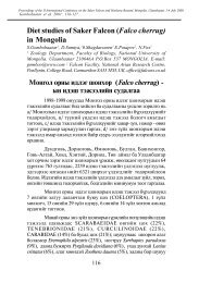
First documented clutch and brood of six in SakerFalcon (Falco cherrug).E. Potapov 1 , D. Sumya 2 , S. Gombobaatar 2 , O.Shagdarsuren 2 , S. Tuya 3 , L. Ochirkhuyag 4 & N. Fox 1 .1. The Falcon Research Institute, PO Box 19,Carmarthen SA33 5YL, UK2. Faculty of Biology, Mongolian State University,Ulaanbaatar, Mongolia3. Center for Environmental Remote Sensing, ChibaUniversity, 1-33 Yayoi-cho, Inage-ku, Chiba, 263-8522Japan, E-mail: s.tuya@lycos.com s.tuya@cr.chibau.ac.jp4. The National Remote Sensing Center, TheInformation and Computer Center, Ulaanbaatar-210646, Mongolia, lochir@yahoo.comIntroduction and methodsThe Mongolian programme of the Falcon ResearchInstitute has been running since 1998. The field team hasbeen led during the 5 field seasons by Prof. D. Sumya andMagister S. Gombobaatar under the supervision of Acad.O. Shagdarsuren. The aim of the scientific programme isto study Saker Falcon biology in Mongolia and to providethe Mongolian government with population data, numbersand breeding rates of the Saker populations to be used inestablishing harvesting quotas for <strong>falco</strong>ns in compliancewith CITES regulations.Within study areas, Saker nests were mapped and theirbreeding performance monitored. There were some nestsoutside the study areas which were visited but they werenot used in calculations of population density. Clutch size,which is considered to be one of the most important factorsdetermining a species breeding rate (Newton 1979), aswell as breeding success was determined in as many nestsas possible. In total we have data on 188 clutches/broodsduring the period 1998-2002.difficult.The symmetry of the clutch size distribution was notconstant across the years (Figure 1). An almost symmetricaldistribution was observed in 2001 and 2002 but wasskewed towards smaller clutches in all other years, mostnotably in 2000. During 2002 we found an unexpectedlyhigh proportion of large clutch sizes.Fig. 1. Clutch size variation across years with S.D. Figureson horizontal axis are sample size.Snow cover in Mongolia showed significant variationbetween 2001 and 2002 (ANOVA: F=9.36, F=0.005) asdid clutch size across the years (ANOVA: F=6.68, P=0.01)(Figure 2).Fig. 2. Clutch size variation plotted against % snow cover.We also examined the possible influence of winter snowcover on clutch size. The snow coverage data was analysedusing GIS methods at the Mongolian National RemoteSensing Center of Mongolia. Snow cover in Mongolia hasbeen monitored since 1999 and the snow cover maps arepublished on the website of the Ministry of Nature andEnvironment of Mongolia. A combination of meteorologicalstation measurements and mapped snow distributionwas used to validate and quantify the satellite data.ResultsClutch size in Mongolian Saker <strong>falco</strong>ns (Figure 1) variessignificantly across years (ANOVA: F=9.59 P=0.0000007).Largest clutch sizes were observed in 1999 and 2002. Bothwinters prior to these breeding seasons were characterisedby low winter snow coverage. The winters of 1999/2000and 2000/2001 are known as severe cold and snowy winterswith so-called “zud” conditions. “Zud” conditionsmeans that the ground was covered by ice formed beforesnow cover, thus making grazing by rodents and livestockIn 2001 there was greater snow coverage and smallerclutch sizes. In 2002 the snow cover across Mongolia wasvery unstable and unusually thin. Clutch sizes for 2002were at a maximum for the whole 5 year period. We haverecorded two clutches of 6: six chicks in down in one nestand 6 eggs at a different location 241 km apart.DiscussionA clutch of 6 is very big for large <strong>falco</strong>ns. References on14
Simple molecular methods for sexing birds1 Centre d’Ecologie Fonctionelle and Evolutive,UPR 9056 CNRS, 1919 Route de Mende, F-34293, Montpellier CEDEX 5, France. Email:gobiology@yahoo.com2 DEEB, Graham Kerr Building, Glasgow University,Glasgow G12 8QQ, U.K.A recent article in Falco documented the problem of correctlyidentifying the sex of birds where it was not possibleto do so from physiological appearance (D’Aloia 2002).Molecular sexing techniques were applied to blood samplestaken from pale chanting goshawks Melierax [canorus]canorus to determine the sex of birds. Although manyraptors can be sexed visually by reversed size dimorphism,this is not always conclusive, especially with young birds.In the case of the goshawks it was desirable to determinethe sex of all the birds so that they could be relocatedin pairs, and positive identification of sex is of obviousimportance for creating potentialbreeding pairs. It may also behelpful in speeding up trade ofbirds so they can settle into newhomes quicker, and when caringfor birds, when one sex may beparticularly susceptible to diseaseor stress. For veterinarians, scientistsand ecologists, identifyingsex is necessary to monitor sexdifferences in development. Forbreeders it can be useful to recordthe sex of nestlings hatched byfemales, to create family treesrecording the success of pairs andtheir young (see also Griffiths 2000a).The last ten years has seen the development of moleculartechniques to allow sexing of birds from DNA samples, sothat invasive methods such as laparotomy and laparoscopy,or expensive and time-consuming cytological sex identificationare becoming obsolete. Sexing from DNA requiresonly a tiny (2-10ml) amount of blood (e.g. Griffiths et al.1998, Kahn et al. 1998) or feathers (Griffiths and Tiwari1995), which appeals to conservationists, breeders and scientistsalike. Furthermore, one quick and simple methodhas been developed which can be applied universally to allbirds excepting ratites. As it is simpler, more efficient, andless expensive than other molecular methods, we explainits functioning here.Molecular sexing methods rely on isolating and identifyingdifferent sized fragments of DNA found in male andfemale birds. The avian genome (see glossary for scientificterms) has a set number of chromosome pairs calledautosomes; for each pair, one is derived from the motherand one from the father. Two other chromosomes, thesex chromosomes, also exist and are derived in a similarmanner. These ‘Z’ and ‘W’ chromosomes determine thesex of the bird, as females have a Z and a W chromosomebut males have two copies of the Z. This scheme is called‘female heterogamety’ because female birds have two differentsex chromosomes.Fragments of DNA from the Z and W chromosome canbe identified from a blood sample, so allowing us todetermine a bird’s sex. A method called PCR is used toidentify a specific piece of a gene, the CHD1 gene, foundon the sex chromosomes. This gene is highly conserved:homologous have been found in mice (Delmas et al. 1993),humans, Drosophila and yeast (Woodage et al. 1997). Itis because of this that the sexing test can be applied to allbirds (Griffiths et al. 1998), as all bird species contain theCHD1 gene (although there have been problems sexingratites).CHD1, like all genes, is made up of ‘exons’ which arefunctional pieces of DNA and consequently whosesequence (form) is highly conserved,and ‘introns’ - DNA withno purpose. In the sexing PCR,a pair of primers (short strands ofDNA) identify the CHD1-W geneon the W chromosome and theCHD1-Z gene on the Z chromosome.The chemical propertiesof DNA allow these primers tomake vast numbers of copies of aspecific fragment of DNA withinthese genes. The primers havebeen designed especially to locatethis exon region and amplify DNAacross an intron. The intron sizevaries between the CHD1-W and CHD1-Z gene and consequentlythe products of the PCR are two different sizedfragments. These fragments can be visualised by standardDNA visualisation techniques that allows discriminationof DNA fragments by size (see box 1). The result is thatfemales have two bands but males only one. An advantageof this test is that it amplifies both CHD1-W and CHD1-Z, which allows positive identification of both sexes. (Ifa PCR amplified only CHD1-W it would be impossibleto designate a null result as a true male or simply a failedPCR reaction).Currently there are three tests designed to identify sexusing this method (see also Grifiths 2000b), two whichamplify the same intron (Griffiths et al. 1998, Kahn et al.1998) and another which amplifies a different intron stillon the CHD1 gene (Fridolfsson and Ellegren 1999). Table1 gives primer sequences and PCR conditions for reactions.The test used by D’Aloia (2002 and also D’Aloiaand Eastham 2000) functions in a similar way, but requiresDNA cutting with a restriction enzyme after PCR amplification,necessitating the identification of a suitable enzymeand adding extra work, time, expense and opportunity for16
error. We therefore suggest this more simple test. Thechoice of primers to use is based initially on study species,and where one test fails another can be performed; for <strong>falco</strong>nspecies the 2550F/2718R (table 1) have been found towork well.TroubleshootingWhen PCR conditions described here are not successfulseveral steps can be taken to remedy the situation. Firstly,the concentration of DNA in the PCR mix may hamperthe reaction (usually too much) and it is advisable to trya dilution series of PCRs. If the CHD1-Z product amplifiesbetter than the CHD1-W, try lowering the annealingtemperature and/or reducing extension time. Where thereis little size difference between the W and Z bands, use apolyacrylamide rather than agarose gel to allow better discriminationbetween band lengths.The sexing methods explained here represent an efficient,cheap and reliable means to sex birds and can be carriedout in any standard molecular biology lab. For those withoutaccess to such facilities, commercial sexing is available,e.g. from University Diagnostics, 90 London Rd,London, SE1 6LN, UK.Glossary:Autosomes: non sex chromosomesGenome: entire genetic complement of a prokaryote(organisms with no nuclie) or virus, or the haploid geneticcomplement of a eukaryoteHaploid: a state of having one copy of each chromosomeper nucleusHeterogametic: the sex with different morphs of sexchromosomes; in cell division different kinds of gametesare produced in regard to these sex chromosomes (e.g. themale state in humans that produces sperm with X and Ychromosomes)Homogametic: the sex with same morphs of sex chromosomesthat produces just one kind of gamete (e.g. femaleshumans that produce ova with X chromosomes)PCR (polymerase chain reaction): method to amplify rapidlyselected DNA segments in cycles of DNA denaturation,primer binding and replication.References:D’Aloia, M. A. (2002) Molecular sexing of Pale ChantingGoshawks. Falco 19: 13-14.D’Aloia, M.A. and Eastham, C. (2000) DNA-based sexidentification of <strong>falco</strong>ns and its use in wild studies andcaptive breeding. Falco 16: 12.Delmas, V., Stokes, D. G. & Perry, R. P. (1993) A mammalianDNA binding protein that contains a chromodomainand an SNF2/SW12-like helicase domain. Proceedings ofthe National Academy of Sciences, USA 90, 2414-2418.of Avian Biology 30: 116-121.Griffiths, R. G. (2000a) Sex identification in birds.Seminars in Avian and Exotic Pet Medicine 9.Griffiths, R. (2000b) Sex identification using DNA markers.In Molecular methods in ecology (ed. A. J. Baker).London: Blackwell Science.Griffiths, R., Double, M. C., Orr, K. & Dawson, R. J. G.(1998) A DNA test to sex most birds. Molecular Ecology7: 1071-1075.Griffiths, R. & Tiwari, B. (1995) Sex of the last wild Spix’smacaw. Nature 375: 454.Kahn, N., St John, J. & Quinn, T. (1998) Chromosomespecificintron size differences in the avian CHD gene providesa simple and efficient method for sex identification.The Auk 115: 1074-1078.Promega. (1996) GenePrint STR Systems TechnicalManual Notice of Protocol Change. Madison, WI:Promega.Woodage, T., Basrai, M., Baxevanis, A., Hieter, P. &Collins, F. (1997) Characterisation of the CHD familyof proteins. Proceedings of the National Academy ofSciences, USA 94, 11472-11477.Fridolfsson, A. & Ellegren, H. (1999) A simple and universalmethod for molecular sexing of non-ratite birds. Journal17
Aspergillosis: Therapy and Prevention in ZooAnimals with Emphasis on RaptorsT. BaileyMSc Programme Wild Animal HealthZoological Society of LondonThe Royal Veterinary College, London,UK1. BackgroundAspergillosis is a disease of increasing importance inhuman medicine because of rising numbers of immunosuppressedpatients vulnerable to infection (Hill et al, 1995).Aspergillosis was the first mycotic disease to be describedin animals, although accounts of an incurable disease ofthe lungs called ‘pantas’ by the <strong>falco</strong>ner Latham (1615)pre-date this. Aspergillosis has implications in avianconservation programmes, the first generation of captivebreeding projects are often derived from the wild,which are stressed and susceptible to infection. In areview of factors involved in the failure of the translocationand breeding programmes undertaken by theNew Zealand Conservation Department of the rarestitchbird (Notiomystis cincta), aspergillosis was thesingle most important cause of mortality (Cork et al 1999).Aspergillosis is the most common systemic avian mycosis.Of 66 case reports published on aspergillosis from 1970-2000 in exotic species (Veterinary Librarian) 54 (81%)were avian species, 11 (17%) were mammals and 1 was areptile. Established infections are challenging to resolveand this paper aims to review current approaches to therapyand prevention of aspergillosis with emphasis on raptors.2. Treatment Considerations2.1. Classification of AspergillosisAvian aspergillosis has been classified by Redig (1993a)as 1) an acute form caused by a single exposure to anoverwhelming number of spores; 2) a tracheal form; 3)localised granulomas in the air sacs and lungs; and 4) aninvasive form which initially affects the respiratorysystem and then spreads haematogenously andthrough the air sacs to the rest of the body. Thesuccess of treatment depends on location and extentof the lesions and effective treatment can usuallyonly be instituted in the tracheal and semi-invasive orlocalised forms. Severe cases, where the patient exhibitscachexia, dyspnea and vomiting, are beyond the point oftreatment (Redig 1993a). Treatment goals for aspergillosisare 1) removal of lesions that restrict airflow, 2) killing andelimination of the fungus and 3) provision of supportivecare (Redig 1981). Unless the lesions are detected early,drug therapy alone is unlikely to be effective because of thethick caseous encapsulation which develops in the airsacs(Forbes 1996). Factors associated with the susceptibilityof animals to aspergillosis are presented in Tables 1 & 2.2.2. Antifungal Susceptibility TestingThere are over 185 species in the Aspergillus genus(Richardson 1998). In avian infections A. fumigatus is themost common etiological agent, followed by A. flavus andA. niger (Bauck 1994). Selection of antimicrobial drugsdepends on knowledge of the susceptibility of the suspectedpathogen (Prescott 2000), but clinical mycology haslagged behind clinical bacteriology with respect to uniformstandardisation of susceptibility testing. In vitro antifungalactivity does not correlate well with in vivo efficacy andwithout readily available susceptibility tests in veterinarymedicine most therapeutic choices are empirically based(Papich et al, 2001). Resistance following antifungal druguse is recognised (Graybill et al 1998). Table 3 presentsthe activity (MIC) of antifungal agents against fungi &Table 4 summarises the susceptibility of Aspergillus sp. toavailable antifungal drugs.18
193. Antifungal Agents Effective Against Aspergillus sp.Amphoteracin BMechanism of action: Amphoteracin B is a fungicidalpolyene antibiotic. It binds ergosterol in the fungal cellmembrane making it more permeable, leading to electrolyteleakage and cell death.Spectrum of activity: Organisms with MIC < 1 mg/ml areconsidered susceptible (Prescott 2000). There is good correlationbetween MIC values & clinical response (Papich etal, 2001). Aspergillus sp. are the most frequently reportedresistant fungi (Prescott 2000).Chemical properties and pharmacokinetics: Its pharmacokineticshave been poorly studied in veterinary species.It is poorly absorbed from the GIT so must be given i.v.,i.o. or i.t. Absorption from the lungs following aerosoladministration is poor and this route is used to treat pulmonaryaspergillosis (Prescott 2000).Adverse effects: The most important clinical toxicosis isnephrotoxicity (Prescott 2000). It is dose-related and isseen clinically as increases in BUN and creatinine in mammals.Dosing every other day, electrolyte loading and slowinfusion of amphoteracin B decrease the severity and rateof development of renal toxicity. Other adverse effectsinclude phlebitis, fever, nausia, vomiting and hypokalemiawith resulting cardiac arrhythmias (Prescott 2000).Measures to prevent vomiting include giving antiemeticdrugs before infusion.Clinical protocols: Dosage regimens are presented in Table5. Treatment protocols should include pre-treatment withsodium chloride and it should be infused at a slow rate. Itcan be mixed with 5% dextrose solution during infusion.Administration of amphoteracin B in 0.45% saline with0.5% dextrose s.c. in dogs/cats is a way of administeringlarge quantities of amphoteracin B without producingthe marked azotemia associated with i.v. injection (Malik1996).New formulations: New less toxic lipid-based formulationshave been used in humans (Walsh et al 2001). Thesedrugs can be given at higher doses with less toxicity. Inhumans daily doses of the lipid-based formulations are 3-5mg/kg daily, whereas the dose of the conventional form are0.5-1 mg/kg every 48 hours (Prescott 2000).FlucytosineMechanisms of action: Flucytosine is a fluorinated pyrimidinewhich is deaminated to 5-fluorouracil and incorporatedinto mRNA in the fungal cell, producing faulty proteins.Spectrum of activity: It has a narrow spectrum of activityand few Aspergillus sp. strains are susceptible. Strainswith MIC < 16mg/ml are susceptible (Prescott 2000). Twothirds of fungal isolates change from susceptible to resistantduring treatment so flucytosine should only be usedin combination with other antifungal agents (Papich et al2001). Combination with amphoteracin B is synergisticbecause amphoteracin B increases fungal permeability toflucytosine (Orosz & Frazier 1995).Clinical protocols: Dosage regimens are presented in Table5 and only occasional side-effects are reported (Papich etal 2001).The Azole Antifungal DrugsThese are broad-spectrum antifungal drugs that exert theirantifungal mechanism on the cell membrane of the fungusby inhibiting synthesis of the sterol of the fungal cell membrane.They are generally fungistatic at concentrationsachieved clinically and are slower acting than the polyenes.EnilconazoleThis drug has excellent antifungal activity. It has aresidual effect after topical application and is used totreat topical dermatophyte infections. Enilconazole is thetreatment of choice for nasal aspergillosis in dogs & localinfusion is used in the treatment of guttural pouch mycosisin the horse (Prescott 2000). It can be given topically ornebulised and the Clinafarm-EC formulation is used in theenvironmental decontamination of poultry facilities (Table5) and equipment to prevent aspergillosis.ClotrimazoleThis azole is inhibitory to Aspergillus sp. and concentrationsabove 10mg/ml are fungicidal (Prescott 2000). It isavailable as a topical product, can be nebulised and fewstrains of fungi are resistant (Prescott 2000).ItraconazoleSpectrum of activity: Itraconazole is the drug of choiceto treat aspergillosis infections (Papich et al 2001).Organisms with MIC < 0.12 mg/ml are susceptible, thosewith MIC 0.25-0.5 mg/ml are susceptible depending ondose and those with MIC > 1 mg/ml are resistant (Prescott2000).Pharamcokinetics: Absorbtion is increased in an acidicenvironment and when taken with meals. Bioavailabilityincreases from 40% after fasting to 99.8% when givenwith food (Papich et al 2001). It is extensively distributedthroughout the body. It is hepatometabolised and mainlyeliminated in the bile. Therapeutically active concentrationsare maintained longer in tissues than in plasma andthe drug can be detected in vaginal epithelium and skin for4 days and 4 weeks respectively (Papich et al 2001).Clinical use: It is used to treat superficial and systemicinfections and to treat and prevent aspergillosis in birds(Table 5). As oral absorbtion is pH dependent dosageadjustments are necessary if gastric pH is increased(Papich et al 2001). Oral capsules should be given with
F10-DisinfectantF10 is a complete spectrum virucidal, bactericidal,fungicidal & sporicidal, but aldehyde freecompound of six synergistic active ingredients(Verwoerd 2001). It has been tested against everysignificant animal/human pathogen and has outperformedother disinfectants during efficacy testing,over a range of temperatures, in the presenceof organic material, at low concentrations, shortcontact times, without any corrosive effects oninfrastructure, metal nozzles or any tissue irritationon workers and animals (Anon 2001). Withthese characteristics, F10 can be used to disinfectanimal environments in their presence, loweringthe environmental pathogen challenge significantly,with no side-effects. Using either a nebuliseror a ‘smogger’ unit, F-10 has been used totreat either individuals or groups of <strong>falco</strong>ns withaspergillosis (Table 4; Bailey and Sullivan, 2001).New Antifungal AgentsVoriconazole & echinocandin are new triazole &lipopeptide drugs respectively which show promiseagainst aspergillosis (Prescott, 2000; Graybill,2001).food, but the oral suspension is better absorbed on anempty stomach. Orosz et al (1995) reported higher tissueconcentrations when itraconazole is dissolved in acidand gavaged with orange juice in pigeons (Columba livia).Treatment of serious infections should be prolonged (>3months) and relapses occur (Prescott 2000). An i.v. formulationis being developed and is effective against experimentaldisseminated aspergillosis in guineapigs (Odds2000).Adverse effects: Itraconazole is better tolerated than ketoconazole.Hepatic toxicosis occurs in 10% of dogs, whilein cats there are dose-related GIT effects of anorexia andvomiting (Papich et al 2001). Long-term administration (3months) to dogs and cats resulted in no side-effects,but maternal toxicity, embryo toxicity and teratogenicitywere observed in rats; therefore, its usein pregnant mammals is not recommended (Prescott2000). Itraconazole is considered to cause anorexia,depression and death in African grey parrots(Psittacus erithacus) at doses above 5 mg/kg bid(Birdmed discussion 2002).Other Antifungal AgentsTerbinafineThis synthetic allylamine drug is highly fungicidal.Allylamines decreases the fungal synthesis of ergestroland cause fungal death by disrupting the cell membrane(Prescott 2000). It is active against Aspergillus sp. but todate clinical use has been limited in veterinary medicine,although preliminary nebulisation results are promising(Dalhausen 2000).4. TherapyCombination TherapyIn vitro synergism of 2 antifungal compounds,seen as a 4x reduction in the MIC when given alone is seenwhen amphoteracin B is given with flucytosine (Papichet al 2001). Combinations of amphoteracin B and azoleshave been less successful and both antagonism and synergismhave been reported (Bajjoka et al 1999). Because ofthe slow onset of action of azole antifungals it is recommendedto use initial amphoteracin B therapy of serioussystemic fungal infections followed by an azole agent(Papich et al 2001). The treatment combinations used atAbu Dhabi Falcon Hospital are presented in Table 6.SurgeryIn advanced cases surgical removal of granulomas from theairsacs, or lesions that are blocking the trachea or syrinxand direct application of antifungal agents to the lesionsare important therapeutic adjuncts in birds (Tully et al2000). Studies in dogs with nasal aspergillosis have shownthat while systemic therapy is successful in 50% of cases,systemic therapy and local irrigation of nasal lesions issuccessful in 90% cases (Clark 1999).Supportive TherapySupportive therapy including fluids (i.v., s.c., i.o.), tubefeeding, antibiotics, antiemetic drugs, immune stimulants20
and vitamin A supplementation should be given accordingto the needs of the case. The use of immunosuppressivedrugs (steroids) in birds is controversial and should beavoided (Chitty 2001). Indeed cortisone-treated animals(rabbits) are more susceptible to invasive aspergillosis(Khosravi et al 1998).Immune StimulationInterferon g augments the ability of human leucocytes todamage fungal hyphae in-vitro and clinical trials of colonystimulating factor have shown that it can be a usefuladjunct to antifungal therapy (Richardson 1998). Immunestimulation may have a role in veterinary species in thefuture.5. Duration and Response to TreatmentDuration of TherapyAntifungal regimens in humans are often extended for 6-12months (Jones et al 2000). For veterinary species treatmentduration recommendations vary from 2 to 6 monthsand one month past the resolution of the disease (Jones etal 2000; Tully et al 2000).1999). Likewise, prophylactic itraconazole (Table 5) isrecommended for captive-held birds undergoing a changein management, especially high risk species (Table 1)and during times of stress such as trauma (Redig 1993a)or as part of the management of oiled susceptible seabirds.Aspergillus spp. can colonise damaged airways(Richardson 1998), so probably prophylactic antifungalmedication should be considered as part of the therapy ofmany respiratory tract infections (e.g. Serratospiculiasis) ofsusceptible species. Daily fogging with F10 of susceptiblebirds is used to lower the environmental load of respiratorypathogens such as Aspergillus sp. in the Middle East.VaccinationVaccination has been proposed as a means of preventingaspergillosis. A heat-killed vaccine reduced mortality ofaspergillosis in waterfowl (Yearout 1988) and a germinatedconidia vaccine reduced turkey poult mortality (Richardet al 1984). Recently, Graczyk et al (1997) demonstratedthat i.m. immunisation of Cape shelducks (Tadorna cana)with Aspergillus spp. mycelial phase cultures providedprotection against experimental challenge. However,despite these reports no aspergillosis vaccinations are commerciallyavailable for any veterinary species.PreventionRecommendations to prevent aspergillosis in captive aviancollections are presented in Table 8.7. ConclusionsAspergillosis is a manageable and treatable disease ifdetected early, but advanced cases are medically challenging.Well-designed studies to determine the correlationbetween the susceptibility of avian isolates and the clinicalresponses to antifungal drugs are needed so that therapeuticregimens can be optimised.Response Rate and Susceptibility to Re-infectionInterestingly, despite numerous treatment protocols (Table5), little data has been published on the response to treatmentso that the efficacy of different regimens can beobjectively assessed. Mortality from invasive aspergillosisin immunocompromised humans is high (Pattersonet al 2000). The average case fatality rate (CFR) of 50published studies (1995-2000) on the treatment of invasiveaspergillosis in humans was 58% (SwuJane 2001). In asurvey of aspergillosis cases in a large <strong>falco</strong>n hospital theCFR for <strong>falco</strong>ns was 37.5% (Table 7). Little has been publishedon the susceptibility of recovered animalsto rechallenge with aspergillosis. Kunkleand Sacco (1999) demonstrated that convalescencefrom pulmonary aspergillosis in turkeysdid not confer protection against rechallengebut instead decreased resistance to subsequentinfection. Recrudescence of aspergillosisinfections in <strong>falco</strong>ns that have recovered from aspergillosisis common in the Middle East and it is likely that the CFRof 37.5% is higher as this does not account for recrudescencebecause of difficulties in case follow-up.6. Prophylaxis, Prevention & VaccinationProphylaxisProphylactic antifungal therapy is recommended for highrisk humans and itraconazole is considered the most effectiveantifungal agent at preventing aspergillosis (Kibbler218. Glossarybid = twice a day; i.o. = intraosseus; p.o. = peros; CFR = case fatality rate, i.t. = intratrachealinjection; s.c. = subcutaneous; GIT = gastrointestinaltract; i.v. =, intravenous injection;sid = once a day; i.m. = intramuscular injection;MIC = minimum inhibitory concentration;tid = three times a day9. ReferencesAnon (2001) F10 disinfectant Product Datasheet.Interhatch, Sheffield, UK.Bailey, T.A. & Sullivan, T. (2001) Exotic DVMMagazine. 3: 17.
Bajjoka, I.E. et al. (1999) Pharmacotherapy. 19: 118-123.Bauck, L. 1994 In: Avian Medicine: principles & application.Ritchie, B.W., Harrison, G.J. & Harrison, L.R. (eds).Wingers Publiching Inc, USA. Pp. 997-1006.Biasia, I. & Giovardi, A.A. (200)1 In: Biology, medicine,& surgery of south American wild animals. (Fowler, M.E.& Cubas, Z.S., eds). Iowa State University Press, USA.Pp. 163-165.Birdmed discussion group. (2002) http://numbat.murdoch.edu.au/birds/birdmed.htmChitty, J.R. (2001) Veterinary Record. 149: 499.Clark, W.T. (1999) In: Textbook of small animal medicine(Dunn, J.K., ed). W.B. Saunders, London. 345-350.Cooper, J.E. (2002) Birds of prey: health & disease.Blackwell Science, Oxford, U.K. pp. 90-93.Cork, S.C. et al. (1999) Journal of <strong>Wildlife</strong> Diseases. 35:481-486.Dalhausen, B. et al. (2000) Proc. Association of AvianVeterinarians, Portland. Pp. 35-39.Dykstra, M.J. et al. (1997) Journal of Zoo and <strong>Wildlife</strong>Medicine. 28:454-463.Forbes, N.A. (1996) In: BSAVA Manual of raptors,pigeons and waterfowl. British Small Animal VeterinaryAssociation, UK. pp. 180-188.Graczyk, T.K. et al. (1997) Mycopathologia 140: 121-127.Graybill, J.R. (2001) <strong>International</strong> Journal of ClinicalPractice. 55: 633-638.Harry, E.G. & Cooper, D.M. (1970) British PoultryScience. 11: 269-272.Heidenreich, M. (1995) Birds of Prey. Medicine andManagement. Blackwell Science <strong>Ltd</strong>, Oxford. pp. 125-128.Hill, P.B., et al. (1995) Veterinary Dermatology. 6: 59-66.Jones, M.P., Orosz, S.E., Cox, S.K. & Frazier, D.L. (200)0Journal of Avian Medicine and Surgery. 14: 15-22.Kibbler, C.C. (1999) Mycoses. 42: 121-124.Khosravi, A.R. et al. (1998) Journal of the Faculty ofVeterinary Medicine, University of Tehran. 53: 83-87.Kunkle, R.A. & Sacco, R.E. (1998) Avian Diseases. 42:787-790.Orosz, S.E. &, S.D. & Frazier, D.L. (1995) Journal ofAvian Medicine and Surgery. 9: 8-18.Orosz, S.E. et al. (1996) Journal of Avian Medicine andSurgery. 9: 255-262.Papich, M.G. et al. (2001) In: Veterinary pharamacologyand therapeutics. (Adams, R.A., ed). Iowa StateUniversity Press, USA. Pp. 918-932.Patterson, T.F. et al. (2000) Medicine (Baltimore). 79:250-260.Prescott, J.F. (2000) In: Antimicrobial therapy in veterinarymedicine (Prescott, J.F., Baggot, J.D. & Walker, R.D.,eds). Iowa State University Press, USA. Pp. 367-395.Redig, P. (1981) In: J.E. Cooper & A.G. Greenwood(eds). Recent advances in the study of raptor diseases.Chiron Publications <strong>Ltd</strong>, UK.. pp. 117-122.Redig, P. (1993a) In: Zoo & Wild Animal Medicine:Current therapy 3. (Fowler, M.E., ed). W.B. SaundersCompany, USA. Pp. 178-181.Redig, (1993b) A collection of notes on selected topics,2nd Edition. The Raptor Center, University of Minnesota,USA. Pp. 115-124.Richard, J.L. et al. (1984) Mycopathologia. 87: 3-11Richard, J.L. (1991) In: Diseases of poultry, 9th Edition(Calnek, B.W., ed). Wolf Publishing <strong>Ltd</strong>, Iowa StateUniversity Press, USA. Pp. 332-334.Richardson, M.D. (1998) In: Microbiology & MicrobialInfections: Volume 4 Medical Mycology (Collier, L.,Balows, A. & Sussman, M.). Arnold, London. Pp. 281-312.Ritchie, B.W. & Harrison, G.J. (1994) In B.W. Ritchie,G.J. Harrison & L.R. Harrison (eds). Avian Medicine:Principles and Application. Wingers Publishing Inc. pp.457-478.SwuJane, L. et al. (2001) Clinical Infectious Diseases.32: 358-366.Tully, T.N. et al. (2000). Avian Medicine. ButterworthHeineman, Boston. Pp. 200-201.Verwoerd, D. (2001 Falco, 17: 15-17.Veterinary Librarian Database, First Move, Littleton, USAWalsh, T.J. et al. 2001 Antimicrobial Agents andChemotherapy. 45: 3487-3496.Yearout, D.R. 1988 Proc. 1988 Annual Meeting of theAssociation of Avian Veterinarians, Houston. Pp. 139-144.Latham, S. 1615 Falconry, or the faulcons lure and cure.Printed by J.B., London.Malik R. (1996) Australian Veterinary Journal. 73: 124.Odds, F.C., et al. 2000 Antimicrobial Agents andChemotherapy. 44: 3180-3183.22
An update on the prevalence of Chlamydiainfections in avians in the U.A.E.U. Wernery, W. F. D’Mello and R. ZachariahCentral Veterinary Research Laboratory, P.O. Box 597,Dubai, U.A.E.Over the last years we have twice reported aboutChlamydia-infections in avians (Zachariah and Wernery,1997 and 1998) in the U.A.E. The reason to update theresults, is a sharp increase of positive cases especially in<strong>falco</strong>ns in 2001 (Table1 and Figure 1).The ag-ELISA does not differentiate between the differentChlamydia species as the antigen used in the test sharesa common antigen with all the different species. The testutilizes a monoclonal antibody to detect a genus-specificchlamydial antigen. Further work should now concentrateon the identification of Chlamydia species in U.A.E. avians.For this purpose a tissue culture laboratory should beestablished. The new Chlamydia classification is seen inTable 2.Table 2.ABCA) Falcon; B) Houbara; C) Pigeon; D) OthersChlamydiosis and Chlamydia-infections in <strong>falco</strong>ns andother avian species are a persistent problem in the U.A.E.,but it is so far not clear why the increase has suddenlyoccurred in 2001. For the diagnosis of an infection, we usethe Chlamydia-antigen ELISA from DAKO, UK (IDEIA,Code No. K601).Swabs are taken from the pharynx and are immediatelyplaced into Chlamydia transport medium. They are thensent to CVRL for testing. Many of the positive birds do notshow any clinical signs. However, they have a Chlamydiainfectionand may suffer from Chlamydiosis when understress.References:R. Zachariah and U. Wernery, 1997. Chlamydiosis in captive<strong>falco</strong>ns. Falcon 9: 11.R. Zachariah and U. Wernery, 1998. Chlamydia infectionin avian species. Falco 11: 10.K.D.E. Everett, R.M. Bush and A.A. Andersen, 1999.Amended description of the order Chlamydiales, proposalof Parachlamydiaceae fam. nov. and Simkaniaceae fam.nov., each containing one monotype genus, revised taxonomyof the family Chlamydiaceae, including a new genusand five new species, and standards for the identification oforganisms. Int. J. Syst. Bacteriol. 49: 415-440.23
What’s new in the literature ?Barton, N.W.H., Fox, N.C., Surai, P.F. and SpeakeB.K. (2002) Vitamins E and A, carotenoids andfatty acids of the raptor egg yolk. Journal of RaptorResearch 36 (1) 33-38.A captive population of <strong>falco</strong>ns was fed a diet containinga known quantity of vitamin A (retinol) and vitamin E(a-tocopherol) for six weeks prior to and during egg-laying.Infertile eggs were analysed for vitamin A, vitaminE, carotenoid and fatty acid composition. Mean, dailyvitamin intake was 29mg Vit E (35IU) and 1157mg Vit A(3363IU). Adjusted mean egg yolk content for infertile,unincubated eggs was 314 mg/g a-tocopherol and 3.06mg/g Vit A. A distinctive feature of the raptor egg yolkis a very high proportion of arachidonic acid which isprobably a reflection of their carnivorous diet. A smallnumber of plasma samples were also available from egglaying<strong>falco</strong>ns. Mean, plasma vitamin E was 32.2 mg/mland plasma vitamin A 1.02 mg/ml.Casado, E., Balbontin, J., Ferrer, M. (2002) Plasmachemistry in booted eagle (Hieraaetus pennatus) duringbreeding season. Comparative Biochemistry andPhysiology. A, Molecular & Integrative Physiology,131, No.2, pp.233-241Most studies that have examined raptor plasma chemistryhave been conducted on birds living in captivity. Inthis study, we describe typical plasma chemistry valuesindicators of body condition in free-living Booted Eagles,Hieraaetus pennatus, from Donana National Park (Spain).A total of 143 young, 55 adults and one bird of unknownage were caught, banded and sampled between 1996 and2000. Values were compared with those of other raptors.Mean concentrations of creatinine, uric acidand urea were lower in adults than in nestlings, whileglucose, DAT and AAT were lower in nestlings than inadults. Interactions of age/sex affected plasma meanlevels of creatine kinase, glucose, AAT, uric acid andurea. Adult females showed significantly lower levels ofcreatine kinase, uric acid and urea than adult males andnestlings. Adult males had significantly higher levels ofAAT than the other groups. The lowest levels of glucoseand the highest levels of uric acid were found in nestlingfemales. We think the differences in blood parameterscan be explained by differences in size of species, ofindividuals (because of both body condition and sexualdimorphism) and diet.Dawson, R. D., Bortolotti, G. R. (2001) Sex-specificassociations between reproductive output and hematozoanparasites of American kestrels. Oecologia, 126,No.2, pp.193-200Parasites have the potential to decrease reproductive outputof hosts by competing for nutrients or forcing hosts toinvest in immune function. Conversely, reproductive outputmay affect parasite loads if hosts allocate resourcesto reproduction such that allocation to immune functionis compromised. Both hypotheses implicitly have a temporalcomponent. Parasites (Haemoproteus spp.) weresampled both before and after egg laying to examine therelationship between reproductive output (indexed using acombined measure of clutch size, egg volume and initiationdate) and blood parasite loads of American kestrels (Falcosparverius). Parasite loads measured prior to egg layinghad no adverse effects on subsequent reproductive output.Females that previously had large reproductive outputs subsequentlyhad lower parasite intensities than those whoseoutputs were smaller, suggesting that females were capableof allocating energy to both forming clutches and reducingparasite loads. Because male kestrels provide most of theirmate’s energetic needs before, during and after egg laying,mate choice by females may have consequences for theirparasite loads. Females choosing high-quality mates maynot only have increased reproductive output, but may alsoobtain sufficient resources from their mates to enable themto reduce their parasite burdens. Males whose mates hadlarge reproductive outputs were more likely to subsequentlybe parasitized and have more intense infections. For individualmales sampled both before and after egg laying,those whose mates had larger reproductive outputs werealso more likely to become parasitized, or remain parasitized,between sampling periods. Increased parasite loads ofmales may be one mechanism by which the costs of reproductionare paid.Dawson, R. D. and Bortolotti, G. R. (2000) Effects ofhematozoan parasites on condition and return rates ofAmerican kestrels. Auk 117, No.2, pp.373-380The relationship between blood parasites and body conditionof American kestrels (Falco sparverius) was evaluatedduring the breeding season. Females that were infectedwith at least one species of parasite were in poorer conditionthan those without parasites during incubation but notprior to egg laying. It is suggested that the relationshipbetween parasitism and condition was masked before layingbecause of large increases in body mass of females duringegg formation. Reduced condition of males during incubationalso was associated with higher intensity of infectionsby Haemoproteus in one of 2 years. The negative associationbetween condition and intensity of infection suggeststhat blood parasites impose costs on kestrels owing tocompetition for nutrients or allocation of energy by hoststo immune function or tissue repair. Alternatively, kestrelsin poor condition may be more likely to have relapses ofchronic infections, or they may be less able to control newinfections because of resource limitations. In contrast toresults during incubation, during the prelaying period theprevalence of parasites tended to be higher, and in one yearinfections were more intense, among males in good condition.One possible explanation is that body condition ofmales during courtship is an important determinant of thequality of mate they are able to obtain, and males may beaccumulating body reserves at the expense of decreasedimmune function. Return rates of female kestrels to thestudy area declined as the intensity of their Haemoproteusinfections increased, suggesting that blood parasitism isassociated with reduced survival or increased dispersalprobability.24
25Eastham, C.P., Nicholls M.K. and Fox, N.C. (2002)Morphological Variation of the Saker (Falco cherrug)and the implications for conservation. Biodiversityand Conservation 11: 305-325.This study assesses the external morphology of the sakerwhich is highly variable. Although most variation wasbetween individuals within regions, the remaining variationin size and plumage characters may be described asa gradual cline between small, dorsally uniform brownsakers from western lowland regions (Kazakhstan andsouth west Russia) to large grey coloured sakers fromeastern highland regions (south east Russia, Mongoliaand China). Between these extremes exists a plethora ofhighly variable and contiguous populations.Krone, O., Priemer, J., Streich, J., Sommer, P.,Langgemach, T., Lessow, O. (2001) Haemosporidaof birds of prey and owls from Germany. ActaProtozoologica 40, No.4, pp.281-2891149 free-living birds of prey from Germany were examinedfor blood parasites. The prevalence of infection was11% (adult birds 18%, immature birds 16%, and nestlings4%). Among the Falconiformes and Strigiformes,11% of 976 and 13% of 173 birds, respectively, wereinfected. Of 17 <strong>falco</strong>niform species, 9 were infected withblood parasites whereas the Eurasian buzzard (Buteobuteo) had the highest prevalence for haematozoa; i.e.Leucocytozoon toddi (31%), the highest prevalence(25%) for Haemoproteus sp. was found in the hobby(Falco subbuteo). Eight species of owls were examinedfor blood parasites; the tawny owl (Strix aluco) had thehighest prevalence with Haemoproteus syrnii (22%).In one pygmy owl (Glaucidium passerinum) examined,Trypanosoma avium and Plasmodium fallax weredetected. The white-tailed sea eagle (Haliaeetus albicilla)was found to be a host of L. toddi for the first time.Differences in the prevalence of blood parasites werefound in the seasons and age classes of the birds.Lierz, M., Gobel, T., Schuster, R. (2002) Review andinvestigations on parasites in birds of prey and owlsfound injured or debilitated. Berliner und MunchenerTierarztliche Wochenschrift 115, No.1/2, pp.43-52In the present paper, a general overview on parasites inbirds of prey and owls is given. This part is followedby a study investigating the prevalences and species ofparasites in free- ranging birds of prey and owls in Berlinand Brandenburg State, Germany. In a period of oneyear (between August 15, 1996 and August 15, 1997),84 birds of prey and owls of the following species wereexamined for the presence of endo- and ectoparasites:Common Buzzard (Buteo buteo; n=32), Kestrel (Falcotinnunculus; n=20), Sparrowhawk (Accipiter nisus; n=9),Goshawk (Accipiter gentilis; n=8), Black Kite (Milvusmigrans; n=4), Peregrine Falcon (Falco peregrinus; n=3),Marsh Harrier (Circus aeruginosus; n=1), White-tailed-Sea Eagle (Haliaeetus albicilla; n=1), Tawny Owl (Strixaluco; n=4), Long-eared Owl (Asio otus; n=1) and BarnOwl (Tyto alba; n=1). In 97.6% of the cases, ectoparasites(feather mites and hippoboscid flies) were found.It was observed that 93.3% of eyasses were positive forhippoboscid flies. Trichomonas was detected in 28.6%of all birds of prey and owls examined. A prevalence of100% was established in the Sparrow Hawks as well asPeregrine Falcons. Leucozytozoon sp. and Haemoproteussp. as blood parasites were found in 26.9% of the birdsin total. Common Buzzards showed the highest prevalence(44.8%). A total of 58.3% of birds examinedwere positive for endoparasites. Flukes were found in16.7%, tapeworms in 14.3%, roundworms in 48.8%and acanthocephales in 2.4% of the cases. Interestingly,Tylodelphis clavata (in a Common Buzzard) andHovorkonema variegatum (in a Goshawk) were found forthe first time in raptors. The results of this study underlinethe importance of a parasitological examination inthe process of raptor rehabilitation.Manvell, R. J., McKinney, P., Wernery, U., Frost, K.(2000) Isolation of a highly pathogenic influenza Avirus of subtype H7N3 from a peregrine <strong>falco</strong>n (Falcoperegrinus). Avian Pathology 29, No.6, pp.635-637A peregrine <strong>falco</strong>n (Falco peregrinus) was presented tothe Al Safa Falcon Clinic in Dubai, United Arab Emiratesunable to stand [date not given]. Four hours after hospitalization,the bird died despite supportive care andcalcium disodiumedetate treatment. The <strong>falco</strong>n had beenon a hunting trip to Syria with its owner of 2 years, priorto its death. The carcass was submitted to the CentralVeterinary Research Laboratory in Dubai where it wassubjected to postmortem examination. Investigationsresulted in the isolation of an influenza A virus subtypeH7N3, which proved to be highly pathogenic for chickens.Parga, M. L., Pendl, H., Forbes, N. A. (2001) The effectof transport on hematologic parameters in trained anduntrained Harris’s hawks (Parabuteo unicinctus) andperegrine <strong>falco</strong>ns (Falco peregrinus). Journal of AvianMedicine and Surgery 115, No.3, pp.162-169Birds of prey are found increasingly in captive situations.When sick, these animals show only very subtle signs ofdisease, making the detection of disease difficult. For thisreason, the analysis of haematological parameters is avery useful technique for the avian veterinarian. However,birds are most frequently transported to the veterinaryclinic for a health check-up. This study investigates theeffects of transport-related stress on heterophil and lymphocytemorphology and haematological parameters ofperegrine <strong>falco</strong>ns (Falco peregrinus) and Harris’s hawks(Parabuteo unicinctus). Twelve birds of each speciesfrom the UK, all adults, mixed males and females, wereanalysed. Each group of 12 was comprised of 6 trained(accustomed to being transported) and 6 untrained birds.Samples of blood were taken from all birds in their placeof origin and again 1 week later after 1 hour (35 km) oftransport. Both samples were taken in similar conditions(eg, time of day, duration after feeding, environmentaltemperature, sample handling) so that any variationwould be caused only by transport-related factors. Bothuntrained groups showed a significant (P
untrained peregrines showed no other significant change,untrained Harris’s hawks had a significant leukopenialymphopaenia and eosinopaenia. Trained Harris’s hawksshowed a significant monocytosis, whereas trained peregrinesshowed no significant change. Transport had noapparent influence on heterophil or lymphocyte morphology.Although the difference between pre- and posttransportwas significant in some parameters, all values in the4 groups remained within the reference ranges for thespecies. Therefore, we can conclude that 1-hour transportfor trained or untrained members of these 2 species toa clinic need not be factor that requires the clinician’sconsideration when interpreting a haematological sample.However, the 2 species reacted differently to transport.Further studies on other species are suggested. The H/Lratio is proposed as the most sensitive measure of stressresponse in the blood picture of raptors and possible usesare suggested.Samour, J. H., Naldo, J. L., John, S. K. (2001) Stainingcharacteristics of the eosinophil in the saker <strong>falco</strong>n.Exotic DVM 3, No.4, p.10The differentiation of eosinophils is one of the most difficulttasks encountered during hematology analysis inavian species, but in particular in birds of prey.1 In addition,the eosinophils and heterophils share many morphologicalcharacteristics2-6 making differentiation betweenthem challenging.5-7 These difficulties can be overcomeby the adequate staining of these cells and by becomingaware of the relevant differences of the normal morphologyof heterophils and eosinophils in the different avianspecies. During routine hematology analysis of samplesfrom saker <strong>falco</strong>ns it was found that the stainingof granules of eosinophils using May GrünwaldGiemsa stain was not satisfactory. After some trialsinvolving different stains, timings, concentrationsand buffers, the best results were obtained using amodified Wright’s-Giemsa stain. The modificationto the Wright’s-Giemsa stain consisted of adding5.0ml of glycerol and including step number fourto the staining procedure. Diff-Quick or similarstains are not recommended for routine hematologyanalysis because these techniques tend to rupturethe granules of the granulocytes. The use of themodified Wright’s-Giemsa stain as described canprobably be applied to other avian species in whichcell differentiation has proved difficult.<strong>falco</strong>n died 9 days after the challenge, with typical lesionsof herpesvirus inclusion body hepatitis. The 3 vaccinated<strong>falco</strong>ns seroconverted and did not show any symptoms.Following the challenge, their antibody titres to <strong>falco</strong>nherpesvirus increased. No herpesvirus was isolated fromany of the cloacal swabs taken during this experiment,indicating that there was no danger for any other birdsfrom DuFaHe. This experiment shows that <strong>falco</strong>ns canbe protected from herpesvirus infection by an attenuatedherpesvirus vaccine. However, it should be stressed thatonly 4 <strong>falco</strong>ns were used for this experiment.Conference announcement6th World Conferenceon Birds of Prey and OwlsBudapestHUNGARY18-25 May 2003Deadline for abstract submissions 1 March 2003For further information contact:World Working Group on Birds of PreyP.O. Box 52, Towcester, NN12 7ZW, EnglandTel/Fax +44 1604 862331Email: WWGBP@aol.com or robin.chancellor@virgin.netWernery, U., Joseph, S., Kinne, J. (2001) Anattenuated herpes vaccine may protect Gyrhybrids from fatal inclusion body hepatitis.A preliminary report. Journal of VeterinaryMedicine. Series B, 48, No.10, pp.727-732Four Gyr<strong>falco</strong>n hybrids were used for this <strong>falco</strong>nherpes vaccine trial. Three <strong>falco</strong>ns were given 1ml of an attenuated <strong>falco</strong>n herpesvirus vaccine(DuFaHe) subcutaneously twice within 14 days,whereas the fourth <strong>falco</strong>n was used as a control. 18days after the booster vaccination, all 4 Gyr hybridswereintranasally and ocularly challenged with a virulentlow- passage <strong>falco</strong>n herpesvirus. The controlGyr/Saker26