Demma, et al: Scrub Typhus, Republic of Palau - Friends of Tobi Island
Demma, et al: Scrub Typhus, Republic of Palau - Friends of Tobi Island
Demma, et al: Scrub Typhus, Republic of Palau - Friends of Tobi Island

Create successful ePaper yourself
Turn your PDF publications into a flip-book with our unique Google optimized e-Paper software.
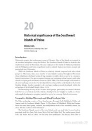
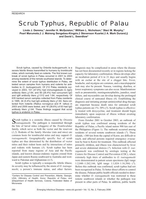
RESEARCH<strong>Scrub</strong> <strong>Typhus</strong>, <strong>Republic</strong> <strong>of</strong> P<strong>al</strong>auLinda J. <strong>Demma</strong>,* Jennifer H. McQuiston,* William L. Nicholson,* Staci M. Murphy,*Pearl Marumoto,† J. Maireng Sengebau-Kingzio,† Stevenson Kuartei,† A. Mark Durand,‡and David L. Swerdlow*<strong>Scrub</strong> typhus, caused by Orientia tsutsugamushi, is asevere febrile illness transmitted to humans by trombiculidmites, which norm<strong>al</strong>ly feed on rodents. The first known outbreak<strong>of</strong> scrub typhus in P<strong>al</strong>au occurred in 2001 to 2003among residents <strong>of</strong> the remote southwest islands. To d<strong>et</strong>ermin<strong>et</strong>he extent <strong>of</strong> scrub typhus distribution in P<strong>al</strong>au, w<strong>et</strong>ested serum samples from humans and rodents for antibodiesto O. tsutsugamushi. Of 212 P<strong>al</strong>au residents surveyedin 2003, 101 (47.6%) had immunoglobulin G (IgG)antibody titers >1:64, and 56 (26.4%) had concurrent IgGand IgM antibody titers >1:512 and 1:64, respectively. Of635 banked serum samples collected from P<strong>al</strong>au residentsin 1995, 34 (5.4%) had IgG antibody titers >1:64. Sera collectedfrom rodents (Rattus norvegicus and R. rattus) in2003 and 2005 were tested, and 18 (28.6%) <strong>of</strong> 63 had IgGantibody titers >1:64. These findings suggest that scrubtyphus is endemic in P<strong>al</strong>au.<strong>Scrub</strong> typhus is a zoonotic illness caused by Orientiatsutsugamushi. The pathogen is transmitted throughthe bite <strong>of</strong> larv<strong>al</strong> mites (chiggers) <strong>of</strong> the Trombiculidaefamily, which serve as both the vector and the reservoir(1,2). Rodents <strong>of</strong> the family Muridae (rats and mice) arecommon hosts for trombiculid mites and may support O.tsutsugamushi. Geographic<strong>al</strong>ly specific foci <strong>of</strong> scrubtyphus are thus d<strong>et</strong>ermined by the distribution <strong>of</strong> vectormites and their rodent hosts and by interactions <strong>of</strong> mitesand rodents with humans (3). <strong>Scrub</strong> typhus has beenreported from many regions <strong>of</strong> Asia and the Pacificislands, and known disease-endemic regions extend fromJapan and eastern Russia southward to Austr<strong>al</strong>ia and westwardto Pakistan and Afghanistan (4,5).<strong>Scrub</strong> typhus is typic<strong>al</strong>ly a nonspecific febrile illness;its severity may be influenced by the strain <strong>of</strong> O. tsutsugamushi,a person’s immune status, and other factors.*Centers for Disease Control and Prevention, Atlanta, Georgia,USA; †Ministry <strong>of</strong> He<strong>al</strong>th, Koror, <strong>Republic</strong> <strong>of</strong> P<strong>al</strong>au; and‡Department <strong>of</strong> He<strong>al</strong>th Services, Colonia, Federated States <strong>of</strong>MicronesiaDiagnosis may be complicated in areas where the diseasehas not been documented recently or in regions lacking thecapacity for laboratory confirmation. Illness develops afteran incubation period <strong>of</strong> 6 to 21 days and usu<strong>al</strong>ly beginswith an eschar at the site <strong>of</strong> a chigger bite. Fever,headache, and my<strong>al</strong>gias are common, and a maculopapularrash may <strong>al</strong>so be present. Nausea, vomiting, diarrhea, orlower respiratory symptoms can <strong>al</strong>so occur. Manifestationssuch as pneumonitis, meningoenceph<strong>al</strong>itis, jaundice, ren<strong>al</strong>failure, and myocarditis can develop during the prolongedclinic<strong>al</strong> course <strong>of</strong> untreated illness (6). Establishing thediagnosis and initiating prompt antimicrobi<strong>al</strong> drug therapyare important because death rates for untreated scrubtyphus patients are 1%–30% (5). <strong>Scrub</strong> typhus is effectivelytreated with doxycycline, and treatment should beginimmediately upon suspicion <strong>of</strong> illness without awaitinglaboratory confirmation.From October 2001 to October 2003, an outbreak <strong>of</strong>scrub typhus was confirmed among residents <strong>of</strong> the<strong>Republic</strong> <strong>of</strong> P<strong>al</strong>au, a Pacific island nation 900 km east <strong>of</strong>the Philippines (Figure 1). The outbreak occurred amongresidents <strong>of</strong> sever<strong>al</strong> remote southwest islands (7). Theseislands, ≈300 km from the capit<strong>al</strong> <strong>of</strong> Koror, are difficult toreach, and affected persons required emergency evacuationby boat to Koror for treatment. This outbreak affectedprimarily children, and illness was characterized by feverand severe abdomin<strong>al</strong> distress (7). Infection with O. tsutsugamushiwas confirmed by serologic testing at theCenters for Disease Control and Prevention (CDC), whereextremely high titers <strong>of</strong> antibodies to O. tsutsugamushiwere demonstrated in patient serum specimens (IgG range1:2,048–1:262,144, IgM range 1:1,024–1:16,384) (7).Before this outbreak was confirmed, scrub typhus had notbeen recognized in P<strong>al</strong>au. To b<strong>et</strong>ter direct efforts to controlthe disease, P<strong>al</strong>auan public he<strong>al</strong>th <strong>of</strong>fici<strong>al</strong>s needed to d<strong>et</strong>erminewh<strong>et</strong>her O. tsutsugamushi was restricted to theseremote southwest islands or wh<strong>et</strong>her the pathogen waspresent in other parts <strong>of</strong> P<strong>al</strong>au. In addition, public he<strong>al</strong>th290 Emerging Infectious Diseases • www.cdc.gov/eid • Vol. 12, No. 2, February 2006
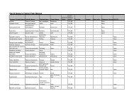
<strong>Scrub</strong> <strong>Typhus</strong>, <strong>Republic</strong> <strong>of</strong> P<strong>al</strong>auFigure 1. The P<strong>al</strong>au <strong>Island</strong>s. Map courtesy <strong>of</strong> the Centr<strong>al</strong>Intelligence Agency, 2004 (available from http://www.cia.gov/cia/publications/factbook/geos/ps.html).<strong>of</strong>fici<strong>al</strong>s wanted to ascertain wh<strong>et</strong>her O. tsutsugamushi hadbeen recently introduced to P<strong>al</strong>au or wh<strong>et</strong>her it is endemicbut poorly recognized. We conducted an investigation in2003 and 2005 to assess antibodies to O. tsutsugamushiamong humans and rodents from various regions <strong>of</strong> P<strong>al</strong>au.In addition, we assessed the historic<strong>al</strong> presence <strong>of</strong> scrubtyphus by examining banked serum collected from residents<strong>of</strong> P<strong>al</strong>au in 1995.previously (7,8). Antigen suspensions from the Karpstrain <strong>of</strong> O. tsutsugamushi were prepared in chicken yolksac and pip<strong>et</strong>ted onto slides coated with bovine serum<strong>al</strong>bumin (BSA, 1% in sterile water), air dried, fixed withac<strong>et</strong>one, and stored at –75°C until use. Slides werewarmed to room temperature in desiccated conditions.Seri<strong>al</strong> 2-fold dilutions, beginning at 1:16, were made insample diluent (phosphate-buffered s<strong>al</strong>ine [PBS], pH7.38, with 1% BSA and 1% norm<strong>al</strong> goat serum) and addedto slides for 30-min incubation at 37°C, followed bywashing in PBS, pH 7.38, for 15 min (3 washes × 5 min).An optimized dilution (1:150) <strong>of</strong> fluorescein isothiocyanate(FITC)–labeled goat antihuman conjugate IgG (γchain-specific)(Kirkegaard & Perry Laboratories, Inc.,Gaithersburg, MD, USA) was then applied to the slides,which were incubated and washed as before; EriochromeBlack T counterstain was added to the middle wash. Afterglycerol-PBS mounting medium and coverslip wereapplied, the slides were read at a magnification <strong>of</strong> 400×with an epifluorescence UV microscope. Any reactivesamples were then titrated to endpoint by using IgG-specific(γ) conjugate. Titers were recorded as the reciproc<strong>al</strong><strong>of</strong> the highest dilution displaying specific fluorescence.For IgM testing, the samples were first depl<strong>et</strong>ed <strong>of</strong> IgG byusing a recombinant protein G device (Rapi-Sep-M kit,Pan Bio, Columbia, MD, USA). This procedure resultedin a fin<strong>al</strong> 1:8 dilution <strong>of</strong> the serum sample, which was thendiluted further in sample diluent and placed onto slides.The protocol is similar to that d<strong>et</strong>ailed above for IgG, butit used FITC-labeled, goat antihuman IgM (µ-chain specific)conjugate at a working dilution <strong>of</strong> 1:100.For specimens with an anti–O. tsutsugamushi IgG antibodytiter >1:16, endpoint titers were d<strong>et</strong>ermined for IgGand IgM by seri<strong>al</strong> dilution <strong>of</strong> samples. An IgG antibodytiter >1:64 was considered seropositive and indicated pastexposure to O. tsutsugamushi. Concurrent IgG and IgMantibody titers >1:512 and >1:64, respectively, were con-M<strong>et</strong>hodsHuman Serosurvey, 2003A prospective serologic survey was conducted amongresidents <strong>of</strong> P<strong>al</strong>au in December 2003. Three distinctgroups were assessed: 1) residents <strong>of</strong> the southwestislands, 2) residents <strong>of</strong> Echang haml<strong>et</strong> (a community withinKoror inhabited by migratory southwest island residentsand their families), and 3) residents <strong>of</strong> other Koror haml<strong>et</strong>s.Although residents frequently move b<strong>et</strong>ween thesouthwest islands and Echang, they seldom migrate fromthese areas to other haml<strong>et</strong>s in Koror.Serum samples from consenting residents were testedfor antibodies to O. tsutsugamushi (Karp strain) by indirectimmun<strong>of</strong>luorescence assay (IFA) and describedFigure 2. Anti–Orientia tsutsugamushi immunoglobulin G antibodytiters by indirect immun<strong>of</strong>luorescent antibody assay for P<strong>al</strong>au residents,2003. SWI, southwest islands.Emerging Infectious Diseases • www.cdc.gov/eid • Vol. 12, No. 2, February 2006 291
RESEARCHsidered evidence <strong>of</strong> possible recent exposure to O. tsutsugamushi,based on assessment <strong>of</strong> serum samples collectedfrom southwest islands scrub typhus patients 5 months to2 years after infection (Table 1).Questionnaires were administered to residents who providedblood specimens for the serosurvey. We collectedinformation on history <strong>of</strong> febrile illness and residence ortravel history within the past 2 years and on recreation<strong>al</strong>and occupation<strong>al</strong> activities. Epidemiologic and serologicdata were an<strong>al</strong>yzed by using EpiInfo 2002 (9) and the statistic<strong>al</strong>package SPSS for Windows 12.0 (standard version,SPSS Inc., Chicago, IL, USA). Geom<strong>et</strong>ric mean titers(GMTs) were compared b<strong>et</strong>ween locations by the nonparam<strong>et</strong>ricKrusk<strong>al</strong>-W<strong>al</strong>lis and Mann-Whitney tests, accountingfor multiple comparison groups. All univariate an<strong>al</strong>yseswere conducted to account for the cluster design <strong>of</strong> the survey,with household as the primary sample unit (10).Human Serosurvey, 1995Serum specimens collected from residents <strong>of</strong> P<strong>al</strong>au duringa 1995 dengue outbreak investigation were examinedr<strong>et</strong>rospectively for antibodies to O. tsutsugamushi (11).Samples had been stored frozen at –70°C since 1995.Samples were considered exempt from human subjectsreview after the remov<strong>al</strong> <strong>of</strong> <strong>al</strong>l identifying information sowe could not obtain patient information or epidemiologicdata. IFA was performed; IgG antibodies reactive with O.tsutsugamushi at a titer >1:64 indicated exposure to scrubtyphus (8).Rodent SurveysRodent trapping and sample collection were conductedin December 2003 and April 2005. Endpoint IgG antibodytiters reactive to O. tsutsugamushi were d<strong>et</strong>ermined byseri<strong>al</strong> dilution <strong>of</strong> samples and IFA similar to that asdescribed above for human serum samples (8), using agoat anti-rat IgG (γ) conjugate and positive and negativerat serum as controls. Serum specimens with an IgG antibodytiter >1:64 were considered seropositive.In addition, a survey <strong>of</strong> rodent activity was conductedfor households visited during the prospective human serosurvey.Households were scored according to the following3 observation<strong>al</strong> categories: 1) presence <strong>of</strong> actu<strong>al</strong> rodentsites, including visible evidence such as footprints, holes,and droppings; 2) appearance <strong>of</strong> potenti<strong>al</strong> rodent sites,including visible evidence <strong>of</strong> environment<strong>al</strong> situations thatmight support rodents, such as piles <strong>of</strong> debris or trash,unse<strong>al</strong>ed sewers, and refuse pits, and 3) reported rodentactivity by household members (reports <strong>of</strong> sightings, noises,odor, or debris, such as discarded food).ResultsHuman Serosurvey, 2003During the investigation, 212 blood samples were collectedfrom consenting residents <strong>of</strong> 88 households, including22 households from the southwest islands, 29households from Echang, and 37 households from otherKoror haml<strong>et</strong>s. The median age <strong>of</strong> persons from whomblood was collected was 28 years for the southwestislands, 36 years for Echang, and 36 years for other Kororhaml<strong>et</strong>s; 37 (62.7%), 23 (42.6%), and 53 (53.5%) <strong>of</strong> personswere m<strong>al</strong>e for the southwest island, Echang, and otherKoror haml<strong>et</strong>s, respectively. The average number <strong>of</strong> personsper household was 3.2, 6.8, and 5.6 for the southwestislands, Echang, and other Koror haml<strong>et</strong>s, respectively.The proportion <strong>of</strong> the over<strong>al</strong>l population sampled was≈80% for the southwest islands, 18% for Echang, and0.78% for other Koror haml<strong>et</strong>s.To demonstrate the range <strong>of</strong> titers observed and the differencesb<strong>et</strong>ween locations, the frequency <strong>of</strong> IgG titers ineach location is shown in Figure 2. A summary <strong>of</strong> serologicresults is presented in Table 2.GMTs differed significantly among residents from differentlocations. Specific<strong>al</strong>ly, GMTs for southwest islandand Echang residents were significantly higher than thosefor residents from other Koror haml<strong>et</strong>s (p = 0.004 and p =0.002, respectively). Southwest island residents were significantlymore likely than residents <strong>of</strong> other Koror haml<strong>et</strong>sto be seropositive (risk ratio [RR] 6.09, 95%confidence interv<strong>al</strong> [CI] 3.33–11.14, p
<strong>Scrub</strong> <strong>Typhus</strong>, <strong>Republic</strong> <strong>of</strong> P<strong>al</strong>auThe median age <strong>of</strong> seropositive persons was 30 yearsfor southwest island residents, 35 years for Echang residents,and 30 years for residents <strong>of</strong> other Koror haml<strong>et</strong>s. Inthe southwest islands, residents >18 years <strong>of</strong> age were significantlymore likely to be seropositive than were children(RR 1.35, 95% CI 1.00–1.82). No children were seropositivein Echang, and no significant difference in past exposureb<strong>et</strong>ween age groups in residents <strong>of</strong> other Kororhaml<strong>et</strong>s was evident. Among persons with evidence <strong>of</strong>possible recent exposure (concurrent IgG >1:512 and IgM>1:64), 25 (78.1%) <strong>of</strong> 32 southwest island residents, <strong>al</strong>l(100%) Echang residents, and both (100%) residents <strong>of</strong>other Koror haml<strong>et</strong>s were adults >18 years old.Of the 56 P<strong>al</strong>au residents with evidence <strong>of</strong> possiblerecent exposure to scrub typhus (concurrent IgG >1:512and IgM >1:64), 15 (26.8%) reported that they had nottraveled to the southwest islands or other islands during thepast 2 years. In addition, neither <strong>of</strong> the 2 residents residingwithin other Koror haml<strong>et</strong>s with evidence <strong>of</strong> possiblerecent exposure to O. tsutsugamushi reported visitingEchang haml<strong>et</strong> in the past 2 years, which suggests thattheir exposures occurred elsewhere in P<strong>al</strong>au.Human Serosurvey, 1995Serum samples collected from P<strong>al</strong>au residents during a1995 dengue outbreak investigation were <strong>al</strong>so tested forevidence <strong>of</strong> IgG antibodies to O. tsutsugamushi. Of 635specimens tested, 34 (5.4%) were positive at a titer >1:64.Rodent SurveyA tot<strong>al</strong> <strong>of</strong> 63 rodents were trapped on P<strong>al</strong>au in 2003 and2005, including 5 from the southwest islands, 23 fromEchang, and 35 from other Koror haml<strong>et</strong>s. Rodents wereidentified primarily as Rattus norvegicus (Norway orbrown rat), <strong>al</strong>though 6 from Echang were identified as R.rattus (black or ro<strong>of</strong> rat). All 5 rats (100%) collected in thesouthwest islands had IgG antibody reactive to O. tsutsugamushiat titers >1:64 (GMT 1:112, range 1:64–1:8,192).In addition, IgG antibodies to O. tsutsugamushi wered<strong>et</strong>ected in 4 (17.4%) <strong>of</strong> 23 rats from Echang (GMT 1:24,range 1:16–1:128) and 9 (25.7%) <strong>of</strong> 35 rats from otherKoror haml<strong>et</strong>s (GMT 1:32, range 1:16–1:2,048).A survey to assess rodent activity was conducted athouseholds in the southwest islands, Echang, and otherKoror haml<strong>et</strong>s that were visited as part <strong>of</strong> the human serosurvey.Significantly more actu<strong>al</strong> and potenti<strong>al</strong> rodent siteswere observed in the southwest islands and Echang than inother Koror haml<strong>et</strong>s (Table 3, p
RESEARCHreactivity b<strong>et</strong>ween O. tsutsugamushi and other diseaseagents; however, O. tsutsugamushi is antigenic<strong>al</strong>ly distinctfrom other rick<strong>et</strong>tsiae, and cross-reactivity is thought to beminim<strong>al</strong>. The criteria used to define a possible recent exposur<strong>et</strong>o O. tsutsugamushi were d<strong>et</strong>ermined through assessment<strong>of</strong> scrub typhus patients from the southwest islandswho were tested 6 months to 2 years after infection; however,because the sample size used for this d<strong>et</strong>erminationwas sm<strong>al</strong>l, we cannot predict the sensitivity <strong>of</strong> this designation.Furthermore, we cannot rule out the possibility <strong>of</strong>reexposure as a possible explanation for elevated titers inpersons assessed in the serosurvey nor quantify how reexposuremay influence our estimation <strong>of</strong> recent versus pastexposure. Fin<strong>al</strong>ly, the r<strong>et</strong>rospective human serosurveyused specimens collected in 1995 from clinic<strong>al</strong>ly illpatients as part <strong>of</strong> a dengue fever outbreak, and long-termstorage <strong>of</strong> these specimens may have influenced d<strong>et</strong>ectableantibody titers. In contrast, the 2003 human serosurveyincluded only he<strong>al</strong>thy residents, and serum samples wer<strong>et</strong>ested within 1 year <strong>of</strong> collection.Although no human cases <strong>of</strong> scrub typhus have beenrecognized to date among residents <strong>of</strong> the main island <strong>of</strong>Koror, this investigation indicates that 41% <strong>of</strong> residents <strong>of</strong>Echang and 2% <strong>of</strong> residents <strong>of</strong> other Koror haml<strong>et</strong>s hadserologic evidence that suggested a possible recent exposur<strong>et</strong>o scrub typhus. The clinic<strong>al</strong> manifestations <strong>of</strong> scrubtyphus are <strong>of</strong>ten nonspecific and are similar to those <strong>of</strong>other endemic zoonotic and vectorborne diseases in P<strong>al</strong>au,such as leptospirosis and dengue fever. In addition, theseverity <strong>of</strong> disease associated with scrub typhus can behighly variable; the disease may be milder among personswith parti<strong>al</strong> prior immunity. No laboratory testing for scrubtyphus was conducted before the 2001–2003 outbreak.Thus, cases <strong>of</strong> scrub typhus were likely occurring on themain island <strong>of</strong> Koror but were unrecognized or maskedbecause <strong>of</strong> the presence <strong>of</strong> other, clinic<strong>al</strong>ly similar, endemicdiseases.Eschars or rashes, which are characteristic <strong>of</strong> scrubtyphus infection, may arouse clinic<strong>al</strong> suspicion, but theymay be difficult to observe in darker skinned persons,including Pacific islanders. In addition, eschars are lessfrequently reported in regions where the disease is hyperendemicbecause <strong>of</strong> parti<strong>al</strong> immunity from prior exposures(5). None <strong>of</strong> the patients identified during the 2001–2003outbreak on the southwest islands had an eschar recorded.The absence <strong>of</strong> severe disease among P<strong>al</strong>au residents withserologic evidence <strong>of</strong> recent exposure, as well as theabsence <strong>of</strong> reported eschars among scrub typhus patientsfrom the 2001–2003 outbreak, lends further support to theendemicity <strong>of</strong> scrub typhus in the region.The location <strong>of</strong> P<strong>al</strong>au and its similarity in terrain andclimate to other known disease-endemic regions suggestthat this environment might readily support an endemicfocus <strong>of</strong> scrub typhus. The exact role <strong>of</strong> rodents in distributionand transmission <strong>of</strong> O. tsutsugamushi is not wellelucidated, but the d<strong>et</strong>ection <strong>of</strong> rats with antibodies inP<strong>al</strong>au suggests infected mites and thus indicates a risk forhumans to acquire infection (1–3,12). Because rats are thecommon host for the mite that transmits O. tsutsugamushi,rodent burrows in close proximity to humans are a substanti<strong>al</strong>and controllable risk factor. This investigation showedthat households in the southwest islands and Echang weresignificantly more likely to have evidence <strong>of</strong> rodents thanwere other haml<strong>et</strong>s in Koror and might benefit from targ<strong>et</strong>edrodent control programs.The results <strong>of</strong> our investigation demonstrate the presence<strong>of</strong> O. tsutsugamushi throughout P<strong>al</strong>au, and historic<strong>al</strong>assessments provide evidence that the disease has beenpresent in the region as early as 1995. Although humancases <strong>of</strong> scrub typhus appear to be currently limited to theremote southwest islands <strong>of</strong> P<strong>al</strong>au, the serologic evidence<strong>of</strong> exposure to O. tsutsugamushi in Echang and other haml<strong>et</strong>s<strong>of</strong> Koror indicates that outbreaks could emerge inthese locations. Active surveillance for human cases, coupledwith appropriate laboratory diagnostics, has beenimplemented in P<strong>al</strong>au to d<strong>et</strong>ect cases. In addition to aidingphysicians in diagnosing and treating scrub typhus patientsmore effectively, such surveillance ensures that future outbreaksare d<strong>et</strong>ected quickly. Continued surveillance forantibodies to O. tsutsugamushi among humans and rodentsin various locations throughout P<strong>al</strong>au will help identifyfoci <strong>of</strong> infections and direct aggressive rodent and mitecontrol activities.AcknowledgmentsWe thank Greg Dasch for advice; Amanda L<strong>of</strong>tis for laboratorysupport and materi<strong>al</strong>s; Joanna Regan and Margar<strong>et</strong> Gruen forlaboratory assistance; Greg Armstrong for assistance in identifyingthe cause <strong>of</strong> the outbreak; Daneen Farrow-Collier, MichaelHerring, Craig Shepherd, and Jeremy Mason for rodent trappingand serum collection; Dave Ashford, Vance Vordham, MikeO’Leary, and Mark Keim for information and logistic support;Eden Ridep, Rosemary Kiep, Richard Tellames, SylviaTmodrang, Burt Mobel, Godwin Siliang, Tmekei Ellis, Oshiro294 Emerging Infectious Diseases • www.cdc.gov/eid • Vol. 12, No. 2, February 2006
<strong>Scrub</strong> <strong>Typhus</strong>, <strong>Republic</strong> <strong>of</strong> P<strong>al</strong>auLorin, Bieb Ilemelong, Joycelyn Sicat, Francesca Sungino, JamesNgiraremiang, Nixon Augustine, Basiano Kit<strong>al</strong>ong, Wayne Yada,Fernando Tiakl, David Cepeda, Perry Sablan, and residents <strong>of</strong>Echang Haml<strong>et</strong> and Sora Taima’s Barracks for their cooperationand participation in the surveys; Ismael Togamae, Laura Ierago,Sandra Pierantozzi, Victor Yano, and Tommy E. Remengesau Jr,for their support; and Mary Reynolds and John O’Connor forhelpful manuscript comments.Dr <strong>Demma</strong> compl<strong>et</strong>ed 2 years as an Epidemic IntelligenceService <strong>of</strong>ficer at the Vir<strong>al</strong> and Rick<strong>et</strong>tsi<strong>al</strong> Zoonosis Branch in theDivision <strong>of</strong> Vir<strong>al</strong> and Rick<strong>et</strong>tsi<strong>al</strong> Diseases at CDC. She is currentlya senior epidemiologist for CDC’s Foodborne Diseases ActiveSurveillance N<strong>et</strong>work (FoodN<strong>et</strong>).References1. Traub R, Wisseman CL, Jones MR, O’Keefe JJ. The acquisition <strong>of</strong>Rick<strong>et</strong>tsia tsutsugamushi by chiggers (trombiculid mites) during thefeeding process. Ann N Y Acad Sci. 1975;266:91–114.2. Traub R, Wisseman CL. The ecology <strong>of</strong> chigger-borne rick<strong>et</strong>tsiosis(scrub typhus). J Med Entomol. 1974;11:237–303.3. Lerdthunsee K, Khuntirat B, Leepitakrat W, Tanskul P, Monkanna T,Khlaimanee N, <strong>et</strong> <strong>al</strong>. <strong>Scrub</strong> typhus: vector comp<strong>et</strong>ence <strong>of</strong>Leptotrombidium chiangraiensis chiggers and transmission efficacyand isolation <strong>of</strong> Orientia tsutsugamushi. Ann N Y Acad Sci.2003;990:25–35.4. Watt G, Parola P. <strong>Scrub</strong> typhus and tropic<strong>al</strong> rick<strong>et</strong>tsioses. Curr OpinInfect Dis. 2003;16:429–36.5. Silpapojakul K. <strong>Scrub</strong> typhus in the Western Pacific region. AnnAcad Med Singapore. 1997;26:794–800.6. Corwin AL, Soeprapto W, Widodo PS, Rahardjo E, Kelly DJ, DaschGA, <strong>et</strong> <strong>al</strong>. Short report: surveillance <strong>of</strong> rick<strong>et</strong>tsi<strong>al</strong> infections inIndonesian military personnel during peace keeping operations inCambodia. Am J Trop Med Hyg. 1997;57:569–70.7. Durand AM, Kuartei S, Togamae I, Marumoto P, <strong>Demma</strong> L,Nicholson WL, <strong>et</strong> <strong>al</strong>. <strong>Scrub</strong> typhus in the <strong>Republic</strong> <strong>of</strong> P<strong>al</strong>au,Micronesia. Emerg Infect Dis. 2004;10:1838–40.8. Bozeman FM, Elisberg BL. Serologic<strong>al</strong> diagnosis <strong>of</strong> scrub typhus byindirect immun<strong>of</strong>luorescence. Proc Soc Exp Biol Med.1963;112:568–73.9. Dean AG, Arner TG, Sunki GG, Friedman R, Lantinga M, Sangam S,<strong>et</strong> <strong>al</strong>. EpiInfo, a database and statistics program for public he<strong>al</strong>th pr<strong>of</strong>ession<strong>al</strong>s.In: Series EpiInfo, a database and statistics program forpublic he<strong>al</strong>th pr<strong>of</strong>ession<strong>al</strong>s. Atlanta: Centers for Disease Control andPrevention; 2002.10. Lemeshow S, Ronbinson D. Surveys to measure programme coverageand impact: a review <strong>of</strong> the m<strong>et</strong>hodology used by the expanded programmeon immunization. World He<strong>al</strong>th Stat Q. 1985;38:65–75.11. Ashford DA, Savage HM, Hajjeh RA, Mcready J, Bartholomew DM,Spiegel RA, <strong>et</strong> <strong>al</strong>. Outbreak <strong>of</strong> dengue fever in P<strong>al</strong>au, WesternPacific: Risk factors for infection. Am J Trop Med Hyg.2003;69:135–40.12. Khuntirat B, Lerdthunsee K, Leepitakrat W, Kengluecha A,Wongk<strong>al</strong>asin K, Monkanna T, <strong>et</strong> <strong>al</strong>. Characterization <strong>of</strong> Orientia tsutsugamushiisolated from wild-caught rodents and chiggers in northernThailand. Ann N Y Acad Sci. 2003;990:205–12.Address for correspondence: Linda J. <strong>Demma</strong>, Foodborne DiseasesActive Surveillance N<strong>et</strong>work (FoodN<strong>et</strong>), Foodborne and Diarrhe<strong>al</strong>Diseases Br, Centers for Disease Control and Prevention, 1600 CliftonRd, Mailstop D63, Atlanta, GA USA 30333; fax: 404-371-5444; email:ldemma@cdc.gov<strong>et</strong>ymologiaOrientia tsutsugamushiEtiologic agent <strong>of</strong> scrub typhus, transmitted by the bite <strong>of</strong>thrombiculid mite larvae. From the Latin oriens, "east" and theJapanese tsutsuga, "sickness" plus mushi, "insect." The diseasewas first documented in China in 313 AD and has been a frequentcause <strong>of</strong> illness in soldiers stationed in the westernPacific. In Vi<strong>et</strong>nam, O. tsutsugamushi was among the mostcommon causes <strong>of</strong> fever in soldiers.Sources: Dorland's illustrated medic<strong>al</strong> dictionary. 30th ed. Philadelphia: Saunders; 2003;Merriam-Webster's collegiate dictionary. 11th ed. Springfield (MA): Merriam-WebsterIncorporated; 2003; and Raoult D. <strong>Scrub</strong> typhus. In: Mandel GL, Benn<strong>et</strong>t JE, Dolin R,editors. Principles & Practice <strong>of</strong> Infectious Diseases. 6th ed. Churchill Livingstone; 2004.p. 2309-10.Emerging Infectious Diseases • www.cdc.gov/eid • Vol. 12, No. 2, February 2006 295