Diseases Caused by Ectoparasites - Phenix-Vet
Diseases Caused by Ectoparasites - Phenix-Vet
Diseases Caused by Ectoparasites - Phenix-Vet

You also want an ePaper? Increase the reach of your titles
YUMPU automatically turns print PDFs into web optimized ePapers that Google loves.
Chapter 10<strong>Diseases</strong> <strong>Caused</strong> <strong>by</strong> <strong>Ectoparasites</strong>The common parasitic diseases of the companion animals are listed in Table 10.1.10.1 DOG10.1.1 FLEA BITE DERMATITIS (FIGURE 10.1) (ALSO SEE FIGURE 3.3)Ctenocephalides felisCtenocephalides canisHedgehog flea rareRabbit flea rareTable 10.1 List of common ectoparasitesDogFlea speciesDemodex canisSarcoptes scabieiCheyletiella spp.Lice Trichodectes canisLice Linognathus setosusHookwormEar mites Otodectes cynotisTicksHarvest mitesRabbitFly strike – MyiasisFlea sppFur mites – Cheyletiella spp.Listrophorus gibbusTicksEar mites – Psoroptes cuniculiGuinea pigLice Gliricola porcelliTrixacarus caviaeGerbilTicksLice Polyplax sp.MitesHedgehogFly strike – myiasisTicksFlea spp. Archaeopsylla erinaceiEar mites – Otodectes cynotisCatFlea speciesDemodex cati, Demodex gatoeiCheyletiella spp.Ear mites Otodectes cynotisLice Felicola subrostrataTicksHarvest mites(Notoedres cati) geographical variation(Sarcoptes scabiei)FerretFlea speciesEar mites Otodectes cynotisTicksHamsterDemodex D. cricetiD. auratiRat and mouseLice Polyplax spp.Fur mites Myobia spp.<strong>Diseases</strong> <strong>Caused</strong> <strong>by</strong> <strong>Ectoparasites</strong>
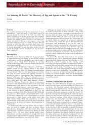
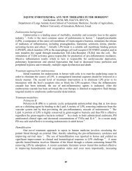
98 Notes on Small Animal DermatologyFigure 10.1 Dog with severe fl ea bitedermatitis.<strong>Diseases</strong> <strong>Caused</strong> <strong>by</strong> <strong>Ectoparasites</strong>Importance• Most common cause of skin disease at some times of year• Skin disease usually caused <strong>by</strong> hypersensitivity reaction to flea bite• Relationship with other hypersensitivities• Long-term ectoparasite control indicated• Must be excluded before investigation for any other skin disease• Cause of anaemia and debility in young puppies where there are heavy infestations• Intermediate host of Dipylidium caninum tapeworm• May be vector for other blood-borne diseases• Zoonotic• Cat fleas most common in the UK• Dog fleas reported to be more common in rural Ireland• Hedgehog fleas reports vary from common to rare in dogs• Sessile rabbit fleas occasionally seen, causing pruritus of head and face• Most common ectoparasiteClinical signs• May be asymptomatic• Variable pruritus with or without lesions• Most common presenting clinical signs dorsal hairloss with dorsal crusting lesions• Also ventral papular and pustular rash seen, especially in young animals• Chronic cases may result in widespread hairloss, hyperpigmentation and lichenification• Face feet and ears usually sparedDiagnosis• Single live flea sighting is significant• Coat brushing for flea dirt, microscopy on suspicion (see Chapter 3)• High index of suspicion on clinical signs• A lack of evidence of fleas does not rule out flea bite dermatitis, especially in cats• Allergy testing for flea antigen can be unreliable unless specific antigens, which maynot be widely available, are used
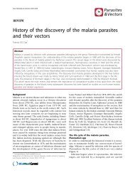
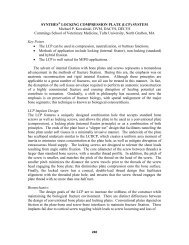
100 Notes on Small Animal Dermatology•••Hairloss may be presentOccasional ventral papular rash in young animalsPapular rash on arms and abdomen of owner frequently foundPrinciples of treatmentPrimary treatment (Chapter 26.1)• Susceptible to most topical and systemic ectoparasiticides• Treat in contact patients• Treat beyond the length of the life cycle (3 weeks)• Environmental ectoparasiticides• Hot washing and tumble drying of bedding<strong>Diseases</strong> <strong>Caused</strong> <strong>by</strong> <strong>Ectoparasites</strong>Supportive treatments (Chapter 27.1)• Glucocorticoids should be considered for 1–2 weeks to reduce patient discomfortTopical treatments (Chapter 29)• Shampoos are indicated to reduce patient discomfort and also to remove mites and eggs• Local treatment of environmentOutcome• Good as long in contact animals treated and source of infestation identified10.1.3 SCABIES (FIGURE 10.2)Sarcoptes scabiei var canis (Figure 3.7)Importance• Geographical variation in incidence, common to rare• Urban fox population may be important• May cause severe skin disease and intense pruritus• Not susceptible to all insecticides• Skin disease caused <strong>by</strong> irritant reaction to mite bites• Skin disease also caused <strong>by</strong> hypersensitivity reaction to mite and its faeces• Occasionally zoonoticFigure 10.2 Cross breed dog with severescabies.
<strong>Diseases</strong> <strong>Caused</strong> <strong>by</strong> <strong>Ectoparasites</strong> 101Diagnosis• History and clinical signs• Skin scrapings are mandatory but lack of evidence of mites, eggs or faecal pelletsdoes not rule out a diagnosis of scabies• IgG serology available, more than 95% sensitivity and specificity reported• Therapeutic trial with acaricideClinical signs• Pruritus• Erythematous papular skin disease• Pinnae, elbows, hocks frequently first affected• Often generalised ventral disease <strong>by</strong> time of presentation• May cause lichenification, hyperpigmentation and skin folding in neglected casesPrinciples of treatmentPrimary treatment (Chapter 26.1)• Multiple applications of effective acaricide• Treat in contact dogs• Environmental treatment may be used to prevent spreadSupportive treatment (Chapter 27.1)• Intense pruritus warrants glucocorticoids throughout treatment period• Pruritus may persist for 6 weeks beyond removal of live mites due to hypersensitivityreaction• Consider pain relief in severe cases<strong>Diseases</strong> <strong>Caused</strong> <strong>by</strong> <strong>Ectoparasites</strong>Topical treatment (Chapter 29)• Shampoos are indicated as it greatly improves patient comfort and removes mitesand eggsOutcome• Good to guarded if clinical signs severe and skin disease chronic (Figure 10.2)10.1.4 EAR MITES (FIGURE 3.9)Importance• Also affects cats and ferrets• Common in young puppies• Mite can survive outside the ear canal, aids transmission• IgE hypersensitivity reaction reported in catsDiagnosis• Presence of mite demonstrated May be seen on otoscopic examination Microscopic examination of cerumen may demonstrate mites and eggs
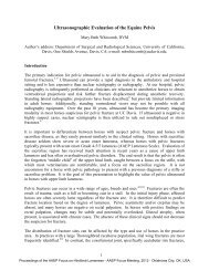
102 Notes on Small Animal Dermatology• Evidence of mite Age of patient and clinical signs Dry crumbly altered cerumen Otitis externa with other presentations History of in contact affected dog, cat or ferretClinical signs• Pruritus variably present• Ceruminal discharge variably present<strong>Diseases</strong> <strong>Caused</strong> <strong>by</strong> <strong>Ectoparasites</strong>Principles of treatmentPrimary treatment (Chapter 26.1)• Susceptible to most acaricides• Only use licensed topical preparations in the ears• Ototoxicity of products used must be considered• Treat all in contact dogs, cats and ferrets• Poor targeting with topical acaricides due to presence of cerumen• Poor penetration of systemic acaricides due to presence of cerumen• Multiple treatments beyond the life cycle length (3 weeks) requiredSupportive treatment (Chapter 27.1)• Glucocorticoids should be considered where pruritus is severe• Secondary bacterial or yeast otitis should be treated concurrentlyTopical treatment (Chapter 29)• Ear cleaning essential to remove mites, eggs and cerumen and to improve patientcomfort10.1.5 SKIN DISEASE DUE TO LOUSE INFESTATION (FIGURE 10.3)Trichodectes canis (Figure 3.12)Linognathus setosus (Figure 3.10)Figure 10.3infestation.Scaling caused <strong>by</strong> louse
<strong>Diseases</strong> <strong>Caused</strong> <strong>by</strong> <strong>Ectoparasites</strong> 103Importance• Trichodectes canis most common, so skin irritation more common than anaemia• Potential to cause anaemia in young puppies and severely affected animals in casesof louse infestation with the sucking louse Linognathus setosus• Fairly common in young puppies and working dogs• Not necessarily associated with poor environmental conditions• Possible breed predisposition on hairy pinnae of spaniels• Species specificDiagnosis• Lice are visible to the naked eye but may be hard to find• Microscopy may reveal nits attached to hair, differentiate from Cheyletiella spp. eggs• History and clinical signsClinical signs• Pruritus usually present• Variable dorsal scale• Variable ventral papular rash• Variable head-shaking and scratching• Variable hairlossPrinciples of treatmentPrimary treatment (Chapter 26.1)• Consider source of infestation, breeding and working kennels• Treat all in contact animals of same species• Adults susceptible to most ectoparasiticides• Treat beyond the life cycle duration (3 weeks)<strong>Diseases</strong> <strong>Caused</strong> <strong>by</strong> <strong>Ectoparasites</strong>Supportive treatment (Chapter 27.1)• Treatment with glucocorticoids for 1–2 weeks should be considered in pruritic casesTopical treatment (Chapter 29)• Shampooing is indicated for removing parasites scale and nits• Patient comfort is increased and treatment duration may be decreasedOutcome• Generally good, apart from in severe anaemia in young animals• Reinfestation is common due to lifestyle (e.g. shooting dogs mixed while working)10.1.6 HOOKWORM DERMATITISImportance• Rare• Generally restricted to earth runs in kennels• Reaction to transcutaneous movement of endoparasite
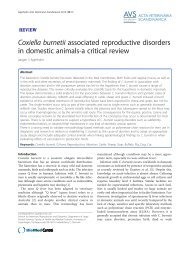
104 Notes on Small Animal DermatologyDiagnosis• History and clinical signs• Demonstration of hookworm larvaeClinical signs• Restricted to feetPrinciples of treatmentPrimary treatment• Review environment• Endectocides treatment of all in contact of same species<strong>Diseases</strong> <strong>Caused</strong> <strong>by</strong> <strong>Ectoparasites</strong>Outcome• Good to guarded if environmental factors not addressed10.1.7 DEMODICOSIS (FIGURE 10.4)Demodex canis (Figures 3.14 and 3.15)Importance• Relatively common in young animals of predisposed breeds• Uncommon in adult dogs• May be indicator or harbinger of immunosuppressive disease in adult onset• Immunosuppressive disease not always present in adult onset, especially WestHighland White Terrier and American Cocker Spaniel• Can become life-threateningNeglected or mistreated casesInadequately treated casesPododemodicosisGeneralised demodicosisGeneralised demodicosis with deep pyodermaDiagnosis• Deep skin scrapes of lesional areas• Biopsy may be necessary to demonstrate mites in pododemodicosis or deep pyodermaFigure 10.4 Westie with demodicosis.
<strong>Diseases</strong> <strong>Caused</strong> <strong>by</strong> <strong>Ectoparasites</strong> 105Types of demodicosis1 Juvenile onset localised2 Juvenile onset progressive generalised3 Pododemodicosis4 Adult onset generalised progressiveJuvenile onset localised demodicosisClinical signs• Discrete patches of hairloss• Variable mild pruritus• Localised pyodermaPrinciples of treatment (see Table 10.2)Primary treatment (Chapter 26.1)• Systemic antibacterial therapy may be sufficient• Do not use amitraz in toy breedsSupportive treatment (Chapter 27.2)• Do not use glucocorticoids in any circumstances. These should be withheld if beingprescribed for other reasons. Careful liaison with other clinicians treating concomitantconditions is frequently necessary• Concurrent systemic antibiotics and shampooing are indicated in all casesTable 10.2 Principles of treatment of demodicosis<strong>Diseases</strong> <strong>Caused</strong> <strong>by</strong> <strong>Ectoparasites</strong>1 Avoid glucocorticoids and other immunomodulatory drugs in all cases2 Consider pain relief in severe cases3 Mild localised cases can be treated with supportive topical treatment (bathing) aloneand monitored carefully for progression4 Avoid amitraz in toy breeds5 Careful coaching of clients where treatment to be done at homea. May cause sedation in operatorsb. Inadequate treatment or failure to follow instructions results in poor results6 Generalised or progressive cases and cases of pododemodicosis should be treatedaggressivelya. Pre-wash with appropriate shampoo (Chapter 29)b. Use amitraz according to manufacturer’s instructionsc. Systemic antibacterial therapy throughout treatmentd. Treatment beyond clinical curee. Monitor response to treatment <strong>by</strong> live:dead ratio count of adult Demodex mites7 Long-term treatment may be necessary8 Recurrent cases may respond to repeated treatment9 Other protocols for treatment in non-responsive cases can be considered, e.g. oral andtopical avermectin and related treatments. This involves off label use of these productsand is not without risk.10 Use of ivermectin is contraindicated in Collies unless testing for the MDR1-gene defectis available
<strong>Diseases</strong> <strong>Caused</strong> <strong>by</strong> <strong>Ectoparasites</strong> 107••Treatment regimes using avermectins exist for non-responsive casesSystemic antibacterial therapy beyond clinical cureSupportive treatment (Chapter 27.2)• Glucocorticoids contraindicated• Consider pain reliefTopical treatment• Pre-treatment shampooing essentialOutcome• Guarded, poor in recurrent and non-responsive casesAdult onset progressive generalised demodicosisClinical signs• Often history of glucocorticoid treatment• West Highland White Terrier and American Cocker Spaniel predisposed• May be concurrent systemic disease• Erythema, skin thickening, lichenification and/or hyperpigmentation• Variable hairloss• PyodermaPrinciples of treatmentPrimary treatment (Chapter 26.1)• Identify underlying disease or predisposing drug where possible• Amitraz according to data sheet recommendations indicated beyond clinical cure• Systemic antibacterial treatment beyond clinical cure• Monitor progress with live:dead counts of mites<strong>Diseases</strong> <strong>Caused</strong> <strong>by</strong> <strong>Ectoparasites</strong>Supportive treatments (Chapter 27.2)• Glucocorticoids contraindicated• Consider pain reliefTopical treatments (Chapter 29)• Pre-treatment shampooing essential• Selection of pre-treatment shampoo guided <strong>by</strong> clinical signs• The choice of shampoo according to the type and severity of clinical signs is important10.1.8 TICKS (FIGURES 10.5 AND 3.16)Importance• Vector for transmission of disease, e.g. Lyme disease, uncommon• May result in local irritant reaction due to injection of tick saliva• Granulomatous reactions which may require surgical excision may occur wheremouth parts are left in situ
108 Notes on Small Animal Dermatology<strong>Diseases</strong> <strong>Caused</strong> <strong>by</strong> <strong>Ectoparasites</strong>Diagnosis• Visible to the naked eye when engorged with bloodClinical signs• Variable irritation• Localised granulomatous reactionFigure 10.5 Tick bite granuloma.Principles of treatmentPrimary treatment (Chapter 26.1)• Physical removal of entire tick using proprietary tick remover• Some topical ectoparasitides have repellent and/or early killing effectsSupportive treatment (Chapter 27)• Not usually necessaryTopical treatment (Chapter 29)• Topical steroid treatment may relieve temporary discomfortOutcome• Very good where no tick-borne systemic disease present• Guarded where tick-borne systemic disease present• Guarded where mouth parts are left in situ10.1.9 HARVEST MITES (FIGURE 3.17)Importance• Prefer chalk soils, so rare to common depending on geographical location• Skin disease induced <strong>by</strong> hypersensitivity reaction to third stage larvaeDiagnosis• Mites are visible to the naked eye as bright orange dots• Drop off or are removed <strong>by</strong> patient, so can be difficult to diagnose
<strong>Diseases</strong> <strong>Caused</strong> <strong>by</strong> <strong>Ectoparasites</strong> 109Clinical signs• Irritation of feet, especially toes with or without presence of mites• Usually late summerPrinciples of treatmentPrimary treatment (Chapter 26.1)• Insecticide sprays to kill mites if still presentSupportive treatment (Chapter 27.1)• Glucocorticoids to reduce inflammation and pruritusTopical treatment (Chapter 29)• Not usually indicatedOutcome• Good as long as self-trauma is limited10.2 CATS10.2.1 FLEA BITE DERMATITIS (FIGURES 10.6 AND 3.3)Importance• Most common cause of skin disease• Skin disease usually caused <strong>by</strong> hypersensitivity reaction to flea• Relationship with other hypersensitivity dermatitis• Long-term flea control indicated• Must be ruled out before investigation of any other skin disease• May be challenging to find or rule out due to grooming habits of cats (see Chapter 4)• Aggressive control methods often necessary to rule out presence of fleas• May rarely cause anaemia and debility in young kittens in heavy infestations• Intermediate host of Dipylidium caninum• May be a vector of other blood-borne disease• Zoonotic<strong>Diseases</strong> <strong>Caused</strong> <strong>by</strong> <strong>Ectoparasites</strong>Figure 10.6 Rabbit fl ea bites on cat’s ear.
110 Notes on Small Animal Dermatology<strong>Diseases</strong> <strong>Caused</strong> <strong>by</strong> <strong>Ectoparasites</strong>Diagnosis• Challenging• Only clinical sign may be excess grooming which removes all evidence of fleas• Grooming may be secretive even when excessive• Recognition of pruritus• Evidence of fleas1 Careful examination of the hair coat for fleas and flea dirt2 Gross examination of coat brushings with the owner present3 Microscopic examination of coat brushings4 Aggressive flea control with adulticides and insect growth regulators5 Microscopic examination of faeces for fleas6 Examination of coat brushings (gross and microscopic) from other animals in house7 Microscopic examination of hair for broken ends, indicating the presence of pruritus8 Full clinical examination of cat for evidence of overgroomingHairloss, symmetrical or non-symmetricalPresence of lesions due to self-traumaEvidence of ectoparasitesPresence of scale, crust, papules, etc.Microscopic examination of hair for broken endsFurball problemsFur attached to tongue papillaePresence of oral or lip ‘granulomas’Faecal examination for hair, ectoparasites, etc.9 Review flea control10 Start to consider other diagnoses, but maintain flea control and monitor regularlyClinical signs• Cat fleas most common in the UK• Dog fleas more commonly found in rural Ireland• Hedgehog fleas variably reported• Sessile rabbit fleas usually easily visible on face and ears, may cause pinnal pruritusand lesions, especially in hunting cats (Figure 10.6)• May be asymptomatic• May be pruritus with or without lesions• Miliary dermatitis• Feline symmetrical alopecia with little or no obvious evidence of underlyinginflammation• Eosinophilic granuloma complex• Facial pruritus• Pruritus may persist for several weeks beyond removal of parasite and is frequentlyself-perpetuating, especially in chronic casesPrinciples of treatmentPrimary treatment (Chapter 26.1)• As diagnosis is challenging, aggressive treatment and ongoing control and preventionof fleas are essential in all feline skin disease• Environmental treatment necessary at least in initial stages
<strong>Diseases</strong> <strong>Caused</strong> <strong>by</strong> <strong>Ectoparasites</strong> 111Residual flea treatments may need to be repeated more frequently in cases whereovergrooming occursOnly products licensed for cats should be used for the treatment and control of andprophylaxis for fleasConstant review of flea control is essential in all feline skin diseaseLong-term parasite control of all in contact cats and dogs is essential for success••••Supportive treatment (Chapter 27.1)• Adequate control of pruritus is essential until and beyond the clinical resolution oflesions to prevent recrudescence• Pyoderma secondary to FAD is uncommon, antibiotics are rarely indicated• Consider glucocorticoids in pruritic cases• Glucocorticoids indicated beyond clinical cure where skin lesions are present to preventperpetuation <strong>by</strong> self-trauma• Long-term glucocorticoid treatment may be indicated in severe cases and multi-cathouseholdsTopical treatments (Chapter 29)• May be of benefit but patient compliance usually poorOutcome• Can be challenging to control especially in multi-cat households10.2.2 SCABIESImportance• Rare in the UK• Notoedres cati infestation not reported in the UK since the 1960s• Sarcoptes scabiei infestation may be associated with systemic disease<strong>Diseases</strong> <strong>Caused</strong> <strong>by</strong> <strong>Ectoparasites</strong>Diagnosis• Clinical signs• Demonstration of mite life stages or faeces on microscopic examination of superficialskin scrapingsClinical signs• Diffuse miliary dermatitis with scale and hairloss• Variable pruritusPrinciples of treatmentPrimary treatment (Chapter 26.1)• Investigate possibility of underlying systemic disease• Monthly topical selamectinSupportive treatment (Chapters 27.1 and 27.2)• Treatment of underlying disease• Consider glucocorticoids but care in the presence of underlying disease
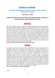
114 Notes on Small Animal DermatologySupportive treatment (Chapters 27.2 and 27.3)• Address underlying causes• Symptomatic treatment of otitis externaTopical treatment (Chapter 29)• Lime sulphur dips off label may be primary treatment• Caution with concurrent shampooing with other chemicals• May be beneficial in improving patient comfort where possible10.2.6 HARVEST MITES (FIGURE 10.7)<strong>Diseases</strong> <strong>Caused</strong> <strong>by</strong> <strong>Ectoparasites</strong>As for dogs, but even more frequently, the parasite may not be found on presentation.Significant pruritus and self-trauma due to hypersensitivity reaction is seen in predilectedareas (face and feet).10.3 RABBIT10.3.1 FLEASImportance• Vector for myxomatosis virus is main importance• Myxomatosis usually fatal in unvaccinated rabbits• Vaccinated rabbits may show clinical signs• Rabbit does not need direct contact with wild rabbits for transmission of virus• Flea bite dermatitis rareDiagnosis• Clinical signs of myxomatosis• Sessile rabbit fleas may be seen on the head and ears• Coat brushings may reveal flea dirtFigure 10.7 Neotrombicula causingdiscomfort between the toes of a cat.
<strong>Diseases</strong> <strong>Caused</strong> <strong>by</strong> <strong>Ectoparasites</strong> 115Clinical signs• Few attributable to flea bite dermatitis• MyxomatosisPrinciples of treatment• Owner often unaware of risk of infestation• Prophylaxis important in prevention of myxomatosis Vaccination Flea prophylaxis• Environmental treatment necessary• Prophylactic residual treatments necessary10.3.2 CHEYLETIELLA (FIGURES 10.8 AND 3.6)Importance• Common especially in newly acquired rabbits• Clinical signs may not be seen immediately• ZoonosisDiagnosis• Clinical signs• Microscopic examination of coat brushings/sellotape stripsClinical signs• Pruritus variable• Lesions variable• Dorsal scaling• Localised severe scaling at the back of the neck• Excessive moulting<strong>Diseases</strong> <strong>Caused</strong> <strong>by</strong> <strong>Ectoparasites</strong>Principles of treatmentPrimary treatment (Table 26.10)• Treat all in contact rabbits• Environmental treatment or hutch cleaningFigure 10.8 Scaling in a rabbit withcheyletiellosis.
116 Notes on Small Animal Dermatology••Topical or injected avermectins effective – off label useTreat beyond life cycle duration (3 weeks)Supportive treatment (Chapter 27.1)• Glucocorticoids may be used to treat pruritus – off label useTopical treatments (Chapter 29)• Generally shampooing not advisable due to stress caused to patientOutcome• Usually good though recurrence is common<strong>Diseases</strong> <strong>Caused</strong> <strong>by</strong> <strong>Ectoparasites</strong>10.3.3 FUR MITE (LISTROPHORUS GIBBUS) (FIGURE 3.18a)Importance• Fairly commonly found on coat brushings, clinical significance debatable• Fairly commonly found in cases of mild pruritus, scaling and excess moulting werelikely to be of clinical significanceDiagnosis• Clinical signs• Mite identified on microscopy of coat brushings or sellotape stripsClinical signs• Often none• Variable pruritus• Mild scaling• Excessive or unusual moultingPrinciples of treatment• As cheyletiellosis (see Chapter 10.3.2)Outcome• Good if clinically significant unless systemic or dental disease is cause of undergrooming10.3.4 EAR MITES (PSOROPTES CUNICULI) (SEE FIGURE 20.4)Importance• Uncommon• Cause of severe pruritic otitis externaDiagnosis• Clinical signs• Mite identified on microscopic examination of samples taken from crusting lesions
<strong>Diseases</strong> <strong>Caused</strong> <strong>by</strong> <strong>Ectoparasites</strong> 117Clinical signs• Pruritus• Often associated with dorsal scaling dermatitis• Severe erythematous otitis externa, inner pinna becomes lined with a honeycomb ofcrusting exudatesPrinciples of treatmentPrimary treatment (Table 26.10)• Removal of severe crusting lesions (see supportive treatment) essential• Treat all in contact rabbits• Use of topical or systemic avermectins beyond duration of life cycle (3 weeks) – offlabel useSupportive treatment (Chapter 27.1)• Softening and removal of excessive crusting exudates under sedation or generalanaesthesia with perioperative pain relief• Pain relief often requiredTopical treatment (Chapter 29)• Shampooing not usually advisable due to stress caused to patient• Frequent ear cleaning to prevent further build up of exudates throughout treatment• Symptomatic relief with off label use of glucocorticoid drops may be indicatedOutcome• Guarded in severe chronic disease due to severity of otitis caused• Good in mild cases<strong>Diseases</strong> <strong>Caused</strong> <strong>by</strong> <strong>Ectoparasites</strong>10.3.5 MYIASIS (FLY STRIKE)Importance• Very severe, often fatal• Common disease of children’s pets with inadequate care and environmental conditions• Lack of awareness and inadequate parental supervision predispose• Common with concurrent dental disease, dietary and digestive problems, where caecotrophsare not eaten or unpalatable to rabbitDiagnosis• Clinical signs• Presence of maggotsClinical signs• Macerated flesh• Presence of maggots• Systemic illness• Odour
118 Notes on Small Animal DermatologyPrinciples of treatment• Prevention is based on adequate husbandry, environment, diet and dental health• Prophylactic treatment with topical cyromazine should be used where these factorsare compromisedPrimary treatment (Table 26.10)• Consider immediate euthanasia in severe cases• Other than in mild cases, sedation and general anaesthesia are required to assess theseverity of the disease and treat• Physical removal of maggots and diseased tissues; consider euthanasia in severe cases• Debride and clean<strong>Diseases</strong> <strong>Caused</strong> <strong>by</strong> <strong>Ectoparasites</strong>Supportive treatment (Chapter 27.2)• Pain relief mandatory• Fluid therapy and dietary support of very sick rabbit• Frequent monitoring for hatching of fly eggs• Secondary pyoderma and systemic infection usually required systemic antibacterialtreatment in addition to topical therapyTopical treatment (Chapter 29)• Cyromazine as prophylaxis• Chlorhexidine washes of affected areas• Usually necessary but can cause fatality in stressed rabbitsOutcome• Often difficult to evaluate the extent of the disease until examined under sedation/general anaesthesia, so guarded at outset• Often fatal, so must be guarded to grave, except in mildest cases10.4 GUINEA PIGS10.4.1 SCABIES (TRIXACARUS CAVIAE INFESTATION) (FIGURE 3.19)Importance• Common• Clinical signs may not occur until some long time after infestation• Clinical signs may be related to stress• Pruritic episodes and examination may induce fits, sometimes associated with collapseand deathDiagnosis• Clinical signs• Demonstration of mite on microscopy of superficial skin scrapesClinical signs• Pruritus• Erythematous excoriated lesions in areas easily scratched or nibbled
<strong>Diseases</strong> <strong>Caused</strong> <strong>by</strong> <strong>Ectoparasites</strong> 119••Generalised hairloss, skin thickening, crustingSevere cases associated with weight loss and inappetencePrinciples of treatmentPrimary treatment (Table 26.10)• Treat all in contact guinea pigs• Topical or injectable avermectins – off label use• Treat beyond life cycle of mite (3 weeks)Supportive treatment (Chapter 27.1)• Glucocorticoids may give short acting relief in severe pruritus• Care when handling to avoid fits or collapse• Shampooing may be beneficial but risk of stress is severeTopical treatments• Topical glucocorticoid preparations are usually removed <strong>by</strong> the patient and result infurther self-trauma• Shampooing should be avoided in pruritic guinea pigs to avoid stress and fitsOutcome• Guarded in severe cases• May be recurrent, especially in groups of guinea pigs10.4.2 SKIN DISEASE DUE TO LOUSE INFESTATION (FIGURE 3.20a)<strong>Diseases</strong> <strong>Caused</strong> <strong>by</strong> <strong>Ectoparasites</strong>Importance• Less common than scabies but may be difficult to differentiate on clinical signs• Often asymptomaticDiagnosis• Lice demonstrated on microscopy of coat brushings, though visible to naked eye• Clinical signsClinical signs• Variable pruritus• Mild to moderate dorsal scaling and hairloss• May be asymptomaticPrinciples of treatmentPrimary treatment (Table 26.10)• Topical or injectable avermectins – off label use• Multiple treatments for 4–6 weeks usually required as biting lice are surface livingSupportive treatment• Often not needed• Consider glucocorticoids to control pruritus in severe cases
120 Notes on Small Animal DermatologyTopical treatments• Not indicated and should be avoided in stressed and pruritic guinea pigsOutcome• Usually good10.5 OTHER MITES AND LICE OF RABBITS MICE AND GERBILS(FIGURES 3.18b, 3.20b AND 3.21)<strong>Diseases</strong> <strong>Caused</strong> <strong>by</strong> <strong>Ectoparasites</strong>Importance• Relatively uncommon as many animals are laboratory bred• Variable clinical significance• May be indicator of systemic disease causing reduced grooming• Rabbit scabies has been reported more frequently in Mediterranean countriesDiagnosis• Clinical signs• Parasite demonstrated on microscopy of coat brushings or superficial scrapesClinical signs• Variable pruritus• Lesions not always present• Mild cases may show mild scaling only• Hairloss scale skin thickening and crustingPrinciples of treatment• Treat all in contact petsPrimary treatment (Table 26.10)• Topical or injectable avermectins – off label useSupportive treatment• Not usually indicated or effectiveTopical treatment• Usually results in increased self-traumaOutcome• Usually good if patient and owner compliance good