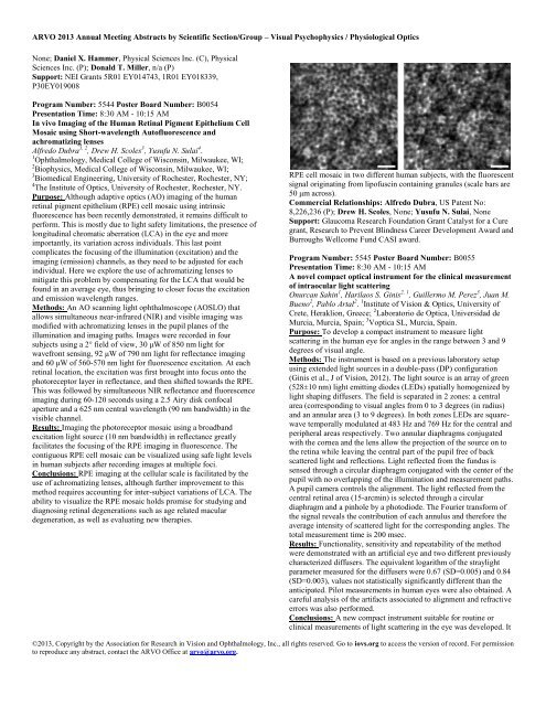
<strong>ARVO</strong> 2013 Annual Meeting Abstracts by Scientific Section/Group – <strong>Visual</strong> <strong>Psychophysics</strong> / <strong>Physiological</strong> <strong>Optics</strong>None; Daniel X. Hammer, Physical Sciences Inc. (C), PhysicalSciences Inc. (P); Donald T. Miller, n/a (P)Support: NEI Grants 5R01 EY014743, 1R01 EY018339,P30EY019008Program Number: 5544 Poster Board Number: B0054Presentation Time: 8:30 AM - 10:15 AMIn vivo Imaging of the Human Retinal Pigment Epithelium CellMosaic using Short-wavelength Autofluorescence andachromatizing lensesAlfredo Dubra 1, 2 , Drew H. Scoles 3 , Yusufu N. Sulai 4 .1 Ophthalmology, Medical College of Wisconsin, Milwaukee, WI;2 Biophysics, Medical College of Wisconsin, Milwaukee, WI;3 Biomedical Engineering, University of Rochester, Rochester, NY;4 The Institute of <strong>Optics</strong>, University of Rochester, Rochester, NY.Purpose: Although adaptive optics (AO) imaging of the humanretinal pigment epithelium (RPE) cell mosaic using intrinsicfluorescence has been recently demonstrated, it remains difficult toperform. This is mostly due to light safety limitations, the presence oflongitudinal chromatic aberration (LCA) in the eye and moreimportantly, its variation across individuals. This last pointcomplicates the focusing of the illumination (excitation) and theimaging (emission) channels, as they need to be adjusted for eachindividual. Here we explore the use of achromatizing lenses tomitigate this problem by compensating for the LCA that would befound in an average eye, thus bringing to closer focus the excitationand emission wavelength ranges.Methods: An AO scanning light ophthalmoscope (AOSLO) thatallows simultaneous near-infrared (NIR) and visible imaging wasmodified with achromatizing lenses in the pupil planes of theillumination and imaging paths. Images were recorded in foursubjects using a 2° field of view, 30 μW of 850 nm light forwavefront sensing, 92 μW of 790 nm light for reflectance imagingand 60 μW of 560-570 nm light for fluorescence excitation. At eachretinal location, the excitation was first brought into focus onto thephotoreceptor layer in reflectance, and then shifted towards the RPE.This was followed by simultaneous NIR reflectance and fluorescenceimaging during 60-120 seconds using a 2.5 Airy disk confocalaperture and a 625 nm central wavelength (90 nm bandwidth) in thevisible channel.Results: Imaging the photoreceptor mosaic using a broadbandexcitation light source (10 nm bandwidth) in reflectance greatlyfacilitates the focusing of the RPE imaging in fluorescence. Thecontiguous RPE cell mosaic can be visualized using safe light levelsin human subjects after recording images at multiple foci.Conclusions: RPE imaging at the cellular scale is facilitated by theuse of achromatizing lenses, although further improvement to thismethod requires accounting for inter-subject variations of LCA. Theability to visualize the RPE mosaic holds promise for studying anddiagnosing retinal degenerations such as age related maculardegeneration, as well as evaluating new therapies.RPE cell mosaic in two different human subjects, with the fluorescentsignal originating from lipofuscin containing granules (scale bars are50 μm across).Commercial Relationships: Alfredo Dubra, US Patent No:8,226,236 (P); Drew H. Scoles, None; Yusufu N. Sulai, NoneSupport: Glaucoma Research Foundation Grant Catalyst for a Curegrant, Research to Prevent Blindness Career Development Award andBurroughs Wellcome Fund CASI award.Program Number: 5545 Poster Board Number: B0055Presentation Time: 8:30 AM - 10:15 AMA novel compact optical instrument for the clinical measurementof intraocular light scatteringOnurcan Sahin 1 , Harilaos S. Ginis 2, 1 , Guillermo M. Perez 3 , Juan M.Bueno 2 , Pablo Artal 2 . 1 Institute of Vision & <strong>Optics</strong>, University ofCrete, Heraklion, Greece; 2 Laboratorio de Optica, Universidad deMurcia, Murcia, Spain; 3 Voptica SL, Murcia, Spain.Purpose: To develop a compact instrument to measure lightscattering in the human eye for angles in the range between 3 and 9degrees of visual angle.Methods: The instrument is based on a previous laboratory setupusing extended light sources in a double-pass (DP) configuration(Ginis et al., J of Vision, 2012). The light source is an array of green(528±10 nm) light emitting diodes (LEDs) spatially homogenized bylight shaping diffusers. The field is separated in 2 zones: a centralarea (corresponding to visual angles from 0 to 3 degrees (in radius)and an annular area (3 to 9 degrees). In both zones LEDs are squarewavetemporally modulated at 483 Hz and 769 Hz for the central andperipheral areas respectively. Two annular diaphragms conjugatedwith the cornea and the lens allow the projection of the source on tothe retina while leaving the central part of the pupil free of backscattered light and reflections. Light reflected from the fundus issensed through a circular diaphragm conjugated with the center of thepupil with no overlapping of the illumination and measurement paths.A pupil camera controls the alignment. The light reflected from thecentral retinal area (15-arcmin) is selected through a circulardiaphragm and a pinhole by a photodiode. The Fourier transform ofthe signal reveals the contribution of each annulus and therefore theaverage intensity of scattered light for the corresponding angles. Thetotal measurement time is 200 msec.Results: Functionality, sensitivity and repeatability of the methodwere demonstrated with an artificial eye and two different previouslycharacterized diffusers. The equivalent logarithm of the straylightparameter measured for the diffusers were 0.67 (SD=0.005) and 0.84(SD=0.003), values not statistically significantly different than theanticipated. Pilot measurements in human eyes were also obtained. Acareful analysis of the artifacts associated to alignment and refractiveerrors was also performed.Conclusions: A new compact instrument suitable for routine orclinical measurements of light scattering in the eye was developed. It©2013, Copyright by the Association for Research in Vision and Ophthalmology, Inc., all rights reserved. Go to iovs.org to access the version of record. For permissionto reproduce any abstract, contact the <strong>ARVO</strong> Office at arvo@arvo.org.
<strong>ARVO</strong> 2013 Annual Meeting Abstracts by Scientific Section/Group – <strong>Visual</strong> <strong>Psychophysics</strong> / <strong>Physiological</strong> <strong>Optics</strong>builds on previous experience with multi-wavelength, highsensitivity,imaging double-pass system for the measurement thewide-angle point-spread function of the eye.Commercial Relationships: Onurcan Sahin, None; Harilaos S.Ginis, Universidad de Murcia (P); Guillermo M. Perez, VOPTICA(E); Juan M. Bueno, None; Pablo Artal, AMO (C), Voptica SL (P),Voptica SL (I), AMO (F), Calhoun Vision (F), Calhoun Vision (C),AcuFocus (C)Support: ITN OpAL (PITN-GA-2010-264605), Ministerio deCiencia e Innovación, Spain (grants FIS2010-14926 and CSD2007-00013), Fundación Séneca (Region de Murcia, Spain), grant4524/GERM/06.Program Number: 5546 Poster Board Number: B0056Presentation Time: 8:30 AM - 10:15 AMThe Effect of AOSLO Image Distortion on Metrics of MosaicGeometryRobert F. Cooper 1 , Zachary Harvey 2 , Michael Dubow 3, 4 , Yusufu N.Sulai 5 , Alexander Pinhas 3, 4 , Drew H. Scoles 6 , Nishit Shah 3 , RichardB. Rosen 3 , Alfredo Dubra 2, 7 , Joseph Carroll 2, 8 . 1 BiomedicalEngineering, Marquette University, Milwaukee, WI;2 Ophthalmology, Medical College of Wisconsin, Milwaukee, WI;3 New York Eye and Ear Infirmary, New York, NY; 4 Mount SinaiSchool of Medicine, Mount Sinai Hospital, New York, NY; 5 TheInstitute of <strong>Optics</strong>, University of Rochester, Rochester, NY;6 Biomedical Engineering, University of Rochester, Rochester, NY;7 Biophysics, Medical College of Wisconsin, Milwaukee, WI; 8 CellBiology, Medical College of Wisconsin, Milwaukee, WI.Purpose: Adaptive <strong>Optics</strong> Scanning Light Ophthalmoscopes(AOSLOs) permit near diffraction-limited imaging of the humanphotoreceptor mosaic, though intraframe eye movements lead toimage distortion. Here, we explore the impact of these distortions ona number of metrics commonly used to characterize thephotoreceptor mosaic.Methods: We acquired 9 image sequences of the parafoveal conemosaic from 7 subjects on 3 similar AOSLOs. In another subject, weacquired an AOSLO and flood-illuminated AO image sequences ofthe same retinal location. To assess the effect of distortions withinAOSLO images, ten averaged images were produced by registeringagainst different reference frames using a previously describedalgorithm. The images were then registered with the same softwarewhile tracking the distortion applied to each image. Thephotoreceptor coordinates from the reference frame were transformedusing this distortion. Voronoi geometry, cone density, nearestneighbordistance (NND), inter-cell spacing (ICS), and regularityindex (RI) were calculated for each set of images. Repeatability wascalculated to assess the effect of intraframe distortion on thesemetrics.Results: Across the AOSLO images, we analyzed 17,942 cones, 75%of which retained the number of sides in the corresponding Voronoidomains across the 10 images (range 56%-90%). Cone density wasfound to have a repeatability of 1.8% (i.e., the difference between any2 measurements on the same subject would be less than 1.8% for95% of observations). NND and ICS had even better repeatability, at1.4% and 0.95%, respectively. In contrast, the NND RI and ICS RIhad a repeatability of 11% and 31%, respectively. Comparing anAOSLO image set to a flood-illuminated AO image, we foundsimilar repeatability (density: 2.7%, NND: 0.7%, ICS: 0.83%, NNDRI: 8.9%, ICS RI: 20.3%) and 83% of cells had retained Voronoigeometry.Conclusions: Global metrics (density and cell spacing) are minimallyaffected by intraframe distortions, whereas local metrics (regularityindex and Voronoi geometry) are more significantly affected.Intraframe distortion in AO scanning instruments limits themeasurement accuracy of mosaic geometry, thus every effort shouldbe made to choose minimally distorted reference frames.Commercial Relationships: Robert F. Cooper, None; ZacharyHarvey, None; Michael Dubow, None; Yusufu N. Sulai, None;Alexander Pinhas, None; Drew H. Scoles, None; Nishit Shah,None; Richard B. Rosen, Opko-OTI (C), Optos (C), Clarity (C),OD-OS (C), Topcon (R), Zeavision (F), Genetech (F), Optovue (C);Alfredo Dubra, US Patent No: 8,226,236 (P); Joseph Carroll,Imagine Eyes, Inc. (S)Support: RPB, NIH (UL1RR031973, EY017607, EY001931),Foundation Fighting Blindness, Alcon Research Institute, BurroughsWellcome FundProgram Number: 5547 Poster Board Number: B0057Presentation Time: 8:30 AM - 10:15 AMPhotoreceptor imaging with in-the-plane adaptive optics opticalcoherence tomography using toroidal mirrorsZhuolin Liu, Omer P. Kocaoglu, Qiang Wang, Donald T. Miller.School of Optometry, Indiana University, Bloomington, IN.Purpose: Recent technological advances in adaptive optics (AO) andhigh-resolution ophthalmoscopy have resulted in sharper images ofthe cellular retina than previously possible. As part of these ongoingdevelopments, we have re-engineered the Indiana AO-OCT system toimprove imaging performance. In this study, we assessed systemperformance for imaging the 3D structure of photoreceptors.Methods: The 2nd generation Indiana AO-OCT system is based on anovel in-the-plane design of an off-axis ophthalmic AO systemrealized with toroidal mirrors. Unlike conventional designs that relyon all spherical mirrors, the inclusion of toroidal mirrors avoidsaccumulation of system astigmatism and unwanted beamdisplacement at pupil conjugate planes. As part of the new design, theAO system was upgraded with a 97-magnetic-actuator ALPAOwavefront corrector to improve stroke and fidelity, and a Shack-Hartmann wavefront sensor configured with an Andor Neo scientificCMOS camera to improve sensitivity and speed. To assessperformance, volume images of the retina with focus at thephotoreceptor layer were acquired at retinal eccentricities rangingfrom the fovea to 6 degrees in human subjects. Power spectra werecomputed of en face images at different depths in the photoreceptorlayer. Each spectrum was radially averaged to increase signal tonoise.Results: Ray trace analysis of the in-the-plane design predictsdiffraction-limited imaging across the entire 3.6°x3.6° field of viewof the AO-OCT system. Beam displacement of less than the pitch ofthe SHWS lenslet array is also predicted, thus enabling the fullsensitivity of the SHWS to high spatial frequencies. Measured beamdisplacement and wavefront root-mean-square error of the systemconfirmed the theoretical predictions. Cone photoreceptors wereroutinely observed at retinal eccentricities as small as 0.2 degrees.This corresponds to cones narrower than that detected with theprevious Indiana AO-OCT system. Analysis of power spectra at theinner segment / outer segment junction, and posterior tip of the outersegment revealed more energy at high spatial frequencies.Conclusions: The 2nd-generation Indiana AO-OCT system providesa more detailed view of the photoreceptor optical signature than theprevious generation system.Commercial Relationships: Zhuolin Liu, None; Omer P.Kocaoglu, None; Qiang Wang, None; Donald T. Miller, n/a (P)Support: NIH R01EY018339, NIH R01EY014743, NIHP30EY019008Program Number: 5548 Poster Board Number: B0058©2013, Copyright by the Association for Research in Vision and Ophthalmology, Inc., all rights reserved. Go to iovs.org to access the version of record. For permissionto reproduce any abstract, contact the <strong>ARVO</strong> Office at arvo@arvo.org.