© 2006 - The Charles H. Tweed International Foundation
© 2006 - The Charles H. Tweed International Foundation
© 2006 - The Charles H. Tweed International Foundation

Create successful ePaper yourself
Turn your PDF publications into a flip-book with our unique Google optimized e-Paper software.
© <strong>2006</strong>
THETWEED PROFILEpublished byTHE CHARLES H. TWEEDINTERNATIONAL FOUNDATION FORORTHODONTIC RESEARCH ANDEDUCATIONVolume #5<strong>2006</strong>
Chairman of the Board Report <strong>2006</strong>Jack Dale“Performance drives us as an organization.We deliver performance that endures.” John DeereSome time ago, I sent our Executive Director, Jim Vaden, a book entitled, “<strong>The</strong> John Deere Way” because Iknew that his passion, other than orthodontics, is collecting and restoring John Deere tractors.Within a few weeks, Jim returned the book to me and he said, “You have to read this book.” I was not too faralong when it occurred to me why he said that. You would think that you were reading about <strong>The</strong> <strong>Tweed</strong> <strong>Foundation</strong>,and I realized that that was the reason Jim, instinctively, or by design, was interested in John Deere. <strong>The</strong> company, asdoes <strong>The</strong> <strong>Tweed</strong> <strong>Foundation</strong>, represents excellence and the highest possible standard of “performance”.John Deere is the world’s largest producer and seller of farm and industrial tractors and equipment. It has beenin business for 169 years, since John Deere, blacksmith, invented the first commercially successful self-scouring steelplow in 1837.Today its prosperity continues with 46,000 employeesin 160 countries and with $20 billion in annual sales. Thisremarkable growth began when John Deere said,“I will not put my name on a product that does nothave in it the best that is in me.”<strong>The</strong> company has enjoyed phenomenal successbecause the driving force behind this dynamic organizationis a set of rules and principles instilled, at its inception,by John Deere himself: commitment, integrity, innovationand quality. <strong>The</strong>se qualities come together to create,“<strong>The</strong> John Deere Way”.One could say, with the same determination and sincerity,that commitment, integrity, innovation and quality have created“<strong>The</strong> <strong>Tweed</strong> Philosophy”.Up on Jimmy’s handiwork.<strong>The</strong> <strong>Tweed</strong> <strong>Foundation</strong>’s insistence on the highest possible standard of care in orthodontics has enabled it toprogress from Dr. <strong>Tweed</strong>’s commencement of practice in 1928 to the present day … 78 years.Two sterling examples of commitment in the <strong>Foundation</strong> are the records of our Course Directors since Dr.<strong>Tweed</strong>’s death in 1970, Levern Merrifield and Herb Klontz. Levern Merrifield did not miss a course or a meeting inthe 47 years that he was involved, and Herb Klontz has not missed a course or a meeting since he became CourseCoordinator in 1976, and then Course Director in 1983 … to mention only two of our many dedicated members.As a result of this commitment, integrity, innovation and strict adherence to quality, <strong>The</strong> <strong>Tweed</strong> <strong>Foundation</strong>concepts and protocol of diagnosis and treatment, working in harmony with <strong>The</strong> <strong>Tweed</strong>-Merrifield EdgewiseMechanotherapy, which is the most precise instrument for the routine correction of major malocclusion that existsin the world today, have influenced millions of patients in more than 75 countries and in every state in America.John Deere’s commitment to innovation: “Innovation means inventing, designing and developing productsand services that customers want to buy from John Deere … constantly striving to delight customers withproducts and services that help them realize their own aspirations.”To say it the <strong>Tweed</strong> way: “Innovation means inventing, designing and developing <strong>The</strong> <strong>Tweed</strong>-MerrifieldEdgewise Mechanotherapy and service that are highly beneficial to orthodontic patients … constantly striving todelight patients with treatment and service that help them realize their aspirations.”4
Many times I have said that there is no better experience than the exhilaration that results when the <strong>Tweed</strong>-Merrifield appliance is removed, revealing a successful result and a beautiful smile from an exceedingly happy andsatisfied patient.John Deere’s commitment to integrity: “Integrity means telling the truth, keeping our word and treatingothers with fairness and respect … honest relationships, effective discussions … unquestioned commitment toethical behavior.Our reputation is based, in large part, on our desire toalways act with integrity and this is one of our most valued assets.Of what does integrity consist?Courtesy … in every word.Honesty … in every transaction.Dignity … in every personal act.Progressiveness … in every thought.Constructiveness … in every criticism.Quality … in every piece of work produced.” Or as we wouldsay in the <strong>Foundation</strong> “Quality in the service of every patient wetreat.”It would be impossible for me to express it any better than theauthor, David Magee. <strong>The</strong>refore, as you read the words written byhim think ‘<strong>Tweed</strong>’. That is what I did throughout the book when I wasreading it.Country auction in Cookeville with Jim andhis beautiful daughter ‘Meg’.John Deere’s commitment to quality: “Stand behind the product … at all costs … we do whatever we haveto do because that is our promise … stick-to-itiveness makes a difference.People believe John Deere will stand behind its products because they have seen it do it year after year …that is <strong>The</strong> John Deere Way’By delivering quality and being proud of it John Deere’s equipment became the farmer’s BMW covetedas the ultimate agricultural machine. <strong>The</strong> brand stood for everything great about the product, and the productsstood for everything great about the brand.“In the future we simply take the John Deere legacy to the next level and the secret ingredient is passion …in remaining true to the core values of quality, innovation, integrity and commitment. People trust us to deliverthe best that is within us. This is not only our legacy, it is our purpose … to create and sustain an exceptionalexperience in genuine value for our customers.” (patients)“Performance drives us as an organization. Success is not just for the moment, but also for the long run.”“We deliver performance that endures.”“Ultimately, we learn and understand that John Deere’s secret to success is its legacy, built in layers, yearafter year and generation after generation. It is how the company became a world leader, delivering performancethat endures through quality, commitment, innovation and integrity: “<strong>The</strong> John Deere Way.”And … so it is with <strong>The</strong> <strong>Tweed</strong> <strong>Foundation</strong>.I could go on and on, and … on, but I have made my point. At the same time, I hope that I have inspired thosewho are reading these words to do better … to “jump start” your determination to provide the highest standard oforthodontic care that is within you … before you put your name on your treatment.Reference: Magee, David: <strong>The</strong> John Deere Way. John Wylie and Sons, Inc. 2005.Jack Dale5
Tribute: Dr. Herbert A. Klontz, <strong>Tweed</strong> Course Director<strong>2006</strong> Albert H. Ketcham Memorial AwardWhen I see the name “Herb Klontz”, I immediately think of enthusiasm, energy, discipline and perfection, andwhen I hear his name spoken, I think of the highest possible standard of orthodontic treatment.Whatever Herb attempts, he will not rest until he challenges his talents and capabilities to the absolute maximum.As a youth, he not only aspired to become a member of a highly respected and admired group of young men: <strong>The</strong> BoyScouts of America, but he would not be content until he reached the very pinnacle of that organization: Eagle Scout.When he was growing up in Iowa, he lived on the golf course. Again, he was not content with just being a golfer,he had a burning desire to be the best golfer he could possibly be. He was constantly driven to reach his potential, toreach his standards. He competed with himself more than with others. But, in his competition with others, he did prettywell … exceedingly well. For instance, he won <strong>The</strong> Iowa State High School Championship twice, <strong>The</strong> Iowa StateJunior Championship, <strong>The</strong> Iowa State Amateur Championship, <strong>The</strong> Chicago District Junior Open Championship,and <strong>The</strong> Western Junior Championship.He qualified four times for <strong>The</strong> National Amateur Championship, once at Grosse Point, Michigan whenArnold Palmer won the tournament and once at Pebble Beach,California when Jack Nicklaus won.During the second year of his orthodontic program atWashington University in St. Louis, Earl Shepard asked Herba very important question: “Are you going out to Californiato play golf or are you going to stay here and pursue yourorthodontic career.” Obviously, Herb stayed in the program and,to this day, he thanks Dr. Shepard for his foresight and guidance.Herb Klontz attacked orthodontics with the same energyand enthusiasm as he did scouting and golf, and … with a bundleof talent. He not only decided to be a general practitioner indentistry, he wanted to be a specialist in orthodontics. He notonly wanted to be an orthodontist, he wanted to be the bestorthodontist in the world. While it is difficult, if not impossible,to say who is the best orthodontist in the world, Herb ranks rightup there with the best of them. I have heard the phrase, “<strong>The</strong> bestin the world” associated with him on many, many occasions.Herb reasoned that if he were going to be the bestorthodontist he could possibly be, he would have to becomeassociated with one of the most prestigious organizations inthe specialty. Thus, he enrolled in <strong>The</strong> <strong>Tweed</strong> Course given by<strong>The</strong> <strong>Charles</strong> H. <strong>Tweed</strong> <strong>Foundation</strong>. <strong>The</strong> year was 1963. Dr.Herb Klontz, Eagle Scout.<strong>Tweed</strong> and his protégé, Levern Merrifield, were Co-Directors. Hetook the course again in 1965 with one of his classmates at WashingtonUniversity, and very dear friend, Ed Noffel. In 1967, Herb was asked to become an Instructor and, followingDr. <strong>Tweed</strong>’s death in 1970, he was appointed Course Coordinator by the new Course Director, Levern Merrifield ... in1976.In 1983, Herb was elevated to <strong>Tweed</strong> Study Course Director when Levern Merrifield became <strong>The</strong> Chairman ofthe Board.Since 1976, Herb has never missed a course … all 65 of them. In this regard, his dedication and loyalty equals thatof his mentor and role model, Levern Merrifield. Levern never missed a course, or a meeting, in the 47 years that he wasinvolved with <strong>The</strong> <strong>Foundation</strong>.One can estimate the direct influence that Herb has had on world orthodontics. He has taught in 65 <strong>Tweed</strong>Courses averaging 75 participants per course. In recent years, over 100 orthodontists and students have participated ineach course. <strong>The</strong>refore, he has directly influenced 4875 (75 x 65) participants from approximately 75 countries and,what is important, that influence has been the highest standard of care.6
Herb receiving trophy as <strong>The</strong> WesternJunior Amateur Champion in 1954.If each of these participants returned to his/her respective countryand practiced, even in part, the <strong>Tweed</strong>-Merrifield Principles according toHerb’s teaching, with a conservative 2000 patients during their careers (acareer being approximately 30 years, with a patient load of 300 patients peryear amounting to 9000 patients in total), Herb would directly influence inthe neighborhood of 8,000,000 patients. I am convinced that his influence isof that magnitude … no doubt in my mind.That figure does not include the students he has taught on a routinebasis in three universities: St. Louis University, six years, Baylor University,ten years and Oklahoma University, 18 years.Nor does it include the universities where he has been a visitinglecturer, such as Iowa, Michigan, Southern California, UMKC, Case WesternReserve, New York, Tennessee, NOVA, Toronto, and Harvard.It does not include the lectures presented in a dozen countries, whichinclude Japan, Korea, Taiwan, China, Guatemala, Mexico, Canada, Italy,France and Brazil, and in at least 25 states in the United States.It does not include the extra courses given in Tucson; it does notinclude the 100 plus lectures that he has given to associations other than <strong>The</strong><strong>Tweed</strong> Course, and it does not include his publications in the literature andhis chapter in the Graber text book. Herb Klontz teaches by example; he doesnot tell students what to do, he shows them. He is constantly in the trenches, in his office, treating patients exactly ashe teaches in Tucson. I cannot remember calling Herb on the telephone in his office when he was not present.<strong>The</strong>re is no question, Herb Klontz has directly influenced a staggering number of colleagues, students andpatients and has served as a role model around the world. He represents the gold standard of precision orthodontics.But … he is more than that.I am particularly aware of Herb’s personal qualities for I have seen him in action … behind the scenes. <strong>The</strong>re isnot a more unselfish, giving and thoughtful person in our specialty. For several years he took the time to travel fromOklahoma City to Toronto to share his special talents with 10 – 12 graduate orthodonticstudents for remuneration that was barely more than his expenses. Howmany teachers of his caliber and reputation would be as considerate, as generousand as caring as that … for the benefit of such a few?Herb with his son Kelly and Arnold Palmer.Herb in 1962 upongraduating as an orthodontistfrom Washington University,St. Louis.7
He was not only generous in Toronto, but he was just as generous in Boston. Each year, for a number of years,Herb was a major contributor to <strong>The</strong> Jack G. Dale Residency in Orthodontics at Harvard University. In thisinstance, he traveled a similar distance to teach approximately 20 Harvard orthodontic students and between 5 to 6Toronto dental graduates aspiring to become orthodontic students. Again, he was barely paid for his expenses.Professor Emile Rossouw in Toronto, Professor Leslie Will at Harvard, the students, both from Toronto and Boston, andI are eternally grateful to Herb for his participation and his contribution. When he presented his beautiful clinicalmaterial, you could hear a pin drop all day long. On top of all that … we had a lot of fun with the students in the evening.“On behalf of both groups, I say ‘ThankYou Herb!’ I know that you have been justas engaging and generous with many othergroups around the world throughout yourincredible career of 44 years.”For those many reasons I am delightedthat <strong>The</strong> American Board of Orthodonticshas honored Herb Klontz with what manyrefer to as the most prestigious award inorthodontics … worldwide: <strong>The</strong> Albert H.Ketcham Memorial Award.For health reasons in our family, I wasunable to attend the AAO Meeting in SanFrancisco last year, but I will leave no stoneunturned in my effort to be present to witnessHerb Klontz receiving this high honor, thisyear, at the AAO Meeting in Las Vegas.All of us in <strong>The</strong> <strong>Tweed</strong> <strong>Foundation</strong>should be very, very proud of our CourseHerb Klontz appointed Course Coordinator, 1976.Director for his wonderful achievement. I urge all our membership to travel to Las Vegas to pay tribute to Herb and toenjoy the Friday night reception in his honor.Herb has received many awards for his leadership throughout his career, and he has been recognized on manyoccasions. He is the first to tell you, and he expresses himself with his characteristic enthusiasm, that this would notbe possible without the constant companionship and support of Karen, his partner in life and wife of 47 years. He iseternally grateful to her for the passionate and loving care of their two children. <strong>The</strong>ir son Kelly, an outstanding youngorthodontist himself, is married to Terrie, and together they are parents of Morgan, age 9 and Lilly age 6. Kathy (Klontz)Nassimbene, their daughter, is in real estate, and she is married to a dentist. <strong>The</strong>y live in Denver, Colorado.Herb elevated to Course Director, 1983.A familiar scene at Tucson.8
One of my favorite photographs.Herb with his mentor and role model, Levern Merrifield, inKorea.Together, they are one big happyfamily, and I wish all of them the mostwonderful life of fulfillment in the yearsahead.“Congratulations, Herb! Drs. <strong>Tweed</strong>and Merrifield, both Ketcham Awardrecipients, would be very proud of you onthis very special occasion”.Jack Dale<strong>The</strong> Ketcham Award was announced for <strong>2006</strong> by the ABO at the AAOmeeting in San Francisco. Herb Klontz is seen here with his treasuredfriends of many years: Don James, John Grubb, Eddie Noffel, EdPolk, Herb, Vance Dykhouse, John Bilodeau, and Jimmy Vaden.9
Director of Education — Herb Klontz<strong>The</strong> <strong>Tweed</strong> Study Course is experiencing great demand. <strong>The</strong> April, 2005 Course was full, and the September, 2005Course had a record number of 116 participants. Of these 116, 74 were students from 14 American universities.After the Course, a Continuing Education Workshop dealt with the extraction/nonextraction dilemma, microscrewimplants, surgical considerations, etc.. Eighty-six people participated in the workshop.<strong>The</strong> educational mission of the <strong>Foundation</strong> is alive and well. My thanks to all of you who have graciously given ofyour time to help with the Course because it cannot continue to thrive and prosper without good instructors who areenergetic and knowledgeable. I sincerely thank you. <strong>2006</strong> is shaping up to be a banner year. Many of you will haveto help us again, and we thank you in advance. Enjoy the course photographs that are interspersed throughout thispublications!Secretary/Treasurer — Jim VadenYour <strong>Foundation</strong> continues to “run” well. Our Endowment Fund, managed by Michael Stolpher who is under thewatchful eye of John Bunk, has done very well in 2005. This Endowment Fund allows us to do some things that wecould not ordinarily do. We must be ever mindful, however, that we have to protect it because it is our future. It willcontinue to grow and allow us to do more and more things “for the cause”.At its September, 2005 meeting, your Board of Directors voted to increase dues for members from $50 to $100.Teaching staff members will pay $250 instead of $200. This action was taken because the <strong>Foundation</strong> continues tospend quite a bit more than it receives in dues income. It was felt by the Board of Directors that the burden of runningthe <strong>Foundation</strong> should be more equally shared by all members of the <strong>Foundation</strong>.All of you have received notification of our <strong>Foundation</strong> mixer which will be held in conjunction with the AAOmeeting in Las Vegas. If you have not returned your registration form, please do so. We must know how many forwhom to plan.Lucy Koffman had to quit her job with us because of a health problem. You will remember that Lucy was hired in thesummer and was working out very well. Lucy took a trip to Oregon, fell and broke her hip, and has been inrehabilitation since in Oregon.We have hired a bright and energetic lady named Nancy Netherton to help Esther. We will do a little “feature” onNancy in the next Profile.10
Scientific Papers11
Orthodontic Treatment of a Low Angle Class II Malocclusion thatRequired an Orthodgnathic Sugical Procedure — John BilodeauINTRODUCTIONA common finding in a low angle Class II malocclusion is that the maxillary incisor is in correct position relativeto the cranial base and the facial profile is normal or recessive. This case report shows one treatment approach to thistype of problem.HISTORY AND ETIOLOGY<strong>The</strong> patient was a 31 year 11 month old white female with an unremarkable medical history. Dental historyrevealed prior orthodontic treatment and regular dental care. She had a Class II malocclusion with a straight and overclosed facial profile. Her chief concerns were her “protruding teeth and facial appearance when smiling”. Primaryetiology is believed to be heredity.DIAGNOSIS<strong>The</strong> facial photographs (Fig.1) demonstrate a straight and slightly over closed facial profile.<strong>The</strong> dental casts (Fig. 2A, 2B) exhibit an Angle’s Class II malocclusion with 7mm of overjet and 100% overbite.All third molars are missing. <strong>The</strong>re is a deep curve of Spee.Fig. 1.Pretreatment facial photographs<strong>The</strong> panoramic radiograph (Fig. 3) reveals no dental pathology. All teeth are present except the third molars.Fig. 2A. Pretreatment dental casts12
Fig. 2B. Pretreatment dental castsmeasurement of 6mm reflects alveolar imbalance.<strong>The</strong> cephalogram and itstracing (Figs. 4A, 4B) illustrate anANB angle of 3 o . <strong>The</strong> SNA angleof 77 o confirms a slight maxillarydeficiency. <strong>The</strong> FMA is 8 o . <strong>The</strong>.86 facial height index of Horn 1 isa confirmation of an imbalance ofanterior and posterior facial height.<strong>The</strong> IMPA angle of 92 o reflectsan acceptable inclination of themandibular incisors. <strong>The</strong> Z angle 2of 88 o confirms a retruded softtissue overlay, however, Wits 3,4TREATMENT OBJECTIVES1. Maintain a normal profile line to nose relationship and a normal Z angle 22. Reduce overbite and overjet3. Obtain normal canine and incisal guidance4. Increase lower anterior facial heightTREATMENT ALTERNATIVES1. Extract the maxillary 1 st premolars andmandibular 2 nd premolars to correct the overjet andoverbite. This option could have a deleterious effect onthe facial profile.2. Extraction of only the maxillary right and left 1 stpremolars to correct the overbite and overjet. This optioncould collapse the upper lip.3. Non extraction 10-2 orthodontic treatment. Thisoption was considered but the amount of cooperationrequired was not appealing to the patient.Fig. 3. Pretreatment panoramic radiograph4. Non extraction treatment and a mandibular advancement with a genioplasty to increase lower facial height.This option would provide an ideal result.Fig. 4A. Pretreatment cephalometricradiographFig. 4B. Pretreatment cephalometric tracingTREATMENT PLANWhen orthodontictreatment withoutsurgical correction isplanned, the diagnosisis predicated on thepretreatment positionand the desired finalposition of themandibular incisors.Merrifield’s totalspace analysis 5, 6 wasused to determinespace requirements.13
Fig. 5. McNamara’s NasionFrankfort PerpendicularFig 6. Delaire Analysis Fig. 7. Legan-Burstone analysisFig. 8. Profile Line and Z angle Fig. 9. Pretreatment McNamara tracing showsPoint A and the Maxilla in correct positionFig. 11A. Presurgical cephalometric radiographFig. 10. Presurgical panoramic radiograph14
Fig. 17. Posttreatment panoramic radiographFig. 18B. Posttreatment cephalometrictracingFig. 19. Composite tracingsFig. 18A. Posttreatment cephalometric radiographheight needed to beincreased and themandible advanced.<strong>The</strong> Z angle was78 o and profileline to nose wasideal according toMerrifield (Fig. 14)and would not bemaintained unless areduction genioplastywas part of thesurgical plan. Thiswould take away theinfluence of the chin,further increase facial height, and provide an ideal profile line to nose relationship. <strong>The</strong> patient would not agree to thegenioplasty because her insurance would not approve it.TREATMENT RESULTS• <strong>The</strong> posttreatment facial photographs (Fig. 15) illustrate a marked improvement in the facial profile on smiling. <strong>The</strong>profile line does not intersect the middle of the nose. <strong>The</strong> chin projection is strong but not objectionable.• <strong>The</strong> posttreatment dental casts (Fig. 16) exhibit a Class I occlusion with normal overjet, overbite, canine and incisalguidance.• <strong>The</strong> posttreatment panoramic radiograph (Fig. 17) exhibits no pathology.• <strong>The</strong> posttreatment cephalometric radiograph and its tracing (Fig. 18A, Fig. 18B) illustrates the changes that wereachieved with treatment. <strong>The</strong> mandibular incisors were uprighted over basal bone to an IMPA angle of 98 o . <strong>The</strong> FMAangle increased to 10 o .• <strong>The</strong> composite cephalometric tracings illustrate the changes achieved (Fig.19).• A Maxillary Hawley and a lower bonded retainer were placed. Total treatment time was 17 months.17
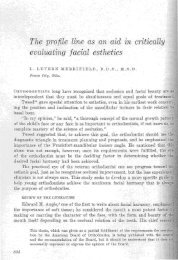
REFERENCES1. Horn AJ. Facial height index. Am J Orthod 1992; 101:180-6.2. Merrifield LL. <strong>The</strong> profile line as an aid in critically evaluating facial esthetics. Am J Orthod 1966; 52,11:804-22.3. Jacobson A. <strong>The</strong> “Wits” appraisal of jaw disharmony. Am J Orthod 1967 (2):125-138, 1975(volume 67,number 2, Feb. 19754. Jacobson A. “Wits appraisal” in Radiographic Cephalometry. Quintessence Publishing Co, Inc :1195:97-112.5. Merrifield LL. Differential diagnosis with total space analysis. J <strong>Charles</strong> <strong>Tweed</strong> <strong>Foundation</strong> 1978; 6:10-15.6. Vaden JL, Dale JG, Klontz HA. <strong>The</strong> <strong>Tweed</strong>-Merrifield philosophy. In: Graber TM, Vanarsdall RL, eds.Orthodontics: current principles and techniques. St. Louis: CV Mosby, 1994:627-84.7. McNamara JA. A method of cephalometric evaluation. Am J Orthod 1984; 86:49-69.8. Delaire J, Schendel SA, Tulasne JF. An architectural and structural craniofacial analysis: a new lateralcephalometric analysis. J Oral Surg 1981; 52:226-38.9. Legan HL, Burstone CJ. Soft tissue cephalometric analysis for orthognathic surgery. J Oral Surg 1980;38:744-51.10. Merrifield LL. Edgewise sequential directional force technology. J <strong>Charles</strong> <strong>Tweed</strong> Found 1986; 14:22-37.11. Burke S, Dent IV, Burch SG, Tetz, JA. Incidence and size of pretreatment overlay and post treatment gingivalembrasure space between maxillary central incisors. Am J Orthod Dentofac Orthop 1994; 105:506-11.12. Kokich, VG. Esthetics: <strong>The</strong> Orthodontic-Periodontic Restorative Connection. Semin Orthod 1996; 2:21-30.13. Kokich VG, Mathews DP. Managing treatment for the orthodontic patient with pseudo dontal problems.Semin Orthod 1997; 3:21-38.18
A Comparative Study of Facial Profiles in Extraction and NonextractionTreatmentRichard Don James, DDS, MSDOklahoma City, Okla.<strong>The</strong> debate concerning the extraction of teeth and its effect on the facial profile began more than 100 years ago.It is still an ongoing topic of discussion. All orthodontists have heard colleagues say, “I don’t extract teeth because itcreates a dished-in face,” or, “I don’t extract teeth because I like a nice full pleasing smile.” Statements like these, madewithout any scientific evidence, led to this study.REVIEW OF THE LITERATUREFacial beauty and harmony were paramount treatment goals of Dr. Edward Angle. He believed that the faceof the Greek god Apollo contained all the essentials of harmony and beauty. From his studies, he developed the profile“line of harmony.” By the time his seventh edition was published, he believed that maintenance of a full complement ofteeth would establish the best harmony and that Nature would allow this to happen through growth, development, andfunction. 1,2<strong>Tweed</strong>, 3 Angle’s disciple, became disappointed with the facial balance found in a great majority of the patientswhom he had treated without tooth removal, but he liked the facial esthetics of patients for whom he was able to uprightthe mandibular incisors so the FMIA value could approach 60° to 68° . His clinical studies led him to retreat more than100 of his “nonextraction” patients with premolar extraction.After many years of studying and comparing untreated normal patients who had pleasing facial balance withpatients he had treated both with and without extraction, he developed the “Diagnostic Facial Triangle” to equate toothposition with facial esthetics.Downs 4 believed that restoring or maintaining balance and harmony of the component parts of the face in manycases required extraction of teeth. His “facial angle” and “angle of convexity” are hard tissue measurements that reflectfacial type and form, but they do not take into account the soft tissue, especially soft tissue chin.Holdaway 5 studied facial balance and developed an 11-mesaurement soft tissue analysis that included “superiorsulcus depth” and the “H line.” He was not fond of the “nasolabial angle” because he believed that it did not adequatelydescribe contour in the subnasal profile.To quantify facial contour, Ricketts 6,7 used a millimeter measurement made from the vermillion border of thelower lip to the “facial esthetic line.” For young patients, he found the measurement to have a mean of 0 mm with arange of ±3 mm. A mean of -4 mm was found in the slightly older patient.Merrifield’s 8 study of facial profiles in a sample of 120 untreated normal patients and treated patients withpleasing facial esthetics led to the development of the “Z-angle” to quantify balance, or lack thereof, of the lower facialprofile. <strong>The</strong> Z-angle is the inner inferior angle formed by the intersection of the Frankfort horizontal plane and the“profile line” (a line that is tangent to the soft tissue chin and the most prominent lip). He found the normal Z-anglerange in his sample to be 72° to 83°.MATERIAL AND METHODS<strong>The</strong> purpose of the study was to quantitatively and visually compare the facial profiles and soft tissue balancefound in patients who underwent premolar extraction with the profiles and facial balance of patients who did notundergo premolar extraction. A sample of 170 consecutively treated patients was collected over a 20-month period.One hundred-eight of these patients were treated after extraction of premolars, and 62 were treated without premolarextraction. <strong>The</strong> patient records used in the study consisted of pretreatment and posttreatment facial photographs,cephalographs, and tracings. <strong>The</strong> photographs were ¼ life size in all three views: profile, fontal, and smile.<strong>The</strong> quantitative measurements selected were (1) an angular measurement – the Z-angle of Merrifield and (2)a linear measurement – the lower lip to the esthetic line of Ricketts, called in thisstudy, the “E-value.”<strong>The</strong> Z-angle and E-value were chosen for their ease and accuracy tomeasure and their relevance to lip position and lip change, thus makingcomparisons relevant and easy to evaluate. As described earlier, the Z-angle is theangular measurement made by the profile line and the Frankfort horizontal plane(Fig. 1). <strong>The</strong> Z-angle in this example is 76°.<strong>The</strong> E-value is the distance in millimeters from the most anterior point of thevermillion border of the lower lip to the esthetic line. If the lower lip is in front ofthe esthetic line, the E-value is positive. If the lower lip is behind the esthetic line,the E-value is negative. <strong>The</strong> E-value in this example is -4mm. (Fig. 2).Fig. 1. Z-angle measurement.20
<strong>The</strong> more protrusive the lip profile, the less the Z-angle. <strong>The</strong> more retrusivethe lip profile, the more the Z-angle. In general, as the lip profile position becomesmore protrusive in its relationship to the chin, the Z-angle will decrease and the E-valuewill become more positive. As the lip profile position becomes more retrusive in itsrelationship to the chin, the Z-angle will increase and the E-value will become morenegative. In this study, pretreatment and posttreatment measurements were comparedwithin the nonextraction and extraction groups and between each of the two groups.THE SAMPLEIn the nonextraction group, the posttreatment Z-angle ranged from 68° to 90°.Fig. 2. E-value measurement.<strong>The</strong> posttreatment E-value ranged from +1.5 mm to -10 mm. Only one patient in thenonextraction group had a positive posttreatment E-value (+1.5 mm). <strong>The</strong> averageposttreatment Z-angle for the nonextraction group was 79.01°. <strong>The</strong> average posttreatment E-value for the nonextractiongroup was -4.05 mm.Several different extraction protocols were used in the extraction group. <strong>The</strong> most common ones were (1)maxillary and mandibular second premolars (14 patients), (2) maxillary first premolars (7 patients), (3) maxillaryfirst premolars and mandibular second premolars (27 patients), and (4) maxillary and mandibular first premolars (36patients). <strong>The</strong> remaining patients (24) in this group required various other extraction combinations.Characteristics of the maxillary and mandibular second premolar extraction subgroup were (1) mild tomoderate arch length discrepancy, (2) good soft tissue facial pattern or mild facial imbalance, and (3) good skeletalpattern. An example of this extraction protocol is shown (Fig. 3A, Fig. 3B).Characteristics of the maxillary first premolar extraction subgroup were (1) Class II dental relationship, (2)maxillary protrusion, maxillary anterior crowding, or both, and (3) good mandibular incisor position with little or nocrowding. An example of this extraction protocol is shown (Fig. 4A, 4B).Characteristics of the maxillary first premolar and mandibular second premolar extraction subgroup were (1)Class II dental relationship and mild to moderate Class II skeletal pattern, (2) mild to moderate mandibular archlength discrepancy, (3) soft tissue facial imbalance, and (4) low to medium Frankfort mandibular plane angle. Anexample of this extraction protocol is shown (Fig. 5A, 5B).Characteristics of the maxillary and mandibular first premolar extraction subgroup were (1) severe archlength discrepancy, dental (bialveolar) protrusion, or both, (2) Class I or Class II dental relationship, and (3) mediumto high Frankfort mandibular plane angle. An example of this extraction protocol is shown (Fig. 6A, 6B).RESULTS<strong>The</strong> facial profiles of the extraction group and the nonextractiongroup were within the normal range of Z-angle and E-values at theconclusion of treatment. On average, the nonextraction group completedtreatment with a slightly more retrusive lip profile position than did theextraction group (Fig. 7A). <strong>The</strong> posttreatment Z-angle average for thenonextraction group was 79.01° (Fig. 7B) compared with 73.74° for theFig. 3. A, Facial photographs: extractionof four second premolars (pretreatment-left;posttreatment-right).Fig. 3. B, Tracings: extraction of four second premolars (pretreatment-left;posttreatment-right).21
extraction group, a 5.27° difference. <strong>The</strong> posttreatment E-value average for the nonextraction group was -4.05 mmcompared with -2.58 mm for the extraction group. <strong>The</strong>refore, the nonextraction group finished treatment with the lowerlip 1.47 mm further behind the esthetic line than did the extraction group (Table I).<strong>The</strong> extraction group began treatment with significantly more lip-chin imbalance; the pretreatment Z-angle ofthe extraction group was 66.61° compared with 73.38° for the nonextraction group, a 6.77° difference. <strong>The</strong>pretreatment E-value for the extraction group was +.74 mm compared with -2.93 mm for the nonextraction group, a 3.67mm difference. A note of interest is the fact that the posttreatment Z-angle and E-values of the extraction group (Z =73.74°; E = -2.58 mm) are very close to the pretreatment Z-angle and E-values of the nonextraction group (Z = 73.38°;E = -2.93 mm). In other words, the extraction group finished treatment approximately where the nonextraction groupbegan treatment.DISCUSSIONWithin the extraction group, the least amount of pretreatment lip-chin profile imbalance occurred in the foursecond premolar extraction subgroup (Z= 71.64°; E = -1.29 mm), and the most pretreatment lip-chin profile imbalanceoccurred in the four first premolar extraction subgroup (Z= 62.78°; E = +2.31 mm). <strong>The</strong>se subgroups had a Z-angledifference of 8.86° and an E-value difference of 3.60 mm (Table II).<strong>The</strong> four second premolar extraction subgroup, the maxillary first premolar extraction subgroup, and themaxillary first premolar – mandibular second premolar extraction subgroup finished treatment fairly close together(Table II). <strong>The</strong> posttreatment Z-angle values were within 1.81° of eachother, and the E-values were within .25 mm. <strong>The</strong>se posttreatment valuesare the result of a predictable diagnostic measurement analysis andtreatment plan.<strong>The</strong> four first premolar extraction subgroup finished treatmentwith a Z-angle value of 70.89° and an E-value of -1.46 mm, becausethe patients in this subgroup started treatment with more arch lengthdiscrepancy, facial imbalance, or both. When the nonextraction groupwas compared with the four first premolar extraction subgroup, theFig. 4. A, Facial photographs: extraction ofmaxillary first premolars (pretreatment-left;posttreatment-right).Fig. 4. B, Tracings: extraction of maxillary first premolars (pretreatment-left;posttreatment-right).22
Fig. 5. B, Tracings: extraction of maxillary first premolars and mandibularsecond premolars (pretreatment-left; posttreatment-right).nonextraction group finished treatment with a Z-angle 8.12° greaterand an E-value 2.59 mm more negative than did the four firstpremolar extraction subgroup.CONCLUSIONS1. Both the extraction and nonextraction group facial profileFig. 5. A, Facial photographs: extraction of maxillaryfirst premolars and mandibular second premo-treatment.value averages were within the normal range at the completion oflars (pretreatment-left; posttreatment-right). 2. <strong>The</strong> average posttreatment lip profile position of thenonextraction group was slightly more retrusive than that of the extraction group.3. <strong>The</strong> extraction group had greater pretreatment facial imbalances.4. <strong>The</strong> extraction group had the greatest improvement in facial balance.5. In all patients in the extraction group, the common denominator “chin prominence” either remained in balance or wasimproved.6. <strong>The</strong> smiles in both groups were balanced and pleasing except for a few patients who had vertical skeletaldisharmonies.7. With a sound diagnostic scheme, treatment plan, and management of extraction space, extraction of the proper teethcan create balanced facial esthetics, not destroy it.Evaluation of facial profiles and facial balance is a constant, continuous, lifelong study and learning process fororthodontists. Tooth movement and proper positioning of the teeth to ensure favorable facial changes and to avoidunfavorable changes should be in the orthodontist’s “diagnostic” mind from the very first examination. To treat patientsnonextraction for the sake of not removing teeth, ease of treatment, or the dictates of an appliance is not sound reasoningand makes as much diagnostic sense as treating all patients with the extraction of all four first premolars. In other23
words, it is just asdiagnostically wrongto treat an extractionpatient nonextractionas it is to treat anonextraction patientwith extractions. <strong>The</strong>truth lies somewhere inbetween and is basedon a sound quantifiedmeasurement analysis,differential evaluation ofthe problem, and clinicalassessment.Fig. 6. A, Facial photographs: extractionof four first premolars (pretreatment-left;posttreatment-right).Fig. 7.A, Facial Photographs: nonextraction,average posttreatment Z-angle (pretreatmentleft;posttreatment-right).REFERENCESFig. 6. B, Tracings: extraction of four first premolars (pretreatment-left;posttreatment-right).Fig. 7. B, Tracings: nonextraction, average posttreatment Z-angle(pretreatment-left; posttreatment-right).1. Angle EH. Malocclusion of the teeth andfractures of the maxillae. 6 th ed. Philadelphia: SSWhite Dental Mfg Co; 1900; p.15-23.2. Angle EH. Malocclusion of the teeth. 7 th ed.Philadelphia: SS White Dental Mfg Co; 1907.3. <strong>Tweed</strong> CH. Clinical orthodontics. Vol 1. St.Louis: Mosby; 1966. p. 31-82.4. Downs WB. Variations in facial relationships:their significance in treatment and prognosis. AmJ Orthod 1948; 34:812-40.5. Holdaway RA. A Soft tissue cephalometricanalysis and its use in orthodontic treatmentplanning. Part I Am J Orthod 1983; 84:1-28.6. Ricketts RM. Cephalometric synthesis. Am JOrthod 1960; 46:647-73.7. Ricketts RM. Perspectives in the clinicalapplication of cephalometrics. Angle Orthod1981; 51:115-50.8. Merrifield LL. <strong>The</strong> profile line as an aid incritically evaluating facial esthetics. Am J Orthod1966; 52:804-22.24
<strong>The</strong> Extraction/Nonextraction Dilemma – <strong>The</strong> Class II SolutionHerb Klontz<strong>The</strong> extraction/nonextraction problem is essentially a 4-7 mm dilemma. If crowding, cephalometric correction,curve of Spee correction or a combination of these things is greater than 7 mm, the patient generally needs extractions.Nonextraction treatment will procline the teeth if space is not available. <strong>The</strong> clinician’s job is to consider all of thediagnostic information and make a decision that is in the best interest of the patient. This decision must consideresthetics, health and function, stability and treatment in harmony with growth. <strong>The</strong> diagnosis which leads the clinicianto a treatment plan must be based upon the facial esthetics of the patient prior to treatment, the skeletal pattern(whether or not the patient has a high or a low mandibular plane angle), the tooth arch discrepancy or excess, and theanteroposterior discrepancy.Facial EstheticsWhen the clinician considers facial esthetics, the desire is either to maintain a very nice face or to improve aface. If the face is to be maintained, the clinician has to determine whether or not there is space in the arches for all ofthe teeth, how much curve of Spee is present, and whether or not the curve of Spee can be leveled without flaring lowerincisors. Flaring lower incisors on a face that is pleasing at the outset of treatment will ruin the face. An anteroposteriordiscrepancy, if it exists, has to be addressed in the posterior part of the mouth.If the face is out of balance due to protrusion, the protrusion must be addressed. <strong>The</strong>se patients generally needextractions so that the teeth can be moved back over basal bone in order to improve facial esthetics.Skeletal PatternHigh Angle – For the patient who has a high angle skeletal pattern, mandibular incisor uprighting is essential.Whether or not the patient has crowding is irrelevant. <strong>The</strong> cephalometric correction must be utilized and respected.Most of these patients must have extractions. If these patients are treated without extraction, the lower lip will beprotruded and facial esthetics will be unacceptable. <strong>The</strong>se patients are not borderline extraction patients. <strong>The</strong>y must beextracted if facial balance is to be improved.Low Angle – <strong>The</strong>se patients generally do not need incisor uprighting, but by the same token, mandibular incisorscannot be proclined. If incisors are proclined, facial balance is harmed and an unstable dentition is created. <strong>The</strong>sepatients generally have an anteroposterior problem as well as a vertical problem.Anteroposterior Discrepancy<strong>The</strong> anteroposterior problem must be resolved in the maxillary arch. One of three diagnostic scenarios is usedto correct anteroposterior problems. Either all the maxillary teeth must be distilized or the patient must be treated toa Class I canine, Class II molar relationship with only maxillary premolar extraction. In some patients the Class II istreated to a Class I canine, Class I molar relationship with extraction of maxillary and mandibular premolars.<strong>The</strong> Class II Solution<strong>The</strong> diagnosis of a patient with a Class II malocclusion or with any sort of an anteroposterior discrepancymust be done with the idea that it requires space to correct the dental relationship. <strong>The</strong> tooth arch discrepancy isconsidered, the curve of Spee is considered, and the antero-posterior discrepancy is considered. <strong>The</strong> anteroposteriordiscrepancy cannot be resolved in the mandibular arch because these patients generally have mandibular teeth thatare in their proper positions or that need very little adjustment. <strong>The</strong> Class II diagnostic decision for these patients istherefore based upon the face and the tooth arch discrepancy - which includes crowding, the curve of Spee correction- and the anteroposterior correction decision. For these patients, one has to distalize the maxillary posterior teeth orremove some teeth in the maxillary arch only or in both maxillary and mandibular arches. Treatment of non premolarextraction patients (Figure 1-9) (Molly Baxter) generally requires for the third molars to be extracted in order to gainspace in the posterior part of the mouth. If only maxillary teeth are removed, it is generally maxillary first premolars ormaxillary second premolars. <strong>The</strong>se patients also require that mandibular third molars be carefully evaluated and quitepossibly removed. Figures 10-17 (Ashley Adams) are illustrative of this type of patient.25
<strong>The</strong>re are times when the diagnosis of these borderline patients requires a treatment plan that necessitates the extractionof the maxillary first premolars and the mandibular second premolars. Figures 18-25 (Tommy Stearman) exhibit thetreatment of a patient with this type of problem. <strong>The</strong> patient had a low mandibular plane angle but the dentition is a fullstep Class II with mandibular crowding. <strong>The</strong> maxillary first premolars and the mandibular second premolars had to beremoved for the patient to have reasonable dentition stability and an esthetic result.Fig. 2. Baxter’s pretreatment dental castsFig. 1. Baxter’s pretreatment facial photographsFig. 3. Baxter’s pretreatment cephalometric radiographFig. 4. Baxter’s pretreatmentcephalometric tracingFig. 5. Baxter’s pretreatment and posttreatment facial photographs26
Fig. 6. Baxter’s posttreatment cephalometric radiographFig. 7. Baxter’s pretreatment and posttreatment dental castsFig. 8. Baxter’s posttreatment cephalometrictracingFig. 9. Baxter’s superimpostionFig. 10. Adams’ pretreatment facial photographsFig. 11. Adams’ pretreatment dental casts27
Fig. 12. Adams’ pretreatment cephalometric radiographFig. 13. Adams’ pretreatment cephalometrictracingFig. 14. Adams’ pretreatment and posttreatment facial photographsFig. 15. Adams’ pretreatment and posttreatment Dental Casts28
Fig. 16. Adams’ posttreatment cephalometrictracingFig. 17. Adams’ superimpostionFig. 18. Stearman’s pretreatment facial photographsFig. 19. Stearman’s pretreatment dental castsFig. 20. Stearman’s pretreatment cephalometricradiographFig. 21. Stearman’s pretreatmentcephalometric tracing29
Fig. 22. Stearman’s pretreatment and posttreatment facial photographsFig. 23. Stearman’s pretreatment and posttreatment cephalometric tracingsFig. 24. Stearman’s pretreatment and posttreatment dental castsFig. 25. Stearman’s superimpostion30
<strong>The</strong> Extraction/Nonextraction Dilemma – <strong>The</strong> Class I Solution<strong>The</strong> Class I solution is based upon the same thingsas the Class II solution except the “borderline” patient whohas a Class I dentition is diagnosed with two considerationsinstead of three. <strong>The</strong>se two considerations are the face andthe tooth arch discrepancy. No anteroposterior problemexists so this problem does not have to be factored into thediagnosis. <strong>The</strong> diagnostic solutions for patients who have aClass I problem are:1. Extract third molars2. Extract second premolars3. Interproximal reduction4. A combination of 1 and 3JimVaden<strong>The</strong> following patient, Figures 1-9 (Caitlin Brady),was treated with the extraction of mandibular third molars andFig. 1.Pretreatment facial photographsFig. 2. Pretreatment dental castsFig. 3. Pretreatment cephalometricFig. 4. Pretreatment cephalometric tracingsome minor interproximalreduction. It must bementioned here that patientswho are treated with thisdiagnosis must wear the highpull J-hook headgear to themandibular anterior teethso that these teeth are notflared and pushed forwardoff basal bone. <strong>The</strong> primarygoal of the orthodontist mustbe to hold the teeth in theirpretreatment position.Fig. 5.Pretreatment and posttreatment facial photographs31
Fig. 6. Pretreatment and posttreatment dental castsIf this is not done, some significantproblems will occur. <strong>The</strong>re aretimes when this approach won’twork. <strong>The</strong> patient might be startedwithout removing teeth, but itsimply doesn’t work. <strong>The</strong>re isnot enough space or the properforce system is not applied. <strong>The</strong>dentition flares forward. Figures10-20 (Lindsey Gallaher) showthe records of a patient whosetreatment was started withoutpremolar extraction, but whosedentition flared forward off basalbone. Secondpremolars wereextracted andthe patient wastreated.Fig. 7. Posttreatment cephalometricradiographFig. 8. Posttreatment cephalometric tracing<strong>The</strong>diagnosis ofthese“borderline”patients and thetreatment planwhich followsmust becarefullyFig. 9. Superimpostion -Bradythought out priorto starting treatment. It is alsoessential that proper forces be appliedto the dentition at the proper time. Ina sense, these patients are much moredifficult to treat because very carefulattention to detail and to preservation oftooth position must be utilized duringthe course of treatment.Fig. 10. Pretreatment facial photographsFig. 11. Pretreatment cephalometric radiograph and tracing32
Fig. 12. Pretreatment dental castsFig. 13. Posttreatment cephalometric radiograph and tracingFig. 14. SuperimpostionFig. 15. Pretreatment and posttreatment facial photographs33
Fig. 16. Pretreatment and Posttreatment dental castsFig. 17. Recovery facial photographsFig. 18. Recovery cephalometricradiographFig. 19. Recovery dental castsFig. 20. Superimpostion34
Microimplant Anchorage (MIA) in OrthodonticsIl-Bong Kim D.D.S., Ph.D.<strong>The</strong> Korean Orthodontic Research Institute Inc.Jae-Hyun, Sung DDS,MSD,Ph.DProfessor Emeritus, Kyungpook National UniversityI. A Brief History of Development of Orthodontic AnchorageLooking back over the last 40 years of my career as an orthodontist has been a constant process of trying to identifywhich technique is the most effective for a certain type of malocclusion correction. Drifting from one technique toanother in pursuit of a better technique, I came to understand how the huge orthodontic community has progressed. Irealized that a careful observation of its evolution would offer answers to fundamental questions. <strong>The</strong>refore, I’d like topresent a summarized development of anchorage concepts. Orthodontics owes much of what it is today to E. H. Angle.Since the 1900s, generational changes seem to have taken place every 30-40 years. <strong>The</strong> development of anchorageconcepts can be divided into 4 distinct periods.I) <strong>The</strong> first generation (1900 – 1930)II) <strong>The</strong> second generation (1930 – 1970)III) <strong>The</strong> third generation (1970 – 2000)IV) <strong>The</strong> fourth generation (2000 – )<strong>The</strong> first generation (1900 – 1930)In the late 1800s, Dr. Edward Hartley Angle became interested in orthodontics and proposed that it should be treatedas a separate science independent of dentistry. In 1900, he established “<strong>The</strong> Angle School of Orthodontics” whereorthodontics was taught as an independent field of dentistry for the first time. He advocated not extracting, butaccommodating the full complement of teeth during orthodontic treatment. <strong>The</strong>re was no concepts of anchorage.2) <strong>The</strong> second generation (1930 – 1970)In 1930, with the death of Dr. Angle and the introduction of the cephalometric headplate by Dr. Broadbent,orthodontics took on a new aspect. Dr. <strong>Tweed</strong> established the diagnostic facial triangle. People agreed that extractionwas often necessary. <strong>Tweed</strong>’s reasons for extraction seem to have prevailed. When extraction is done, one is inevitablyreminded of Newton’s third law “For every action, there is an equal and opposite reaction.”In orthodontics this means that when retracting front teeth, one will be faced with the problem of having to minimize thetendency of the posterior teeth to moveforward. <strong>The</strong> minimization of such forceis called “Anchorage”. <strong>The</strong> strengtheningof anchorage is referred to as “anchoragepreparation”. Dr. <strong>Tweed</strong> introducedenmasseanchorage preparation with secondorder bends.3) <strong>The</strong> Third generation (1970 – 2000)Dr. Levern Merrifield refined <strong>Tweed</strong>’s enmassanchorage preparation by using J-hookhigh pull headgear and by proposingsequential anchorage preparation.4) <strong>The</strong> fourth generation (2000 – )<strong>The</strong> problem that is sometimes encounteredwith the <strong>Tweed</strong> Merrifield technique isrelated to J-hook high pull headgear wear.Since this procedure requires co-operationfrom patients, this technique cannot be fullyeffective when patients do not follow through.Edward H. Angle(1855~1930)First Generation (1900~1930)“Old Glory” which Angle used toexemplify nomal occlusion35
<strong>Charles</strong> H. <strong>Tweed</strong>(1895~1970) <strong>Tweed</strong>’s Diagnostic Facial TriangleEn-masse anchorage preparationNewton’s third law “Everyaction has its equal andopposite reaction”Second Generation (1930~1970) <strong>Tweed</strong> Anchorage PreparationMicro implant can substitute“J” hook high pullheadgearVarious types of micro implantsLevern L. Merrifield(1921~2000)Directional forces techniqueusing high-pull “J”hooksFourth Generation (2000~)Fig. 1. Brief history of orthodontic anchorageThird Generation (1970~2000) PreparationSequential Anchorage36
Recently, with the rapid development of implantology, the orthodontic field has moved to incorporate suchtechnology into its existing treatment (Fig. 1). <strong>The</strong>re are several kinds of skeletal anchorage systems,1) Prosthetic implant2) Orthodontic implant3) Onplant4) Miniplate5) MiniscrewII. What is a microimplant (MI)?A microimplant is a kind of mini-screw made for orthodontic use. It has a special button-like head with a small holethat accepts ligatures and elastomers. It has a hexagonal shaft for the screwdriver which is used to install it and asmooth shaft for soft tissue mucosa or gingiva as well as the thread part that is implanted into the bone. (Fig.2) OurMIs are 1.2-1.7mm in diameter, so we use the term micro instead of mini. When a foreign object is retained in thehuman body for more than one month, it can be classified in the implant category, so we named it microimplantinstead of miniscrew. We have developed a new miniscrew which is designed for orthodontic use. It is namedthe Absoanchor Microimplant. <strong>The</strong>re are many different diameters, lengths and head shapes of MI for differentorthodontic purposes.III. Why do we use MI as orthodontic anchorage?First, It is very simple to install and remove. Every orthodontist can do it in his or her office in a few minutes.Second, you can install a MI at almost any site you want. <strong>The</strong>re is only minimal anatomic limitation because it is verysmall.Third, immediately after installation, force can be applied with a Niti coil spring, power chain, rubber band or elasticthread etc.Fourth, it is very cheap when compared to dental implants or mini plates.Fifth, MIs provide absolute anchorage. We will show two patients who were treated by using microimplant anchoragein the maxilla. Superimposition of pretreatment and posttreatment cephalograms showed tremendous retraction of themaxillary anterior teeth, a change of ANB, and slight molar distal movement rather than anchorage loss as is seen inconventional treatment. That means MI provides absolute anchorage for these patients (Fig.3).Sixth, we adopt SWA and sliding mechanics using MIA in order to simplify treatment protocols. Depending on theparticular case and the doctor’s preferred technique, the position of the MI, the height of hook, the wire size andcompensating curve, and type of force can be determined.Seventh, it provides a treatment alternative for previously unsolvable or difficult orthodontic problems. Some patientswho were treated with MI will be shown. <strong>The</strong>se patients were very difficult to treat before MI.Molar uprighting without extrusion is one of the challenging movements in adults. However, a microimplant in theretromolar area provides a stable way to upright and intrude molars (Fig.4).Intrusion of an extruded molar on the opposite side of an edentulous area is one of the difficult problems inorthodontic treatment. But now, without any unfavorable reaction of other teeth, one can easily intrude molars usingMI. On the other hand, when intrusion of lower molars for prosthetic space like this example is needed, two MI canbe installed at the buccal and lingual areas and a force applied. Afterwards there is enough space (Fig. 5A,B.).Asymmetric arch expansion or constriction cannot be obtained by using conventional methods because the number of37
Fig. 2. Microimplants: small titaniumminiscrew type (1.2-1.7mmdia.) It has button like head withhole, hexagonal shaft, smoothshaft, and screwFig. 3. Class II Div I case and Class I bimax. case; absolute anchorage; seetremendous retraction of anterior teeth and reduction of ANB, distalmovement of molars.teeth used for anchorage and the number moved are exactly equal. <strong>The</strong> left buccalsegment shows buccal crossbite because of an asymmetrical maxillary arch. <strong>The</strong>microimplant is the choice of treatment (Fig. 6).Molar protraction is one of the most difficult orthodontic movements. In this patient we decided to protract the leftlower 2nd molar to the 1st molar position. We needed very complicated biomechanics for this case even though we usedMI. Finally, a good result was achieved (Fig. 7).One can use intermaxillary elastics (class III or class II elastics) from the MI as shown in this patient. It took only sixmonths for this patient. Class III elastics and MI can be used during the denture preparation stage instead of HPJH (Fig 8).Midline shift is a common problem in orthodontics. Correction of this problem without unfavorable reaction such asocclusal canting is very difficult. This patient shows both the upper and lower midline shifts to the right of the facialmidline. Four first molar extractions were done. We put the microimplant on the left side of the maxilla andtried to shift the maxillary midline to the left used Class III elastics. Class III elastics were simultaneously used toshift the mandibular midline to the left. After treatment, the facial midline and both dental midlines coincided (Fig. 9).Canting of the maxillary occlusal plane is a very difficult problem inorthodontics. <strong>The</strong>re was no solution in the past except surgery. MIprovides a new alternative treatment method. MI was put on the maxillabetween the 1st and 2nd premolars and intrusive force was applied withelastic thread. After intrusion of the maxillary segment, criss crosselastics were placed for uprighting of mandibular premolars. <strong>The</strong> finishedphoto shows a quite good correction of occlusal canting by MIorthodontics (Fig. 10).Fig. 4. Molar uprighting; MI wasinstalled at retromolar area and bondedlingual button on the mesial surface ofthe 2nd molar and applied power chainor elastic thread to upright.Intrusion of incisors: Class II div. 2 patients have a deepbite and lingualtipping of incisors. An MI placed between the central incisors and candeliver intrusive force (around 100gmm) (Fig. 11).Correction of open bite. <strong>The</strong> posterior segment in both arches wasintruded using four MIs. We got good results such as bite closure anddecrease of lower facial height due to intrusion of molars (Fig. 12).IV. <strong>The</strong> installation of microimplants.IV – 1 Installation siteWe routinely install the microimplant for directional force between the 2nd premolar and 1st molar in the maxillaand between the 1st molar and 2nd molar in the mandible. However, any other interdental space or acceptable area38
Fig. 5. Molar intrusion: A) Maxillary 2nd molarintrusion B) Mandibular 1st and 2nd molarintrusion.Fig. 6. Asymmetric arch constriction; MI installed onthe midpalatal area.Fig. 7. Molar Protraction: MI Installed between 3 and4. Protraction force applied from MI to lever arm of2nd molar using NiTi coil spring.Fig. 8. Intermaxillary elastics from MI on maxilla tolower dention, to upright lower posterior teeth. Itmay be used for denture preparation in <strong>Tweed</strong>-Merrifield Technology.Fig. 9. Midline Correction: both the upper andlower midline shift to right to face midline shiftboth midline to left by using MI on the left buccalside.Fig. 10. Correction of occlusal plane canting: MI was installedbetween 4 and 5 on maxilla to intrude 4 and 5.39
Fig. 11. Intrusion and Torque Control of Incisors; MIwas installed between 2 central incisors and appliedintrusive force using elastic thread.for the technique can be used. To intrude upperincisors, the micro-implant can be installed in theinterdental space between the two central incisors.Fig. 12. Correction of open bite: Intrusion of molars using microimplants.Consider the anatomy of the interseptal bone and CR of the denitition for selection of the implant site. In typical patients,the height of placement should be around 8-10 mm from the arch wire. However, the vertical height of an MI can bedetermined by the force direction to be used.IV - 2 Choice of implantsSeveral sizes of MIs with diameters from 1.2 mm to 1.8 mm for different tasks and sites have been developed.Generally, a bigger diameter can withstand greater force. However, the diameter of 1.3 mm can withstand up to 450 gmof orthodontic force. Most orthodontic force is less than 300 grams. <strong>The</strong> tapered type seems to offer more “tightness”initially than does the cylinder type.So, the first choice for an MI is the diameter of 1.3 to 1.5 mm tapered type. However, the diameter of 1.5-1.7 mm can beused when there is enough space between the roots and greater force is needed. When intermaxillary elastics are used, weprefer to use the 1.5 mm diameter and circle type head because it is easier to wear elastics and is strong.V. Success and failure.<strong>The</strong> failure of an orthodontic microimplant means that the microimplant becomes loose during treatment. This usuallyhappens within the first three months following placement. If mechanical stability is not achieved immediately afterplacement, do not expect the microimplant to be stable. Initial mechanical stability is important, therefore, to the successof microimplant anchorage. In our experience, the microimplant almost always fails if it touches a root during placement.Thus, to prevent a microimplant from contacting the roots of adjacent teeth, the morphology of the roots and of theinterradicular space in which the microimplant is to be placed must be understood. In addition, the location, path andinsertion area of the microimplant, as well as the size of the microimplant, must be considered. At this point in time, wecannot achieve a 100% success rate when we use MI for temporary orthodontic anchorage. However, MI does have ahigh success rate of approximately 90%, a rate that is similar to that of miniplates and large titanium screws. A MI canbe used as temporary anchorage immediately after placement for any type of orthodontic tooth movement.SummaryWe are convinced that this anchorage technique is a powerful tool for significantly changing the orthodonticparadigm, although we need more study and experience.40
Two Phase Treatment for Deep Bite PatientsIsabelle Thiers-Jegou, DCD, CECSMOMontfort l’Amaury, FranceAs the faces Mona Lisa and Napoléon-Bonaparte exhibit, (Fig 1a/1b), “low angle” patients present similar faces:a short or decreased anterior facial height, a prononced chin, thin lips, a straight or concave profile with an increasedvalue of the Z angle and a poor smile. Very few teeth are visible in the smile. Diagnostic decisions must pay attention tothis type of face and how it will look in the future.What about the dental component of themalocclusion? Most of the time, there is minorcrowding and a mild curve of Spee. <strong>The</strong> upperincisor is well positioned in the profile and in thesmile. As there is no significant pathology in thelower arch, one can often decide not to consider achange in lower incisor position. Because muscleinsertions are perpendicular to the occlusalplane, occlusal forces make any individual toothmovement difficult. So, premolar extraction isgenerally contraindicated in the lower arch.<strong>The</strong> last but not least problem is how tocorrect the skeletal “class II” without unfavorablyimpacting the concave face. This problembecomes more critical in the class II patient whenFig. 1a/1b. Historical Low angle personalitiesthe mandibule is retrognatic and the maxilla is wellbalanced. All these variables have to be factored into an equation to find the best treatment plan that will not accentuatethe low angle characteristics.Our treatment goals are:• To maintain or even to increase the lower vertical dimension to harmonize the profile.• To respect the upper incisor position as a guide• To keep the lower incisor in its pretreatment position (IMPA)• To correct the class II relationship by “stimulating” mandibular growth without retracting the maxilla while keepingin mind the <strong>Tweed</strong>- Merrifield dental principles.How do we do this?• No premolar extractions in order to avoid any alveolo-dental contraction that might decrease the vertical dimension.Use the freeway space which is really excessive in hypodivergent patients.• Promote an increase in anterior face heightSome ideas:• Management in mixed dentition to prevent the worsening of the malocclusion.• Facilitate the opportunity for mandibular growth to express itself.• Consider two phase treatment.• Use functional and orthopedic forces in young patients in the late mixed dentition• Finalize treatment with a nonextraction <strong>Tweed</strong>-Merrifield approach before second molar eruption.<strong>The</strong> following patient record will illustrate this approach. This young boy presents with a class II malocclusion inthe late mixed dentition. <strong>The</strong> pretreatment photographs (Fig. 2) show the upper lip protrusion, an accentuated chin anda hypodivergent face. <strong>The</strong> smile is “well placed” in the face. <strong>The</strong> pretreatment cephalometric tracing (Fig. 3) confirmsa skeletal class II pattern with mandibular retrusion. <strong>The</strong> SNB angle of 76° and the ANB of 6°confirm the Class IIproblem. <strong>The</strong> vertical values confirm a hypodivergent patient: FMA is 23° and the vertical index is .81. <strong>The</strong> occlusalplane is 6°. <strong>The</strong> Z angle of 66° confirms an unbalanced face which is based on a retrognathic chin. <strong>The</strong> crano-facialdifficulty of 56 is mainly due to the horizontal deficit. <strong>The</strong> pretreatment casts (Fig. 4) show a full class II relationshipon both sides, a large overbite and an overjet of 12 mm. <strong>The</strong>re is no crowding. <strong>The</strong> curve of Spee is mild. Arch formsare different because of an old habit of thumb sucking FMIA is 59° and IMPA is 98°. <strong>The</strong> objective is to keep the42
Fig. 2. Pretreatment facial photographsFig. 3 Pretreatment cephalometricsmandibular incisor in itspretreatment position. <strong>The</strong>total difficulty index of 90confirms a difficult problemfor a deep bite Class II. Wedecided to manage thispatients’ treatment at 11years of age and use <strong>Tweed</strong>Merrified mechanics as thesecond phase of a two phasetreament without premolarextraction.Fig. 4. Pretreatment dental castsFig. 5a. Orthopedic appliancePHASE 1First, we planned to treat the horizontal skeletalproblem in the late mixed dentition. An orthopedic appliance(Fig.5a) was worn at night for 6 months. This monoblochad a functional action on the tongue position and preventedthe thumb interposition. Its special design locked the lowerincisor in order to maintain its angulation on the mandibularplane. <strong>The</strong> acrylic bite plane was reduced in height in theposterior areas to allow lower molar eruption. Of note isthe improvement in the posterior occlusion (Fig. 5b). <strong>The</strong>reis a class 1 relationship in the cuspid and bicuspid aeras.<strong>The</strong> overjet and the overbite have decreased. <strong>The</strong> facialphotographs (Fig. 6) show improvement of the face dueto a forward translation of the mandible. <strong>The</strong> Zangle (70°)increased, theAOBO (1mm)decreased due toan increase of theSNB angle (80°).<strong>The</strong> ANB angleFig. 5b. Post orthopedics (phase I)is now 2°. In thevertical, we notethe closure ofthe FMA from 23 to 22 degrees. IMPA remined at 98 degrees. <strong>The</strong> occlusal plane is stable (Fig. 7a). <strong>The</strong> generalsuperimposition (Fig. 7b) shows the growth “acceleration” of the lower jaw while maintaining the maxilla. <strong>The</strong>rewas no change in the lower and upper incisor position. <strong>The</strong> upper molar has been maintained in its vertical andhorizontal dimension. <strong>The</strong> lower molar has moved up and forward, carried by the forward movement of the lowerjaw and the necessity to fill in the free space.This first step allowed us to correct the class II skeletalproblem with a minimum of cooperation. This protocol might movesome extreme low angle malocclusions out of a surgical plan.Fig. 6. Postorthopedic facial photographs (II)PHASE 2A set of records taken in the early permanent dentitionenabled us to evaluate the residual occlusal deficit (Fig. 8). <strong>The</strong>patient is now easy to diagnose: a class 1 maloclusion with amoderate overjet and overbite, a mild curve of Spee, and some spaceson the upper arch. Our objective is to correct the incisor position43
Fig. 7a. Post orthopedic cephalometric tracing (II)Fig. 7b. Pretreatment and post orthopedicgeneral superimpositionin the smile. Now the craniofacial analysis shows a CFIndex of 33. <strong>The</strong> patient will be treated without premolarextraction. A <strong>Tweed</strong> multibonded appliance is placed. <strong>The</strong>first step is to level and idealize the lower arch. Later, theupper arch is bonded and the classic levelling and closingarchwires are used (Fig 9a/9b/9c). Treatment time of thissecond phase has been greatly reduced (10 months).Because of their position, the four third molars wereremoved in the year following the end of treatment.Fig. 8. Postorthopedic dental castsAt the end of treatment, posttreatment facial photographs(Fig. 10) illustrate a well balanced face, a nice profile and apleasant smile. <strong>The</strong> posttreatment casts (Fig. 11) show a goodclass I relationship. Overjet and overbite have been corrected. <strong>The</strong>cephalometric values (Fig. 12) are ideal for a low angle patient-IMPA is 98°and FMA is 18°. <strong>The</strong> lower incisor remains in itspretreatment position. <strong>The</strong> FMA angle decreased from 23° to 18°.<strong>The</strong> vertical index increased from .81 to .89. <strong>The</strong> occlusal planeremained stable at 6°.<strong>The</strong> general superimposition (Fig. 13) shows the totalmandibular response, in height and length due to orthopedics and<strong>Tweed</strong> Merrified therapy. Local superimpositions (Fig. 14)illustrate the tooth movement, especially the increase of the lowerposterior facial height, which is favorable in hypodivergentpatients, but over all, the maintenance of the lower incisor positionwas very positive.Fig. 9a Finishing (phase II)Fig. 9b. Space closure (phase II)Fig. 9c. Posttreatment44
Fig. 11. Posttreatment dental castsFig. 10. Posttreatment facial photographs (III)Fig. 13. SuperimpositionFig. 12. Postreatment cephalometrics (III)Fig. 14. Local superimpositionFig. 15. Facial change compositeCONCLUSIONIt seems apparent that the patients’ facial pattern is a strong determining factor in the treatment decision. Aclosed facial type is going to age faster because of a decrease in the vertical plane in the lower face. It is whyextractions in low angle patients are counterindicated. <strong>The</strong> objective for these patients is to increase the vertical facialheight. <strong>The</strong> total facial change (Fig.15) for this patient was very positive.45
References:1. Dale J.G.. Longitudinal growth and develoment studies and prediction. J <strong>Tweed</strong> 1975; 3:22.2. Dale J.G. Interceptive guidance of occlusion with emphasis on diagnosis. Ch. 6, p.291-379 in Graber TM;Vanarsdal RLJr. Orthodontics; current principles and techniques (2 nd ed., 965 P.) St Louis: Mosby, 1994.3. Horn A.J. Facial height index. Am J Orthod 1992; 102:180-6.4. Horn A.J., Jégou I. La philosophie de <strong>Tweed</strong> aujourd’hui. Rev Orthop Dento Faciale 1993; 27:163-181.5. Horn A.J., Jégou I. Une nouvelle technique: l’Edgewise <strong>Tweed</strong>-Merrifield. Rev Orthop Dento Faciale1995;29:511-27.6. Jégou I. Aménagement de l’occlusion: les thérapeutiques d’accompagnement et d’interception en denture mixte.J Edg 1994; 30:37-56.7. Jégou I. L’Orthodontie chez l’enfant. Diagnostic et gestes utiles en denture mixte. Réal Clin 1997; 8:243-53.8. Martin M. Variante en technique de <strong>Tweed</strong>, dans les cas de classe II traités sans extractions. J Edg 1997;36:47-59.9. Noffel, S.E.: Danger signs of the occlusion face. J <strong>Tweed</strong> 1986;14: 50-96.10. Richier D, Horn-Pantaloni C. Quand et comment utiliser le moteur de la croissance. J Edg 1996; 33:11-23.11. Decker, A.: Traitement s de classe II sans extractions de prémolaires. J Edg 20:89-114.46
F.A.C.DThree of our members, Ken Rowan, Mike Steffen and David Williams were honored with their...Fellowship in the American College of Dentists... at the annual meeting of the college in October 2005.PRESIDENT-ELECTS for <strong>2006</strong>Vann Greer, Southwestern Society of Orthondontists and David Williams, Southern Associationof OrthodontistsCONGRATULATIONS!!