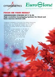
Milestones in Chromosome preparation-DEF X WEB - Euroclone
Milestones in Chromosome preparation-DEF X WEB - Euroclone
Milestones in Chromosome preparation-DEF X WEB - Euroclone

Create successful ePaper yourself
Turn your PDF publications into a flip-book with our unique Google optimized e-Paper software.
<strong>Milestones</strong> <strong>in</strong> <strong>Chromosome</strong> Preparation• 1955.Until the mid 50s’, it was commonly thought that the number of human chromosomeswas 48.The 22nd of December 1955, a Ch<strong>in</strong>ese scientist called Tjio, occasionally hosted by theLaboratory of Genetics at the University of Lund (Denmark) directed by ProfessorLevan, was observ<strong>in</strong>g “cultures of human embryonic lung fibroblasts, grown <strong>in</strong> bov<strong>in</strong>eamniotic fluid” , that were show<strong>in</strong>g an high number of cells <strong>in</strong> mitosis. <strong>Chromosome</strong><strong>preparation</strong>s were made by Tjio accord<strong>in</strong>g to the protocol suggested by Hsu (anAmerican scientist work<strong>in</strong>g at the University of Galveston,Texas - USA), slightlymodified, i.e. after the treatment with colchic<strong>in</strong>e (12-20 hours) to stop the cell cycle,the cells were exposed to hypotonic solution but reduc<strong>in</strong>g the total time to only 1-2m<strong>in</strong>utes. He then proceeded to ”fixation <strong>in</strong> 60% acetic acid twice exchanged <strong>in</strong> order towash out the salts left from the culture medium and from the hypotonic solution…”.F<strong>in</strong>ally he applied the “squash<strong>in</strong>g” technique, followed by orce<strong>in</strong> sta<strong>in</strong><strong>in</strong>g.To his own big surprise, he could observe chromosomes much better spread and biggercompared to the ones obta<strong>in</strong>ed by Hsu (see figures 1 and 2).Figure 1: Typical metaphase obta<strong>in</strong>ed by HsuFigure 2: Typical metaphase obta<strong>in</strong>ed by TjioOn top of that, when Tjio and his colleagues carefully counted the chromosomes, <strong>in</strong> 261metaphases out of 265 the number was 46 and not 48 as universally taught andclaimed by Hsu himself.This discovery, considered to represent the start<strong>in</strong>g po<strong>in</strong>t of the human cytogenetics,was the subject of a scientific paper published <strong>in</strong> January 1956. (1)Although absolutely dictated by casualty, the <strong>in</strong>troduction of acetic acid <strong>in</strong> aqueoussolution was the pr<strong>in</strong>cipal reason of those unexpected results, and, as a matter of fact,s<strong>in</strong>ce then the acetic acid will never leave the stage.Accord<strong>in</strong>g to the recent scientific f<strong>in</strong>d<strong>in</strong>gs, the acetic acid <strong>in</strong> aqueous environment is theessential condition to obta<strong>in</strong> those phenomena of spread<strong>in</strong>g and swell<strong>in</strong>g ofchromosomes, which are fundamental for the chromosome analysis.• 1959.Jerome Lejune et al. noticed the presence of a small extra-chromosome <strong>in</strong> all mitosisobta<strong>in</strong>ed from cultures of fibroblasts belong<strong>in</strong>g to n<strong>in</strong>e <strong>in</strong>dividuals affected by Down’ssyndrome (trisomy of chromosome 21) (2) .
In the same year, Patricia Jacobs observed the presence of an extra-X chromosome <strong>in</strong>male patients suffer<strong>in</strong>g from the Kl<strong>in</strong>efelter’s syndrome (47, XXY); cultures, <strong>in</strong> thiscase, were set up from bone marrow cells obta<strong>in</strong>ed by sternal puncture. (3)Based on these results, the researchers became more and more oriented towards thecl<strong>in</strong>ical application of Cytogenetics.However, the techniques used did not become popular due to both the nature ofsamples used (solid tissues) and the high variability of the quality of chromosomesobta<strong>in</strong>ed.• 1960 - 1970While attempt<strong>in</strong>g to f<strong>in</strong>d cellular substrates that might help reduc<strong>in</strong>g the variability ofresults, and at the same time mak<strong>in</strong>g easier the access to the samples, Moorheadstarted to use PHA (phytohaemagglut<strong>in</strong><strong>in</strong>) stimulated lymphocytes. (4)A significant improvement <strong>in</strong>troduced by Moorhead <strong>in</strong> the chromosome <strong>preparation</strong>sprocedure was a new k<strong>in</strong>d of fixative, now formulated as a mixture of methanol andacetic acid (3:1). Thanks to this new formulation, (along with the air dry<strong>in</strong>g of theslides, which can also be quickly passed on a flame), it was possible to replace theaqueous acetic acid and the squash<strong>in</strong>g technique previously used <strong>in</strong> the f<strong>in</strong>al step of thechromosome <strong>preparation</strong>s.These improvements proved to be so significant that this technique is still usednowadays <strong>in</strong> cytogenetic laboratories.In these years Caspersson (QFQ) (5) , and Seabright (GTG) (6) <strong>in</strong>troduced the band<strong>in</strong>gtechnique, allow<strong>in</strong>g a real analysis of the chromosome structure as well as anunambiguous pair<strong>in</strong>g of chromosomes’ couples (see figures 3-4).Figure 3: Example of QFQ band<strong>in</strong>gFigure 4: Example of GTC band<strong>in</strong>gThese works encouraged the development of other sta<strong>in</strong><strong>in</strong>g techniques, such as RBA,Da-DAPI, Ag-NOR.Yunis et al. then designed the high resolution band<strong>in</strong>g technique (7) , made possible bycell cycle synchronizationConcern<strong>in</strong>g this, it is particularly noticeable the paper of Webber and Garson“Fluorodeoxyurid<strong>in</strong>e Synchronization of Bone Marrow Cultures”, 1983) (8) . This newsynchronization technique was very efficient and simple, to the extent that manylaboratories started to use it rout<strong>in</strong>ely for lymphocyte cultures, too (see figure 5-6).Figure 5-6: Metaphase obta<strong>in</strong>ed from synchronized culture of lymphocytes
Later on (1987), Gibas will adopt this approach to synchronize cytotrophoblast cellsfrom CVS (Chorionic Villi Samples)( see figure 7).Figure 7: Examples of Metaphase obta<strong>in</strong>ed from CVS us<strong>in</strong>g FdU synchronization method.• 1960 – 1970At the end of the 60’ it was discovered that cells of fetal orig<strong>in</strong>, that can be found <strong>in</strong>amniotic fluid samples, showed the ability to form clones produc<strong>in</strong>g a number of cells <strong>in</strong>mitosis (9) .The steps of the chromosome <strong>preparation</strong> (colchic<strong>in</strong>e, hypotonic treatment, fixation <strong>in</strong>methanol-acetic acid 3:1, air dry<strong>in</strong>g, differential sta<strong>in</strong><strong>in</strong>g) were essentially the same asthose rout<strong>in</strong>ely embraced for the chromosome <strong>preparation</strong>s from PHA stimulatedlymphocytes.This was <strong>in</strong>deed the start of the pre-natal diagnosis.• 1980At the beg<strong>in</strong>n<strong>in</strong>g of the decade a new semisynthetic culture medium formulated byChang became available: this medium enabled the laboratories to reduce considerablythe time of culture (from 20-30 to 8-12 days) and to significantly <strong>in</strong>crease the numberof clones. (10)• 1983On February 3 rd <strong>in</strong> the Cytogenetic Laboratory of the Ist Cl<strong>in</strong>ica Ostetrico-G<strong>in</strong>ecologica“Mangiagalli” (University of Milan), a trisomy 21 was diagnosed from cells <strong>in</strong>spontaneous mitosis obta<strong>in</strong>ed from a sample of Chorionic Villi ( CVS) taken dur<strong>in</strong>g thefirst trimester of pregnancy (11) . S<strong>in</strong>ce then, a new material such as the Chorionic Villi(allow<strong>in</strong>g early diagnosis, i.e. dur<strong>in</strong>g the first trimester of pregnancy) was madeavailable.Moreover a new, or rather a different technique to obta<strong>in</strong> analyzable chromosomes, was<strong>in</strong>troduced. Interest<strong>in</strong>gly, the cytotrophoblast cells <strong>in</strong> spontaneous mitosis wereobta<strong>in</strong>ed us<strong>in</strong>g 60-70% aqueous acetic acid (12) .Although this “direct technique” allowed a quicker diagnosis, yet the quality of thechromosomes was not satisfactory.This is the reason why long term cultures of mesenchymal cells took off, as they madepossible the chromosome band<strong>in</strong>g with a quality level comparable to the one achievedwith cultures of cells from amniotic fluid. (13) .As mentioned before, was only <strong>in</strong> 1987 that Gibas et al. succeeded to synchronize thecytotrophoblast cells, hence he could obta<strong>in</strong> a good quality band<strong>in</strong>g with the directtechnique, too (“A simple technique for obta<strong>in</strong><strong>in</strong>g high chromosome <strong>preparation</strong> fromchorionic villus samples us<strong>in</strong>g FdU synchronization”) (14) .• 1985In a “Letter to the Editor”, published <strong>in</strong> “Prenatal Diagnosis”, Lundsteen (15) reported theresults of his experiments deal<strong>in</strong>g with the role of temperature and relative humidity. Itwas clear from his f<strong>in</strong>d<strong>in</strong>gs that these two parameters do have a significant impact on
the chromosome <strong>preparation</strong>s. From a technical standpo<strong>in</strong>t, this translated <strong>in</strong> a veryimportant concept, namely that by properly controll<strong>in</strong>g temperature and relativehumidity, one can expect that the quality of chromosome <strong>preparation</strong>s becomesconsistently good – regardless of seasonal changes and environmental factors.• 1988At the XXI Symposium on Cytogenetics, held <strong>in</strong> Praga <strong>in</strong> August of that year, asequence of pictures (shot dur<strong>in</strong>g the f<strong>in</strong>al evaporation of the fixative) was presentedby G. Terzoli. For the first time it was shown <strong>in</strong> a very evident way that the spread<strong>in</strong>g ofchromosomes does occur dur<strong>in</strong>g that phase; at the same time, the chromosomesdimensions expand <strong>in</strong> a considerable and unexpected way (see figure 8).Figure 8: Sequence show<strong>in</strong>g how the spread<strong>in</strong>g of chromosome occurs dur<strong>in</strong>g the f<strong>in</strong>al evaporation of fixative(aqueous acetic acid).• 1990From the end of this decade, the image analysis computerized systems started to bemore and more widely used <strong>in</strong> the cytogenetics labs. This technical advancesubstantially contributed to improve the resolution of chromosome band<strong>in</strong>g, offer<strong>in</strong>g aswell the possibility to perform caryograms quicker and easier, so that the wholechromosome analysis became much more reliable.In an article published <strong>in</strong> 1996 Spurbeck et al. (16) demonstrated that dur<strong>in</strong>g the f<strong>in</strong>alevaporation of the fixative the chromosome spread<strong>in</strong>g <strong>in</strong>creases as the environmentalrelative humidity <strong>in</strong>creases.• 2002“Demystify<strong>in</strong>g chromosomes <strong>preparation</strong>…”: this work, written by Claussen (17) ,although not so well known, resulted <strong>in</strong> a real milestone among the studies regard<strong>in</strong>gthe chromosome <strong>preparation</strong> steps.This article, published <strong>in</strong> “Cytogenetic and Genome Research”, is so important thatdeserves a summary of the key po<strong>in</strong>ts:1) Dur<strong>in</strong>g the hypotonic treatment the chromosomes move from the center of mitoticcells (which are spherically shaped) towards the cell’s membrane2) When the fixative reaches the cells, the outer and <strong>in</strong>ner membranes are disrupted3) In presence of water and acetic acid, both the cytoplasmatic and the chromosomalprote<strong>in</strong>s swell, clearly and <strong>in</strong>dependently4) When the f<strong>in</strong>al evaporation of fixative occurs <strong>in</strong> an environment without water, theswell<strong>in</strong>g of the cytoplasmatic prote<strong>in</strong>s is <strong>in</strong>hibited and the same happens to the swell<strong>in</strong>gof the chromosomes, so that there is no <strong>in</strong>crease <strong>in</strong> their dimensions5) When the ambient humidity <strong>in</strong>creases, the spread<strong>in</strong>g and the sizes of thechromosomes <strong>in</strong>crease, too.The conclusion was that the swell<strong>in</strong>g of cytoplasmatic and chromosomal prote<strong>in</strong>s <strong>in</strong>mitotic cells dur<strong>in</strong>g evaporation of the fixative is, <strong>in</strong> reality, ‘based on an <strong>in</strong>teraction
etween acetic acid, water and cell prote<strong>in</strong>s’. Def<strong>in</strong>itely a giant move towards the endof “magics” <strong>in</strong> cytogenetic laboratories, and a great contribution to the beg<strong>in</strong>n<strong>in</strong>g of“science”!• 2006On occasion of the IX Congress of the Italian Society of Human Genetics (SIGU), held <strong>in</strong>Venezia-Lido 8-10 November, follow<strong>in</strong>g the speech of Dr. Claussen deal<strong>in</strong>g with thetopic mentioned above, G. Terzoli showed and commented a short video filmed at themicroscope.For the first time the cytogenetists attend<strong>in</strong>g this presentation could actually see whathappens to the mitosis dur<strong>in</strong>g each step of the chromosome <strong>preparation</strong>. It was f<strong>in</strong>allyclear <strong>in</strong> front of them the behavior of cells and chromosomes dur<strong>in</strong>g the hypotonictreatment, <strong>in</strong> the fixative solution and (mostly important) dur<strong>in</strong>g the f<strong>in</strong>al evaporationof the fixative <strong>in</strong> different conditions of relative humidity.Dur<strong>in</strong>g the same presentation, it was proposed that some mechanism <strong>in</strong>volv<strong>in</strong>g prote<strong>in</strong>hydration could be the cause of the swell<strong>in</strong>g of both cytoplasmatic and chromosomeprote<strong>in</strong>s. Based on this hypothesis, the hydration produces the swell<strong>in</strong>g of thecytoplasmatic prote<strong>in</strong>s, that <strong>in</strong> turn gives raise to the chromosome spread<strong>in</strong>g. In thecase of the chromosomes, the same hydration process causes the swell<strong>in</strong>g of theprote<strong>in</strong>s that <strong>in</strong>creases the three-dimension size of the chromosomes; consequently,more or less condensed areas along the chromosome itself (that after sta<strong>in</strong><strong>in</strong>g will beseen as bands) are orig<strong>in</strong>ated.BIBLIOGRAPHY1) Tjio, J.H., Levan, A.: The chromosome number of man. Hereditas 42, 1-6 (1956).2) Lejeune J., Gautier M., Turp<strong>in</strong> R.: Etude des chromosomes somatiques de neuf enfantsmongoliens. C.R. Acad. Sci. Paris 248, 1721-1722, (1959).3) Jacobs P.A., Strong J.A.: A case of human <strong>in</strong>tersexuality hav<strong>in</strong>g a possible XXY sexdeterm<strong>in</strong><strong>in</strong>g mechanism. Nature 183, 302-303, (1959).4) Moorhead P.S., Nowell P.C., Mellman W.J. et al.: <strong>Chromosome</strong> <strong>preparation</strong>s of leucocytescultured from human peripheral blood. Exp. Cell. Res. 20, 613-616, (1960).5) Caspersson T., Zech L., Johansson C.: Differential band<strong>in</strong>g of alkylat<strong>in</strong>g fluorochromes <strong>in</strong>human chromosomes. Exp. Cell. Res. 60(3), 315-319, (1970).6) Seabright M.: A rapid band<strong>in</strong>g technique for human chromosomes. Lancet II:971-972(1971).7) Yunis et al.: High resolution of human chromosomes. Science (1976) 191: 1268-1270.8) Webber L.M. and Garson O.M.: Fluoredeoxyurid<strong>in</strong>e synchronization of bone marrowcultures. Cancer and Cytogenetics (1983) 8: 123-132.9) Genetic disorders and the fetus. Ed. A. Milunsky 3 rd edition (1992).