Cervical radiculopathy - enotes - Q-Notes for Adult Medicine
Cervical radiculopathy - enotes - Q-Notes for Adult Medicine
Cervical radiculopathy - enotes - Q-Notes for Adult Medicine

Create successful ePaper yourself
Turn your PDF publications into a flip-book with our unique Google optimized e-Paper software.
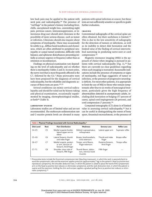
The new england journal of medicinelow back pain may be applied to the patient withneck pain and <strong>radiculopathy</strong>. 24 The presence of“red flags” in the patient’s history (including fever,chills, unexplained weight loss, unremitting nightpain, previous cancer, immunosuppression, or intravenousdrug use) should alert clinicians to thepossibility of more serious disease, such as tumoror infection. Clinicians should also inquire aboutsymptoms of myelopathy. These may occasionallybe subtle (e.g., diffuse hand numbness and clumsiness,which are often attributed to peripheral neuropathyor carpal tunnel syndrome; difficulty withbalance; and sphincter disturbances presenting initiallyas urinary urgency or frequency rather than asretention or incontinence).Findings on physical examination vary dependingon the level of <strong>radiculopathy</strong> and on whetherthere is myelopathy (Tables 1 and 2). In most series,the nerve root that is most frequently affected is theC7, followed by the C6. 2 Many provocative testshave been proposed <strong>for</strong> the diagnosis of cervical<strong>radiculopathy</strong>, but the reliability and diagnostic accuracyof these tests are poor. 19,25Several conditions can mimic cervical <strong>radiculopathy</strong>and should be ruled out by history takingand physical examination, occasionally supplementedby imaging, electrophysiological studies,or both 26 (Table 3).laboratory studiesLaboratory studies are of limited value and are notrecommended. The erythrocyte sedimentation rateand C-reactive protein levels are elevated in manypatients with spinal infection or cancer, but thesetests are not sufficiently sensitive or specific to guidefurther evaluation.imagingConventional radiographs of the cervical spine areoften obtained, but their usefulness is limited. 31This is due to the low sensitivity of radiography<strong>for</strong> the detection of tumors or infections, as wellas its inability to detect disk herniation and thelimited value of the finding of cervical intervertebralnarrowing in predicting nerve-root or cordcompression. 32Magnetic resonance imaging (MRI) is the approachof choice when imaging is pursued in patientswith cervical <strong>radiculopathy</strong> (Fig. 1), 33 butthere are currently no clear guidelines regardingwhen such imaging is warranted. Reasonable indicationsinclude the presence of symptoms or signsof myelopathy, red flags suggestive of tumor orinfection, or the presence of progressive neurologicdeficits. For most other patients, it is appropriateto limit the use of MRI to those who remain symptomaticafter four to six weeks of nonsurgical treatment,particularly given the high frequency ofabnormalities detected in asymptomatic adults, includingdisk herniation or bulging (57 percent ofcases), spinal cord impingement (26 percent), andcord compression (7 percent). 34Computed tomography (CT) alone is of limitedvalue in assessing cervical <strong>radiculopathy</strong>, 35 but itcan be useful in distinguishing the extent of bonyspurs, <strong>for</strong>aminal encroachment, or the presence ofTable 1. Physical Findings Associated with <strong>Cervical</strong> Radiculopathy.*Disk Level Root Pain Distribution Weakness Sensory Loss Reflex LossC4–C5 C5 Medial scapular border, Deltoid, supraspinatus, Lateral upper arm Supinator reflexlateral upper arm toelbowinfraspinatusC5–C6 C6 Lateral <strong>for</strong>earm, thumband index fingerBiceps, brachioradialis,wrist extensorsThumb and indexfingerBiceps reflexC6–C7 C7 Medial scapula, posteriorarm, dorsum of<strong>for</strong>earm, third fingerC7–T1 C8 Shoulder, ulnar side of<strong>for</strong>earm, fifth fingerTriceps, wrist flexors,finger extensorsThumb flexors, abductors,intrinsic handmusclesPosterior <strong>for</strong>earm,third fingerTriceps reflexFifth finger —* Provocative tests include the <strong>for</strong>aminal compression test (Spurling maneuver), in which the neck is passively bent towardthe symptomatic side and the examiner applies pressure (approximately 7 kg) to the patient’s head (a positive testreproduces symptoms); the shoulder abduction test, in which the patient is asked to place the hand of the symptomaticarm on the head (a positive test reduces or eliminates symptoms); and the neck distraction test, in which the patient issupine and the examiner, holding the chin and occiput, applies a gradual pulling <strong>for</strong>ce (a positive test reduces or eliminatessymptoms). 25394n engl j med 353;4 www.nejm.org july 28, 2005Downloaded from www.nejm.org at KAISER PERMANENTE on July 28, 2005 .Copyright © 2005 Massachusetts Medical Society. All rights reserved.